
Weighing proteins and seeing their shapes
Nobel Prize in Chemistry, 2002: John B. Fenn, Koichi Tanaka, and Kurt Wüthrich
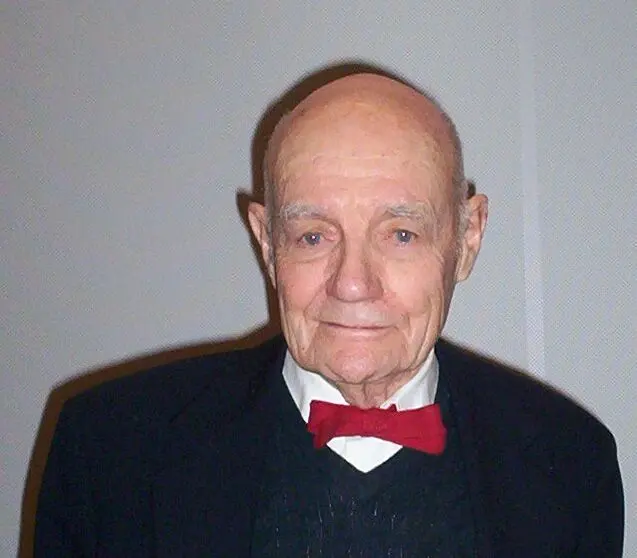
John B. Fenn
1917–2010, born in New York City, United States
His university made him retire at 70. He kept working, and the prize came for papers he wrote after that.
Photo: Wikimedia Commons, Public domain
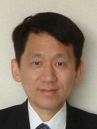
Koichi Tanaka
born 1959 in Toyama, Japan
A company engineer with a first degree in electrical engineering and no PhD. He was 25 when a mistake he refused to waste gave him his result.
Photo: Japan Academy, CC BY 4.0
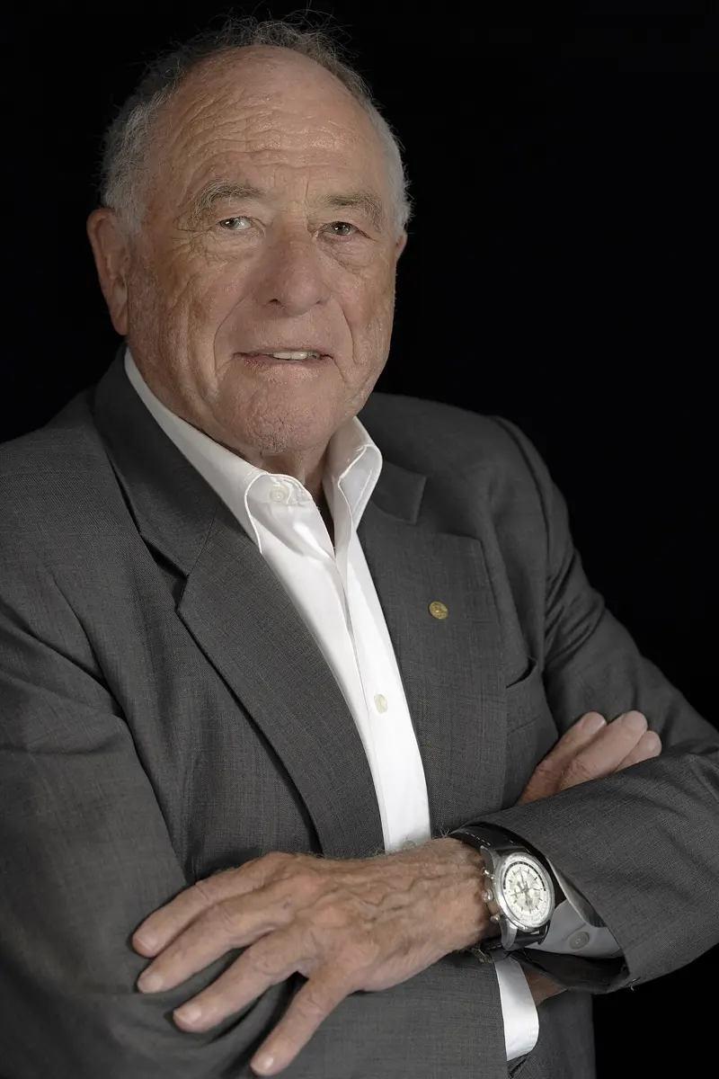
Kurt Wüthrich
born 1938 in Aarberg, Switzerland
He trained as a sports teacher and a ski instructor as well as a chemist, and played competitive football past the age of 50.
Photo: Christopher Michel, CC BY-SA 4.0
You go to a hospital with a fever that won’t go away. The doctor sends a sample of your blood to the laboratory, and germs grow from it overnight. Which germ is it? In many hospitals today, a technician puts a speck of it into a machine, and in a few minutes the machine names it. The machine weighs the germ’s proteins and checks the result against a library of thousands of germs.
Weighing a single protein sounds impossible. One molecule of hemoglobin, the protein that carries oxygen in your blood, has a mass of about 10⁻¹⁹ g: a ten-millionth of a millionth of a millionth of a gram. The 2002 Nobel Prize in Chemistry went to two people who found ways to weigh proteins, John Fenn and Koichi Tanaka, and to a third, Kurt Wüthrich, who found a way to see their shapes.
The problem: the machines of life
Every cell in your body is full of proteins (proteins do the work). Some carry oxygen, some digest your dal bhat, some fight germs, some make your muscles pull. A protein is a long chain of smaller molecules called amino acids, often hundreds of them, folded up into a tight ball like a tangle of wool. What a protein does depends on two things: which protein it is, and its shape.
Scientists wanted to answer two questions about any protein: “What is it, and how much is there?” and “What does it look like?”
For small molecules there was already a good tool for the first question. A mass spectrometer weighs molecules, and from the weight tells you what they are. J. J. Thomson, who discovered the electron, made the first measurements with one in 1912. By the 1970s mass spectrometers were used every day for small molecules: in drug tests, in checking food, in measuring pollution.
For the second question there were X-rays. Since 1957, scientists had worked out the shapes of proteins by shining X-rays through crystals of them. But many proteins will not form crystals at all. And a protein in a crystal is packed tightly among its neighbors, not floating in water as it does in a cell.
Why did scientists want a way to find a protein’s shape?
Pick an answer to see why.
How a mass spectrometer weighs a molecule
A molecule is far too small to put on a balance. A mass spectrometer weighs it in a cleverer way.
First the molecule must become an ion: a particle with an electric charge Grade 9: ions. It must also be floating freely, as a gas, not stuck to other molecules. Then an electric field gives every ion the same push, and they fly down an empty tube with all the air pumped out, a vacuum.
Think of giving the same hard kick to a table-tennis ball and to a football. The light ball flies off fast; the heavy one moves slowly. In the same way, light ions race down the tube and heavy ones lag behind. A detector at the far end notes the moment each one arrives, and from its time of flight a computer works out its mass. (Strictly, it works out mass divided by charge: an ion with two charges gets twice the push.)
Why it was hard
Making an elephant fly
For small molecules, the first step is easy: heat the sample until it turns to gas, then knock an electron off each molecule with a beam of electrons. But do that to a protein and it falls to pieces. Heat cooks a protein: think of what happens to a clear egg white in a hot pan. The long chain breaks, and you end up weighing the broken bits instead of the protein.
Proteins are also enormous. Hemoglobin has a molecular mass Grade 9: molecular mass of about 64,000, compared with 18 for water. Getting a whole protein to float free in a vacuum, in one piece and with a charge, was like trying to make an elephant fly. John Fenn later called his Nobel lecture “Electrospray wings for molecular elephants”.
Too many signals
For seeing shapes, chemists had another tool, NMR (nuclear magnetic resonance). It worked well for small molecules, but for a protein it gave so many signals, all crowded together, that nobody could tell which signal came from which atom. The Nobel Prize’s own explanation compares the result to a lawn of grass seen from the side: thousands of blades, all looking alike.
Why couldn’t chemists simply heat a protein to turn it into a gas, as they did with small molecules?
Pick an answer to see why.
What they did, step by step
Fenn: spray it
John Fenn was a chemist at Yale University in the United States who had spent years studying beams of molecules flying through a vacuum. Around 1984 he and a colleague, Masamichi Yamashita, took up an old idea: spraying a liquid from a needle at a high voltage.
- 1984, the spray. They pushed a solution through a very thin metal needle held at several thousand volts. The liquid came out as a fine mist of tiny droplets, each one electrically charged, a little like the mist from a perfume spray, but far finer.
- The shrinking droplets. As the water evaporates from each droplet, the droplet shrinks, but its charge stays. The charges, all of the same sign, push on each other more and more strongly until the droplet bursts into still smaller droplets. This happens again and again until only bare molecules are left, floating free, each carrying several charges. They have never been heated.
- 1987, retired. Yale had a rule that professors must retire at 70. Fenn reached it in 1987 and lost most of his laboratory space and helpers. He carried on.
- 1988, proteins. Fenn and his team published two papers. The first showed that the spray could handle large molecules. The second showed it working on whole proteins. The method is called electrospray ionization (ESI).
At first the results were confusing: one protein gave not one peak but a whole row of them. Each copy of the protein had picked up a different number of charges. That turned out to be useful: the row of peaks lets a computer work out the mass very exactly.
Tanaka: blast it with a laser
At the same time, in Kyoto, Japan, a young engineer was trying something quite different. Koichi Tanaka had joined the instrument company Shimadzu in 1983, aged 23, straight after his first degree. His team wanted to lift proteins off a surface with a short flash of laser light. The danger was the same: too much energy and the protein breaks.
Tanaka’s job was to find a matrix, a material to mix the protein into that would soak up the laser’s energy and pass on just enough of it, gently. He tried hundreds of substances, day after day, without success.
- February 1985, the mistake. Tanaka meant to use a very fine cobalt powder, but by mistake he mixed it with glycerine, a thick, sticky liquid. His grandmother had brought him up never to throw anything away. Mottainai! (“what a waste!”), he thought. He decided to dry the mixture in the vacuum of the machine, switched on the laser to speed up the drying, and kept the instrument running while he waited. A signal appeared that he had never seen before, and it came back every time he repeated the test.
- 1987, the result. Shimadzu’s team announced it at a meeting in Kyoto in May 1987, with molecules of mass above 48,000. In September, at a meeting in Takarazuka, Tanaka presented it in English for the first time, and it was published in 1988.
- The machine. Shimadzu built an instrument on it and put it on sale in 1988, but it was expensive and not yet good enough. Only one was ever sold.
Tanaka’s idea, a matrix that soaks up the laser’s energy, is the basis of today’s MALDI machines (matrix-assisted laser desorption ionization). In Germany, Michael Karas and Franz Hillenkamp had been working on the same idea with a different matrix, a solid made of small organic molecules, and in 1988 they used it to fly a protein of mass 67,000. Their kind of matrix is the one used today, and many scientists felt they deserved a share of the prize.
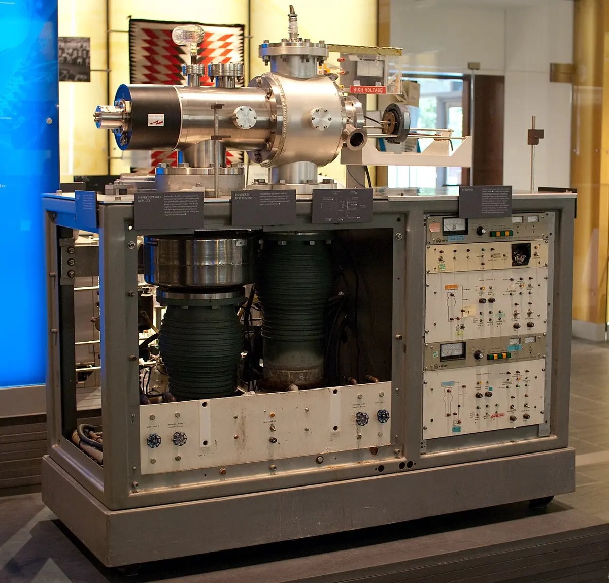
Wüthrich: listen to the hydrogen atoms
Kurt Wüthrich, at the ETH (Swiss Federal Institute of Technology) in Zürich, worked on the second question: what does a protein look like, floating in water as it does in a cell?
He used NMR. The nucleus at the center of a hydrogen atom behaves like a tiny bar magnet (the molecular theory of magnetism is a similar idea, for iron). Put a sample in a very strong magnet, and these tiny magnets line up. Then send in radio waves Grade 9: radio waves. Each hydrogen nucleus soaks up radio waves of one exact frequency and sends back an answer. That frequency depends a little on the atoms around it, so different hydrogens give slightly different signals. Two physicists, Felix Bloch and Edward Purcell, discovered this in the 1940s, and Richard Ernst made NMR far more sensitive from 1966 onwards.
A protein can have thousands of hydrogen atoms, so it gives thousands of signals. In the early 1980s Wüthrich solved the puzzle in two steps.
- Naming every signal. He found a step-by-step way to match each signal to its hydrogen atom, working along the protein chain one amino acid at a time. This is called sequential assignment, and every NMR study of a protein still starts with it.
- Measuring who is close to whom. Two hydrogen atoms that are very close in space, less than about half a nanometer (0.5 nm) apart, disturb each other’s signals in a way NMR can detect. This can happen even when the two atoms are on amino acids far apart along the chain, because the chain has folded back on itself. Wüthrich measured hundreds of these close pairs.
- 1985, the first shape. With mathematicians, he used a computer to find the one fold that fits all the close pairs at once. In 1985 his group published the first complete shape of a protein worked out this way.
The Nobel committee compared it to a house: if you know enough measurements between the parts of a house, you can draw the whole house, even without ever seeing it.
How it works now
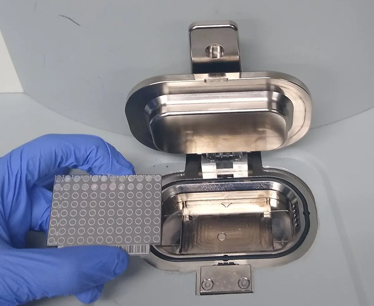
Mass spectrometers that use electrospray or MALDI are now in laboratories all over the world. They can find which of thousands of proteins are in a drop of blood, and how much of each. Joined to other machines that sort a mixture first, they can test hundreds of possible medicines in a day.
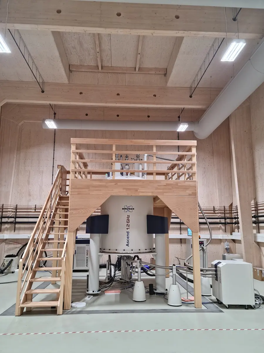
NMR of proteins uses some of the strongest magnets on Earth. The most powerful are cooled with liquid helium to about −269 °C and are hundreds of thousands of times stronger than the Earth’s magnetic field. The protein sits in a thin glass tube of water, as it would in a cell. Of all the protein shapes scientists have published, about one in twenty was worked out with NMR (as of 2026); in 2002 it was 15–20%. Most of the rest come from X-rays, and more and more from electron microscopes.
NMR can show something X-rays can’t: how a protein moves. Wüthrich used it to study the prion protein, which can cause mad cow disease. He found that half of the chain is folded into a firm shape, while the other half flops about freely in the water. In a crystal, that floppy half would hardly show up at all.
A protein will not form crystals. Which of the three methods in this prize could still show its shape?
Pick an answer to see why.
In your life today
- In hospitals. Many hospital laboratories now name the germ in a patient’s sample in minutes with a MALDI machine, instead of a day or more of tests. In many countries, a drop of blood from every newborn baby’s heel is checked with a mass spectrometer for dozens of rare diseases that can be treated if caught early.
- In sport. The tests that catch athletes using banned drugs are done with mass spectrometers.
- In your food. Food is checked for pesticides and for harmful substances that can form in cooking, such as acrylamide in fried and baked foods.
- In new medicines. Drug companies use NMR to see whether a small molecule sticks to a protein they want to block, and mass spectrometers to check what they have made.
- MRI scanners. The hospital MRI scanner, found in many hospitals in Nepal, uses the same effect as NMR: the hydrogen nuclei in your body, in a strong magnet, answering radio waves.
What came next
Since 2002, the tools of this prize have become the everyday work of biology. Scientists can now list most of the thousands of proteins in a cell and see how their amounts change in an illness. Electron microscopes became sharp enough to see proteins too (freezing molecules to take their picture), and in 2020 a computer program, AlphaFold, learned to predict a protein’s shape from its chain of amino acids (the 2024 prize).
But many proteins never settle into one shape. Like the prion’s floppy half, parts of them keep moving, and they do their jobs that way. NMR is one of the few ways to watch them. How these restless proteins work, and how often they go wrong in diseases such as Alzheimer’s, is still an open question.
In your science books
- Grade 8: atomic weight and molecular weight and the molecular theory of magnetism.
- Grade 9: ions, molecular mass and radio waves.
- Grade 10: the electromagnet.
- Going further: molecules have shapes, food is made of big molecules, proteins do the work, counting atoms by weighing and evaporation.
Learn more
- The Nobel Prize’s own explanation: Revolutionary analytical methods for biomolecules (nobelprize.org).
- The Nobel lectures: Fenn, Tanaka and Wüthrich.
- Koichi Tanaka’s own story of his life and his “monumental blunder”: Tanaka, biographical.
The official reason
The prize was given “for their development of soft desorption ionisation methods for mass spectrometric analyses of biological macromolecules.”
The prize was given “for his development of nuclear magnetic resonance spectroscopy for determining the three-dimensional structure of biological macromolecules in solution.”