
The kiss of death for proteins
Nobel Prize in Chemistry, 2004: Aaron Ciechanover, Avram Hershko, and Irwin Rose
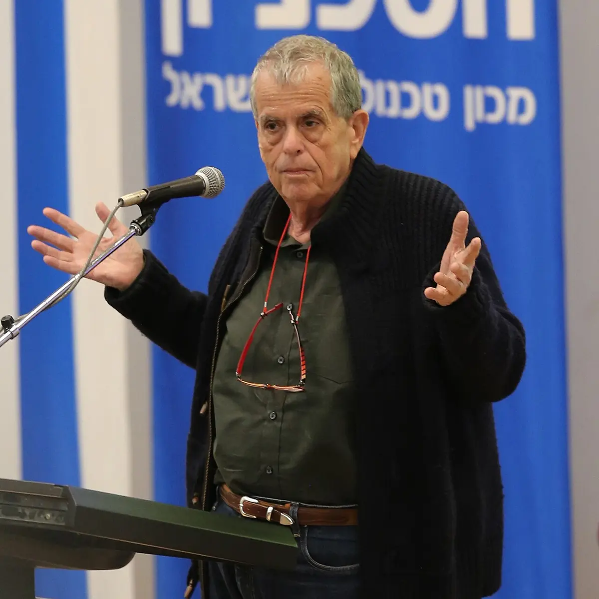
Aaron Ciechanover
born 1947 in Haifa (now in Israel)
Trained as a doctor and served as a navy doctor, then chose the laboratory. He was Hershko’s graduate student when they made the discovery.
Photo: DanielTahar, CC BY-SA 4.0
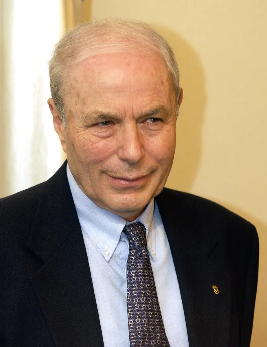
Avram Hershko
born 1937 in Hungary
As a boy of six he survived the Second World War in a forced-labor camp in Austria with his mother and brother. He later became a doctor and a biochemist in Israel.
Photo: Amos Ben Gershom (Government Press Office, Israel), CC BY-SA 3.0
-
Irwin Rose
1926–2015, born in the USA
An expert on how enzymes work, at a cancer research center in Philadelphia. He welcomed Hershko and Ciechanover to his laboratory, summer after summer.
Crack an egg into a hot pan. The clear, runny part turns white and solid in seconds. That white part is mostly protein, and heat has changed its shape for good.
Your body is built and run by proteins. Your muscles, your hair, the hemoglobin that carries oxygen in your blood and the enzymes that digest your food are all proteins. A cell makes them all the time. But a cell must also get rid of proteins: old ones, broken ones, and ones whose job is done. How does a cell decide which proteins to destroy, and how does it destroy them?
In 2004 the Nobel Prize in Chemistry went to three scientists who found the answer: Aaron Ciechanover, Avram Hershko and Irwin Rose.
The problem: throwing away needs energy?
Breaking proteins in your gut
A protein is a long chain of small building blocks called amino acids, folded into a shape (Proteins do the work). There are 20 kinds of amino acid, and the order of them in the chain decides what the protein does. The chains are made in the cell by ribosomes Grade 8: Ribosomes.
When you eat dal, eggs or meat, enzymes in your stomach and small intestine cut the protein chains back into single amino acids (Taking food apart). One of these enzymes, trypsin, works in the small intestine. Cutting a chain needs no energy: it is a bit like letting a stretched spring go.
Cells have their own “stomach” too: small bags of digestive enzymes called lysosomes Grade 8: Lysosomes. They break down things the cell has swallowed from outside, and they need no extra energy either.
A strange result
In the 1950s, scientists found something odd. When liver cells broke down their own proteins, they used energy, from the cell’s energy molecule, ATP (ATP). Stop the ATP, and the breakdown stopped too.
That made no sense. Why should a cell pay to throw something away? Imagine a shop that charges you money to take away your rubbish, when you could simply drop it in a pit for free. For more than twenty years, nobody could explain it. Most scientists thought that making proteins was the exciting question; breaking them down seemed dull, and few people worked on it.
Why was it a puzzle that cells use ATP to break down their own proteins?
Pick an answer to see why.
Why it was hard
A living cell is crowded with tens of thousands of different chemicals, all reacting at once. To find out what does a particular job, a biochemist breaks cells open and separates their contents, part by part, until a job can be done in a test tube with only the parts it needs.
But a cell’s lysosomes break proteins down too, and they would hide anything else that was going on. Hershko and Ciechanover needed cells without lysosomes.
They found them in immature red blood cells Grade 10: Red blood cells. As a red blood cell matures, it destroys most of its insides, lysosomes included, so it must have another powerful way of breaking down proteins. In 1977 Alfred Goldberg and his team in the USA had made a juice from such cells, called an extract, that broke down proteins in a test tube, using ATP. That was the starting point.
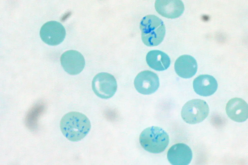
There was one more problem. The extract was full of hemoglobin, which got in the way of every attempt to separate it.
What they did, step by step
Hershko and Ciechanover in Haifa
Avram Hershko was a biochemist at the Technion, a university in Haifa, Israel. In 1976 Aaron Ciechanover, who had trained as a doctor, joined him as a graduate student.
- 1977, two halves. To get rid of the hemoglobin, they passed the extract through a column that holds on to some proteins and lets others through. This split the extract into two parts, called fraction I and fraction II. Neither part could break down proteins on its own. But mixed together again, with ATP, they could. So the job needed at least two different parts working together.
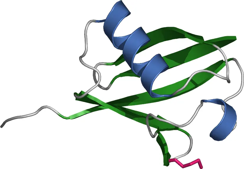
- 1977–78, the heating trick. Fraction I was mostly hemoglobin, and they could not separate the useful part from it. A colleague in the laboratory, Michael Fry, suggested something that sounded crazy: heat it. At 90 °C, the hemoglobin clumped together, Ciechanover later wrote, “like mud”, just as an egg white sets in a pan. But the useful part survived the heat. It was a small, very tough protein. They called it APF-1 and reported it in 1978.
With Rose in Philadelphia
Irwin Rose was an expert on how enzymes work, at the Fox Chase Cancer Center in Philadelphia, USA. Hershko spent a year there from 1977, and from then on he and Ciechanover returned to Rose’s laboratory again and again in their summer breaks. Much of the work was done there.
- 1980, the label. In two papers, the three showed what APF-1 does. It binds firmly to other proteins, with a strong chemical bond. And many APF-1 molecules can stick to the same protein, one after another, in a chain. That same year, scientists in Rose’s laboratory and elsewhere found that APF-1 was a protein already known: ubiquitin.
- 1981–83, three enzymes. They found the three enzymes that fix the label on, which they named E1, E2 and E3. E1 uses ATP to pick up ubiquitin; E2 carries it; E3 holds the protein that is to be destroyed and makes sure the label goes onto the right one.
Ubiquitin had been discovered in 1975, but nobody knew what it was for. Its name comes from the Latin ubique, “everywhere”, because it is found in the cells of animals, plants and fungi, everywhere except bacteria. It is made of only 76 amino acids, and it has hardly changed in hundreds of millions of years: your ubiquitin and a yeast cell’s differ in only 3 of the 76.
What was special about the protein APF-1 that Hershko and Ciechanover found in fraction I?
Pick an answer to see why.
How it works now
The label and the shredder
Here is the whole system, as it is understood today.
The energy pays for the choosing. A human cell has only one or two kinds of E1 and a few tens of E2, but several hundred different E3s. Each E3 recognizes only a few particular proteins. So the cell can destroy exactly the protein it wants, at exactly the moment it wants, and leave everything else alone. That was the answer to the puzzle of the 1950s.
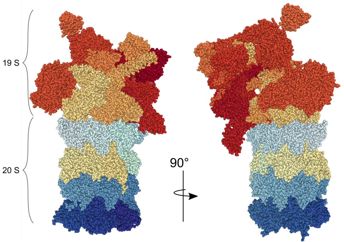
The labeled protein is destroyed in a proteasome, the cell’s shredder. It is a hollow barrel with a lid at one end or both. The lid recognizes the ubiquitin chain, removes the labels so they can be reused, unfolds the protein and feeds it into the barrel, where it is cut into short pieces. The pieces are broken down into amino acids, which the cell uses again to build new proteins. A human cell has about 30,000 proteasomes.
More than rubbish collection
At first the scientists thought this was only a way to get rid of faulty proteins. It is much more. By destroying a protein at the right moment, the cell can switch a job off, like a switch on a wall.
- Cell division. When a cell divides into two Grade 10: Mitosis, its chromosomes are copied and then pulled apart. At the right moment, an E3 labels the protein that holds each pair of copies together, and the copies can separate. A mistake here can give a cell the wrong number of chromosomes.
- Guarding DNA. A protein called p53 checks for damage to a cell’s DNA and can stop a damaged cell from dividing. Normally p53 is labeled and destroyed almost as fast as it is made. When the DNA is damaged, the labeling stops, p53 builds up, and the cell pauses to repair itself.
- Plants. Many plants use the system to reject their own pollen, so that they are pollinated by another plant of their kind.
In your life today
- Cancer medicine. In 2003 a medicine called bortezomib was approved for multiple myeloma, a cancer of blood cells in the bone marrow. It jams the proteasome. Myeloma cells make huge amounts of protein and depend on their proteasomes more than healthy cells do, so they are harmed most. Bortezomib is now on the World Health Organization’s list of essential medicines.
- A virus that causes cancer. The HPV virus, which causes cervical cancer, makes a protein that tricks one of the cell’s own E3s into labeling p53. With its guard destroyed, the infected cell can no longer stop itself from dividing out of control (see the 2008 Nobel Prize in Medicine). Nepal now vaccinates girls against HPV.
- Inherited diseases. In cystic fibrosis, a common change in a gene gives a slightly misfolded protein. The cell’s quality control labels it and destroys it, even though it could still partly work. The disease comes partly from the cell’s own strictness.
What came next
- The whole machine, atom by atom. By 2019, scientists had pictures of the human proteasome, atom by atom, caught in the act of pulling in a labeled protein.
- Medicines that use the label. Instead of blocking the shredder, chemists are now making molecules that bring a chosen E3 together with a harmful protein, so that the cell itself labels the protein and destroys it. Several of these are being tested in patients for cancer.
- A label with many meanings. Ubiquitin chains linked in different ways turn out to carry different messages: some mean “destroy”, others mean “repair DNA” or “move to another part of the cell”. More than a thousand proteins take part in the system, and what most of them do is still being worked out.
In your science books
- Grade 8: the cell’s ribosomes, which make proteins, and lysosomes, its other way of breaking things down.
- Grade 10: red blood cells, mitosis, and RNA, which carries the instructions for making proteins.
- Going further: how a gene makes a protein, how food is taken apart and ATP, the cell’s energy molecule.
Learn more
- The Nobel Prize’s own explanation: The Nobel Prize in Chemistry 2004, popular information (nobelprize.org).
- The laureates’ Nobel lectures: Ciechanover, Hershko and Rose.
The official reason
The prize was given “for the discovery of ubiquitin-mediated protein degradation.”