
Put a spoonful of plain boiled rice in your mouth and chew it slowly, without swallowing, for a minute or two. It begins to taste a little sweet. Nobody added sugar: your saliva made it. Saliva contains amylase, a tool so small that a million of them in a row would be about as long as a grain of rice. It cuts the starch in the rice into sugar.
Amylase is a protein, and the instructions for building it are in your DNA, written in an alphabet of four letters, A, T, G and C. So are the instructions for the hemoglobin that carries oxygen in your blood, the keratin in your hair, and about 20,000 other proteins. How does a cell turn a row of letters into a working tool? And why can a single wrong letter, out of billions, make a person ill? In parts of Nepal’s Terai, where many families know the disease called sickle-cell anemia, that second question is not just for the classroom.
Proteins do the work
You know from Grade 10 that a gene is a stretch of DNA that carries the code for a character, and that the cell uses it to make a protein Grade 10: DNA. Why a protein? Because proteins do almost all the work in a living thing.
| Protein | Where | What it does |
|---|---|---|
| amylase | saliva, and juice from the pancreas | cuts starch into sugar |
| hemoglobin | red blood cells | picks up oxygen in the lungs and lets it go in the body |
| keratin | hair, nails, the outer skin | tough fibers |
| collagen | skin, bone, tendons | strong ropes that hold the body together; about a quarter to a third of all your protein |
| actin and myosin | muscle cells | slide past each other to make a muscle pull |
| insulin | made in the pancreas, carried in the blood | a hormone: tells cells to take in sugar |
| antibodies | made by white blood cells | grab germs so that they can be destroyed |
A protein that speeds up a chemical reaction, like amylase, is called an enzyme. Thousands of different enzymes run the chemistry in every cell.
The proteins a cell makes decide what kind of cell it is. A red blood cell is a bag of about 270 million hemoglobin molecules Grade 10: Red blood cells. A muscle cell is packed with actin and myosin. The cells at the root of a hair pump out keratin, about a centimeter of hair a month. Yet almost every cell in your body has the same DNA, the same genes. What makes a cell a muscle cell is which of its genes it uses. Cells of one kind work together as a tissue, and tissues as organs and systems Grade 8: Cell, tissue and organ: your body works because its specialized cells make the right proteins.
A chain that folds
A protein is a long chain of small molecules called amino acids, joined end to end like the beads of a mala. Living things use 20 kinds of amino acid. A protein chain is usually a few hundred long: one of hemoglobin’s chains has 146; the longest human protein, titin, in muscle, has over 34,000.
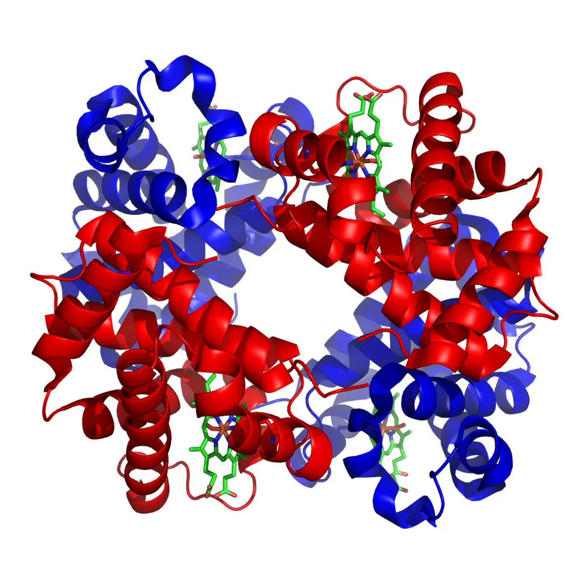
What makes one protein different from another is the order of its amino acids. Some amino acids are attracted to water and some avoid it; some carry an electric charge, positive or negative. So as soon as a chain is made, it folds up by itself into a particular shape: the parts that avoid water tuck inside, the charged parts stay outside, and opposite charges pull together. The same order always folds into the same shape. And the shape decides the job: amylase has a groove that a starch chain fits into, hemoglobin has pockets that hold oxygen, collagen chains twist together into ropes. You met the same idea with spider silk, whose repeating order of amino acids makes it strong (Inside a spider’s thread).
Two proteins are made of exactly the same number of each kind of amino acid. Will they have the same shape?
Pick an answer to see why.
Four letters, twenty amino acids: the code
So a gene must hold an order of amino acids. But DNA has only four different bases, A, T, G and C Grade 10: DNA, and there are twenty amino acids to choose from. How can four letters name twenty things?
Not one letter per amino acid: that names only 4. Two letters per amino acid gives 4 × 4 = 16 pairs (AA, AT, AG, AC, TA…), still not enough. Three letters give 4 × 4 × 4 = 64, more than enough. And that is how the cell does it: the gene is read in groups of three bases, and each group, called a codon, stands for one amino acid. The rule that says which codon means which amino acid is the genetic code.
The code is read not on the DNA itself but on an RNA copy of it, so it is usually written with U (uracil) in place of T, as RNA has it Grade 10: RNA. Here it is: all 64 codons, with each amino acid’s three-letter short name (Ala for alanine, Gly for glycine…; the full names are listed below the table).
| First letter | Second letter U | Second letter C | Second letter A | Second letter G |
|---|---|---|---|---|
| U | UUU Phe | UCU Ser | UAU Tyr | UGU Cys |
| UUC Phe | UCC Ser | UAC Tyr | UGC Cys | |
| UUA Leu | UCA Ser | UAA Stop | UGA Stop | |
| UUG Leu | UCG Ser | UAG Stop | UGG Trp | |
| C | CUU Leu | CCU Pro | CAU His | CGU Arg |
| CUC Leu | CCC Pro | CAC His | CGC Arg | |
| CUA Leu | CCA Pro | CAA Gln | CGA Arg | |
| CUG Leu | CCG Pro | CAG Gln | CGG Arg | |
| A | AUU Ile | ACU Thr | AAU Asn | AGU Ser |
| AUC Ile | ACC Thr | AAC Asn | AGC Ser | |
| AUA Ile | ACA Thr | AAA Lys | AGA Arg | |
| AUG Met, Start | ACG Thr | AAG Lys | AGG Arg | |
| G | GUU Val | GCU Ala | GAU Asp | GGU Gly |
| GUC Val | GCC Ala | GAC Asp | GGC Gly | |
| GUA Val | GCA Ala | GAA Glu | GGA Gly | |
| GUG Val | GCG Ala | GAG Glu | GGG Gly |
The amino acids: Ala alanine, Arg arginine, Asn asparagine, Asp aspartic acid, Cys cysteine, Gln glutamine, Glu glutamic acid, Gly glycine, His histidine, Ile isoleucine, Leu leucine, Lys lysine, Met methionine, Phe phenylalanine, Pro proline, Ser serine, Thr threonine, Trp tryptophan, Tyr tyrosine, Val valine.
Look at the table and you will notice three things.
- 61 codons name amino acids, and 3 mean “stop”: UAA, UAG and UGA end the protein.
- AUG means “start here”. Every protein begins with methionine (Met); many have it cut off later.
- Most amino acids have more than one codon, often differing only in the third letter: GUU, GUC, GUA and GUG all mean valine. Only methionine and tryptophan have one each. So some changes in the third letter make no difference at all.
Why can’t the genetic code use two-letter codons?
Pick an answer to see why.
Step 1: copying the gene
Your DNA stays in the nucleus, like a precious book that never leaves the library. But proteins are built outside it, in the cytoplasm, by ribosomes Grade 8: Ribosomes. So the cell sends out a copy of the one gene it needs, as messenger RNA (mRNA). Making that copy is transcription.
- An enzyme, RNA polymerase, sits on the DNA at the start of the gene and unzips a short stretch of the double helix, breaking the weak hydrogen bonds between the base pairs.
- One of the two strands, the template strand, is used as the pattern. RNA nucleotides floating in the nucleus pair with its bases, one by one, by the pairing rule: A with U, T with A, G with C, C with G.
- The enzyme joins them into a chain and moves along the gene. Behind it the new mRNA peels away, and the DNA zips up again.
- At the end of the gene the finished mRNA is let go. It leaves the nucleus through a nuclear pore Grade 8: Nucleus and goes to the ribosomes.
Because the mRNA pairs with the template strand, it ends up with the same letters as the other strand of the DNA, the coding strand, except that it has U wherever the coding strand has T.
Step 2: reading the message
At a ribosome the mRNA is read, three letters at a time, and turned into a chain of amino acids. This is translation: from the language of bases into the language of amino acids.
The amino acids are brought by transfer RNA (tRNA) Grade 10: RNA. A tRNA is a short RNA, folded into an L shape. At one end it carries one particular amino acid; at the other it has three bases, its anticodon, which pair with a codon. So the tRNA carrying valine has an anticodon that pairs with a valine codon, and nothing else. (Many tRNAs pair loosely at the third letter, so one tRNA can read two or three codons for the same amino acid.) The tRNA is the dictionary: it is what links each codon to its amino acid.
- The small part of a ribosome attaches near the start of the mRNA and slides along it to the first AUG. A tRNA carrying methionine pairs with it, and the large part of the ribosome joins on, clamping the mRNA between the two.
- The next codon sits in the ribosome. The tRNA whose anticodon fits it arrives with its amino acid.
- The ribosome joins the new amino acid to the chain. The chain now hangs from the newest tRNA, and the ribosome moves along the mRNA by three letters. The empty tRNA leaves, to pick up another amino acid.
- This repeats, codon after codon, until a stop codon comes into the ribosome. No tRNA fits a stop codon. The finished chain is let go, and folds into its shape.
In a human cell a ribosome adds roughly 5 to 10 amino acids a second; in bacteria, up to about 20. Many ribosomes read one mRNA at the same time, one behind the other, so a single message makes many copies of its protein.
A codon on the mRNA reads GCA. Which amino acid will the ribosome add, and what carries it there?
Pick an answer to see why.
Where in a cell does each step happen?
Pick an answer to see why.
One wrong letter
In Grade 9 you met mutations: sudden changes in the genes that can be passed on, like the change that causes sickle-cell anemia Grade 9: Mutation. Now you can see exactly what one does.
Look again at the start of the β-globin gene. The amino acid at position 6 (counting from the valine after the methionine, which is cut off) is glutamic acid, coded by GAG. In the gene for sickle hemoglobin, the A in the middle of that codon is a T: GAG has become GTG, and in the mRNA, GUG. GUG codes for valine. One letter of the gene’s 444, one amino acid of the protein’s 146.
But they are very different amino acids. Glutamic acid carries a negative charge and likes water. Valine has no charge and avoids water: it leaves a small greasy patch on the outside of the hemoglobin molecule. While the hemoglobin holds oxygen, it does little harm. When the hemoglobin has given up its oxygen, in the small blood vessels of the body, the greasy patch of one molecule sticks into a pocket on the next. The molecules join into long, stiff fibers, which stretch the red blood cell out of its soft disc shape into a stiff, curved sickle.
Stiff sickle cells can’t squeeze through the narrowest blood vessels the way soft discs do Grade 10: Red blood cells. They jam, block the flow, and cause bouts of terrible pain and damage to organs. They also break up after 10 to 20 days instead of 120, so the body runs short of red cells: anemia. That is the whole chain of cause and effect: a change in the DNA changes a protein’s structure, the structure changes what it does, and that changes the cell and the body.
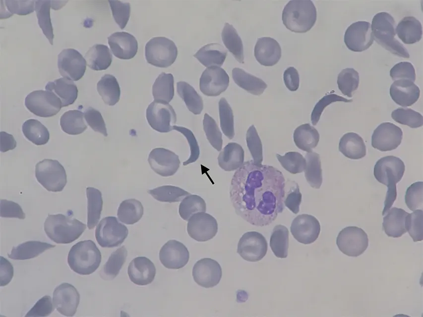
Everyone has two copies of the β-globin gene, one from each parent Grade 10: Mendel’s laws. A person with two sickle copies has sickle-cell disease. A person with one sickle copy and one usual copy, a carrier, makes both kinds of hemoglobin, and is almost always healthy. Carriers have an advantage, too: they are much less likely to die of severe malaria, because the malaria parasite lives inside red blood cells and does badly in those that contain sickle hemoglobin. So where malaria killed many people for many generations, the sickle gene became common: in Africa, in India, and in parts of Nepal’s Terai, whose forests were full of malaria until it was brought under control in the 1950s and 60s. Surveys in western Nepal since 2017 have found carriers in roughly one person in ten to one in seven in some villages. Nepal’s National Public Health Laboratory and its partners tested 6,000 people in Bardiya in 2017–18, and Lumbini Province has offered free screening since 2022.
A carrier of the sickle-cell gene has one usual and one sickle copy of the β-globin gene. What is in their red blood cells?
Pick an answer to see why.
Kinds of change
Not every change in a gene does harm, and not every change does it the same way. Try some real changes in the β-globin gene, all known from real people.
The changes fall into a few kinds:
- A silent change: a letter changes, but the new codon means the same amino acid (CAT and CAC both mean histidine). The protein is exactly the same. Many people carry one or other of these two versions, with no difference.
- A change of meaning: the new codon means a different amino acid, as in sickle hemoglobin (valine) and hemoglobin C (lysine) at the same spot. How much it matters depends on where the change is and how different the new amino acid is: many such changes do little; some, like sickle hemoglobin, change the protein’s behavior completely.
- A new stop: the new codon is a stop codon. The chain ends too soon and the protein is useless. A stop at codon 17 of β-globin is one cause of β-thalassemia, a disease in which the body can’t make enough hemoglobin.
- A letter added or lost: the cell reads in threes from the start, so every codon after the change is read wrongly. This is a frameshift. The chain becomes nonsense until, by chance, a stop codon turns up. An extra G between codons 8 and 9 is one of the commonest causes of β-thalassemia in northern India.
Most changes in DNA are copying mistakes, or damage by ultraviolet light, X-rays and some chemicals Grade 9: Mutation. The cell repairs nearly all of them. Of the few that last, most are harmless, some are harmful, and a very few turn out useful. They are the raw material of evolution.
Gene to trait: the whole chain
Put the pieces together and you have an explanation, step by step, of how DNA decides what a living thing is like:
- The order of bases in a gene is copied into mRNA (transcription).
- The ribosome reads the mRNA three bases at a time and builds a chain with the matching order of amino acids (translation), using the genetic code.
- The order of amino acids decides how the chain folds, and so the protein’s shape.
- The shape decides the protein’s job: carrying oxygen, cutting starch, pulling a muscle.
- Specialized cells make the proteins their job needs, and work together in tissues, organs and systems.
- So a change in the order of bases can change a protein, a cell and the whole body: a trait.
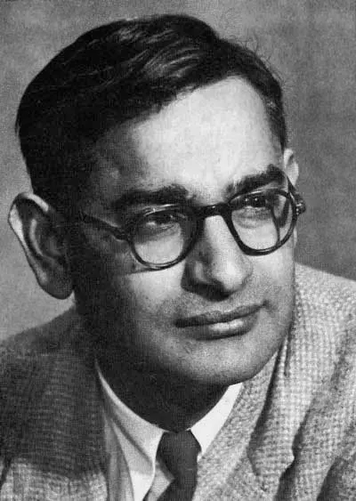
The evidence for each link comes from experiments you have read about: Sanger showed that each protein has an exact order; Nirenberg and Khorana read the code; Ingram found that one changed amino acid makes sickle hemoglobin; and bacteria given a human gene make the human protein.
Questions
A gene has 900 bases that code for its protein, from the start codon to the end of the stop codon. About how many amino acids does the protein have?
Pick an answer to see why.
Which of these changes in the coding letters of a gene is most likely to leave the protein unchanged?
Pick an answer to see why.
Explain why a cell needs mRNA at all. Why doesn’t the ribosome read the DNA directly?
Answer
The DNA stays inside the nucleus, safely packed in the chromosomes, while proteins are made at ribosomes in the cytoplasm. Sending out mRNA copies of a gene, through the nuclear pores, keeps the precious master copy safe and unchanged, while many short-lived copies of just the gene that is needed go out to the ribosomes. Many copies of the mRNA, each read by many ribosomes at once, also let the cell make a lot of one protein quickly; and when the cell needs less, it stops copying the gene and the old mRNAs are broken down.
Part of the template strand of a gene reads TAC AAA CCG TTA ACT.
- i.Write the mRNA.
- ii.Write the order of amino acids.
- iii.What would happen if the fourth codon’s template letters TTA changed to ATT?
Answer
- i.mRNA: AUG UUU GGC AAU UGA.
- ii.Met–Phe–Gly–Asn, then UGA is a stop codon, so the chain ends.
- iii.ATT on the template gives UAA in the mRNA, which is also a stop codon. So the chain would end one codon earlier: Met–Phe–Gly, without the asparagine. (A made-up stretch, for practice.)
Hemoglobin C is caused by a change in the same codon as sickle hemoglobin: GAG becomes AAG.
- i.Which amino acid replaces glutamic acid?
- ii.Glutamic acid has a negative charge; lysine has a positive charge. Pauling’s method separated molecules by their electric charge. Would it tell hemoglobin C apart from usual hemoglobin? Explain.
Answer
- i.AAG codes for lysine (Lys).
- ii.Yes. Usual hemoglobin has a negatively charged glutamic acid at that spot, and hemoglobin C has a positively charged lysine there, so the whole molecule of hemoglobin C carries a different charge (more positive), and in an electric field it moves differently. (It does: hemoglobin C moves even less than sickle hemoglobin towards the positive side, because valine, in sickle hemoglobin, has no charge at all.)
A student says: “A muscle cell and a red blood cell must have different DNA, because they make different proteins.” Use what you know to say whether the student is right.
Answer
The student is wrong. Almost every cell in the body came from the same fertilized egg by mitosis, so they have the same DNA, with the same genes (mature red blood cells actually lose their nucleus, but the cells they grew from had the full set). What differs is which genes each cell uses: a developing red blood cell transcribes the globin genes over and over and makes huge amounts of hemoglobin; a muscle cell transcribes the genes for actin and myosin. The different proteins make the cells different in shape and job.
Use the evidence about sickle-cell anemia to construct an explanation of how the structure of DNA determines the structure and function of a protein, and so the working of a whole body. Your answer should go from the DNA to the person.
Answer
- DNA: in the gene for β-globin, the codon for the sixth amino acid is GAG; in the sickle gene, one base is different, GTG.
- mRNA: transcription copies the difference: GAG becomes GUG.
- Amino acid order: by the genetic code, GAG means glutamic acid and GUG means valine, so the ribosome puts valine in place of glutamic acid; Ingram found exactly this, and nothing else, different in sickle hemoglobin.
- Protein structure: glutamic acid is charged and attracted to water; valine is uncharged and avoids water, so sickle hemoglobin has a greasy patch on its surface (Pauling found its charge was different). When it has given up its oxygen, the patch sticks to the next molecule and the molecules join into long stiff fibers.
- Cell: the fibers stretch the red blood cell into a stiff sickle shape.
- Body: sickle cells block small blood vessels, causing pain and organ damage, and they break down early, causing anemia. A person with two sickle copies has the disease; with one copy, enough usual hemoglobin is made to stay healthy.
So the order of bases in DNA sets the order of amino acids, which sets the protein’s shape and how it behaves, and that decides how the cells and the body work.
Human insulin is now made by bacteria that have been given the human insulin gene. What does this tell us about the genetic code? Why is it useful?
Answer
The bacteria read the human gene and build exactly the same chain of amino acids as a human cell does, so they must read each codon as the same amino acid: the genetic code is the same in bacteria and in humans (it is almost universal across all life). It is useful because bacteria can be grown in huge tanks to make large amounts of insulin with exactly the human sequence, cheaply and reliably, instead of extracting it from the pancreas of pigs and cows, whose insulins differ from ours by one or three amino acids.
Where to go next
- How Does One Cell Become a Whole Person? Every cell has the same genes. How cells become skin, muscle or brain by using different ones.
- Why Aren’t You Exactly Like Your Brother or Sister? Where the variety in genes comes from: the shuffling of chromosomes, and mutations like the ones in this topic.
- How Does DNA Show That All Life Is Related? One genetic code for bacteria, rice and you, and what the small differences between our proteins tell us about our relatives.