
Freezing molecules to take their picture
Nobel Prize in Chemistry, 2017: Jacques Dubochet, Joachim Frank, and Richard Henderson
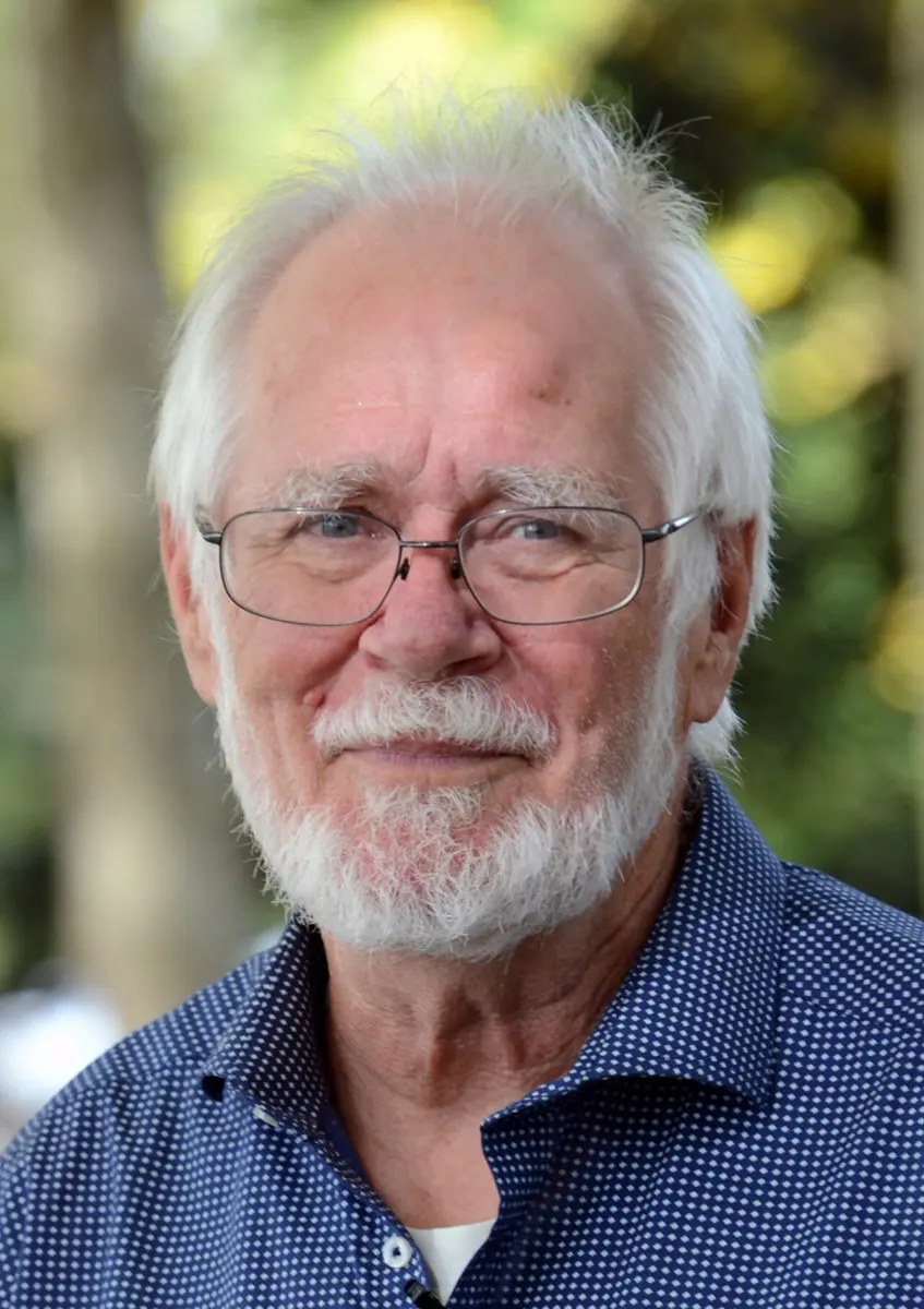
Jacques Dubochet
born 1942 in Switzerland
At school he was the first child in his part of Switzerland officially recognized as dyslexic. He says it “licensed me to be bad at everything”.
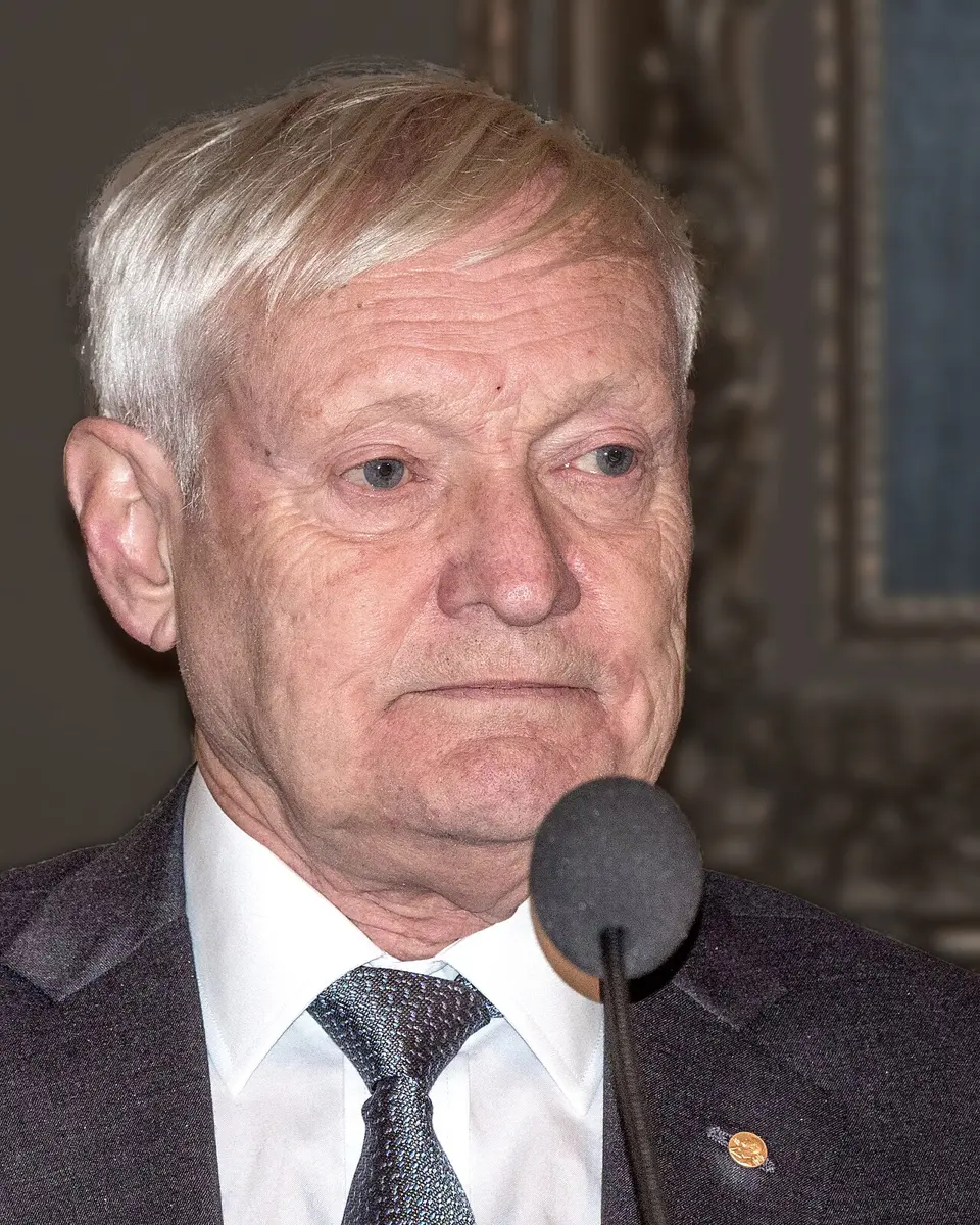
Joachim Frank
born 1940 in Germany
He grew up among bombed ruins after the Second World War, and became a physicist who wrote computer programs to see what the eye can’t.
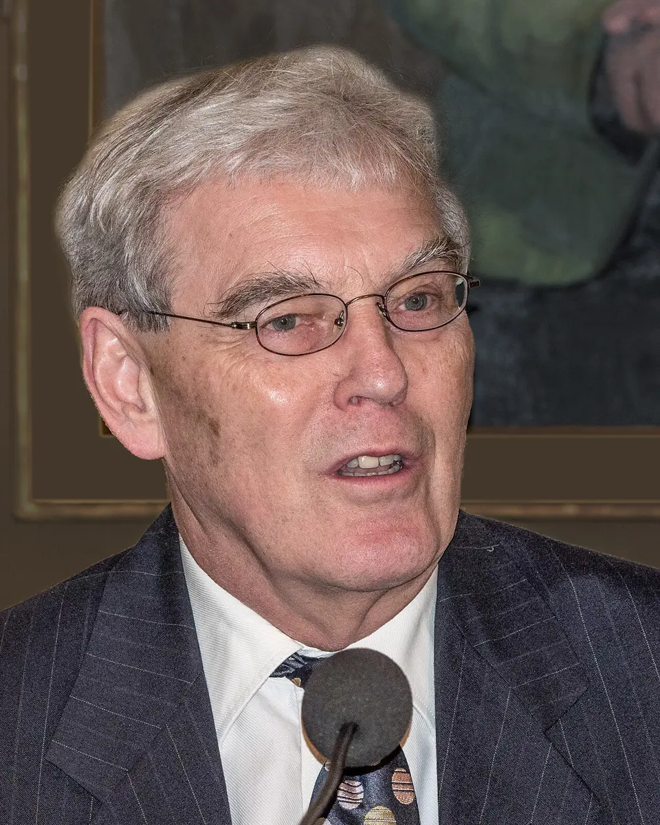
Richard Henderson
born 1945 in the United Kingdom
A baker’s son from a Scottish village school, who spent fifteen years making one picture sharper.
In January 2020, a new coronavirus was spreading in China. Within about a month of its genes being published, scientists in the United States had a picture of the spike that the virus uses to get into our cells: a 3D model detailed enough to show how its long chain of atoms is folded. Scientists making vaccines and medicines used it at once.
That picture was taken with a cryo-electron microscope. “Cryo” means very cold. The method froze the molecules so fast that the water around them had no time to turn into ice crystals. In 2017 the Nobel Prize in Chemistry went to the three scientists who made it work.
The problem: the machines of life are too small to see
Every living cell is full of proteins (how a gene makes a protein). Some carry oxygen in your blood, some digest your dal bhat, some build new proteins, some pump things in and out of the cell. A protein works like a tiny machine, and what it does depends on its shape: which parts stick out, where the pockets are, how it moves.
To understand a protein, or to design a medicine that blocks it, you need to see its shape. But proteins are tiny. You could line up about ten thousand ordinary proteins across the width of one hair.
An ordinary light microscope can’t show them. Light is a wave Grade 9: wavelength, and a microscope can’t show details much smaller than about half the wavelength of the light it uses, about 0.2 micrometers. That is enough to see a cell and some parts inside it, but a protein is about twenty to forty times smaller.
The old ways: crystals
Since the 1950s, scientists had found the shapes of proteins with X-rays. X-rays have a very short wavelength, but they can’t be focused into a picture, and one molecule scatters far too few of them. So you first have to grow a crystal of the protein: millions of copies packed in a perfect, repeating pattern, like the tiles on a floor. X-rays bouncing off the crystal make a pattern of spots, and from it a computer works out where the atoms are. This method, X-ray crystallography, has won many Nobel Prizes.
But many proteins refuse to form crystals. Proteins that sit in the oily skin of a cell, the cell membrane, are especially hard, and they are the ones many medicines act on. Some protein machines are too big and floppy. And even when a crystal forms, it shows the protein frozen in one pose, squeezed among its neighbors, not as it moves in the cell.
Why can’t an ordinary light microscope show the shape of a protein?
Pick an answer to see why.
How an electron microscope works
In the 1930s, a German engineer, Ernst Ruska, built a microscope that looks with electrons instead of light. Electrons behave like waves too, and when they are speeded up by a high voltage, their wavelength is more than a hundred thousand times shorter than light’s. Instead of glass lenses, magnets bend the beam of electrons and bring it to a focus. In a transmission electron microscope, the beam passes through a very thin sample and makes a picture on a screen or a camera underneath. Such microscopes can show single atoms in a metal.
So why not just put a protein in one and look?
Why it was hard
There were three big problems.
- The vacuum. Electrons are stopped by air, so the inside of an electron microscope is a vacuum: almost all the air is pumped out. In a vacuum, water boils away even at room temperature Grade 10: boiling below 100 °C. A protein in a cell is surrounded by water and needs it to keep its shape. Dried out, it collapses like a tent without poles.
- The burning beam. A strong beam of electrons smashes the bonds in a protein. By the time a sharp picture is taken, the protein has been destroyed. Scientists believed the electron microscope was only good for dead, dry things.
- The fuzz. With a weak beam, the protein survives, but the picture is so faint and grainy that you can hardly see the molecule at all: like a photo taken at night without a flash.
People tried freezing the sample, because ice doesn’t boil away in a vacuum as fast as water. But ordinary freezing makes ice crystals, which scatter the electrons and spoil the picture, and their sharp growth can tear the molecules.
Why do proteins usually have to be frozen before they go into an electron microscope?
Pick an answer to see why.
What they did, step by step
Henderson: a protein that lines itself up (1975–1990)
Richard Henderson was trained in X-ray crystallography at Cambridge, in the United Kingdom. In the 1970s he wanted the shape of a membrane protein, but it wouldn’t form crystals. So he turned to the electron microscope.
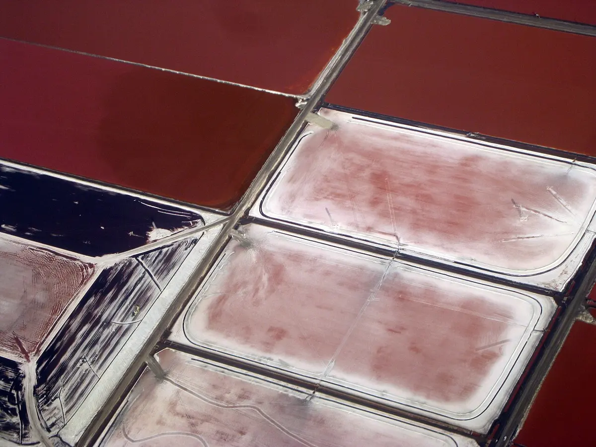
He chose a lucky protein: bacteriorhodopsin, a purple protein in the skin of a microbe that lives in very salty water. The microbe uses it to catch the energy of sunlight. Its skin is packed with this protein in neat rows, all pointing the same way, almost like a crystal already.
- 1975: a rough model. With his colleague Nigel Unwin, Henderson put whole pieces of the purple skin under the microscope, covered with a sugar solution so that they didn’t dry out. They used a weak beam, so the protein survived. Each picture was fuzzy, but because thousands of identical molecules sat in neat rows, a computer could combine them. Tilting the sample and taking pictures from many angles, they built a 3D model. It showed the protein’s chain crossing the membrane seven times. It was the best picture of a protein ever made with an electron microscope, but it showed details only down to 0.7 nanometers, not single atoms.
- 1975–1990: sharper and sharper. Henderson wanted to see atoms. Over fifteen years, as microscopes got better and samples were kept cold with liquid nitrogen, he traveled to the best electron microscopes in the world, using each one for what it did best.
- 1990: atoms. He published a model of bacteriorhodopsin as detailed as X-ray crystallography could give. The electron microscope could show a protein’s atoms after all. But it had worked for a protein that lines itself up. Could it work for proteins scattered every which way?
Frank: a computer that sorts fuzzy pictures (1975–1990s)
Joachim Frank, a physicist from Germany, worked in Albany, in the United States, at a laboratory of the New York State health department. He had an idea, which he first wrote down in 1975: use many copies of one molecule, lying scattered in every direction, and let a computer do the rest.
- Find them. In a fuzzy picture, the computer finds the faint shapes of the molecules.
- Sort them. Each molecule lies at a different angle, so they look different. The computer sorts all the shapes into groups that look alike: all the “side views” together, all the “top views” together, and so on.
- Average them. Inside each group, it lays the pictures on top of each other and averages them. The noise (the random grain in each picture) is different every time, so it cancels out; the molecule is the same every time, so it stays. Out of hundreds of fuzzy pictures comes one sharp one. Frank’s programs for this were ready in 1981.
- Build in 3D. Then the computer works out at which angle each sharp view was seen, and combines them into a 3D shape, rather as a doctor’s CT scanner builds up a 3D picture from X-ray pictures taken all round the body. Frank published this in the mid-1980s and used it on the ribosome, the cell’s protein-building machine Grade 8: Ribosomes.
Dubochet: water that freezes into glass (1978–1984)
In 1978 Jacques Dubochet went to the European Molecular Biology Laboratory in Heidelberg, Germany, with one job: to find out how to keep water in the electron microscope without it boiling away or turning into crystals.
When water freezes slowly, its molecules have time to line up into the neat, repeating pattern of an ice crystal. Dubochet’s idea was to cool the water so fast that the molecules have no time to line up. They would be stuck where they were, jumbled as in liquid water, but solid. A solid whose particles are jumbled instead of lined up is called a glass. Window glass is one; this would be glassy water (scientists call it vitreous ice, from the Latin for glass).
- Liquid nitrogen didn’t work. Liquid nitrogen is very cold, −196 °C. But when a warm sample is dipped in it, the nitrogen next to it boils at once, and the sample is wrapped in a layer of gas that slows the cooling. (It is the same reason a drop of water skates around on a very hot tawa instead of boiling away at once: it sits on a cushion of its own steam.) The water had time to form crystals.
- Early 1980s: liquid ethane. Dubochet’s team used liquid ethane instead, a gas from natural gas, cooled by liquid nitrogen around it to about −180 °C. Ethane doesn’t boil as easily, so it pulls the heat out at once. Under the microscope they saw a smooth frozen drop unlike anything before. When they let it warm up a little, it suddenly turned into ordinary ice crystals. They had made glassy water.
- 1984: the method. They spread a drop of a sample on a tiny metal grid, blotted it to a film so thin that electrons could pass through it, and shot it into the liquid ethane, freezing it in less than a thousandth of a second. That year they published sharp pictures of viruses frozen in glassy water. The method is still used, almost unchanged, in every laboratory.
Dubochet froze water so fast that it became a glass. What is different about glassy water compared with ordinary ice?
Pick an answer to see why.
How cryo-electron microscopy works today
The three ideas fit together, and today every cryo-EM laboratory does the same steps:
- Freeze. A tiny drop of protein in water goes on a metal grid smaller than a lentil. It is blotted thin and plunged into liquid ethane (Dubochet).
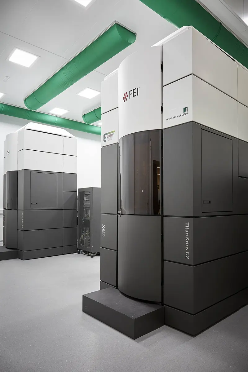
- Photograph. The grid goes into the microscope, kept at about −180 °C. A weak electron beam takes thousands of pictures, each with hundreds of molecules lying in every direction.
- Sort, average, build. Computers find hundreds of thousands of molecules, sort them, average them and build a 3D map (Frank).
- Fit the atoms. Scientists fit the protein’s chain of atoms into the map.
For years the 3D maps were blurry: scientists joked that cryo-EM was “blobology”. Henderson kept pushing to improve every part of the microscope. The last big step came in 2013, with a new kind of camera that counts single electrons, as fast as a video camera, so that even the slight jiggle of the sample in the beam can be corrected. Suddenly cryo-EM could show atoms. In the years after, it became one of the most used ways of finding protein shapes.
In your life today
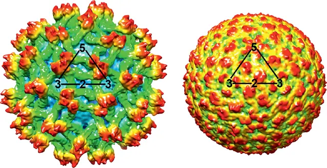
- New viruses, fast. In 2016, when the Zika virus was suspected of causing babies to be born with small heads in Brazil, cryo-EM showed its structure in atomic detail within a few months. In 2020 the coronavirus spike followed within weeks, which helped scientists design vaccines and antibody medicines.
- Dengue. The dengue virus, spread by Aedes mosquitoes, now makes tens of thousands of people in Nepal ill in a bad year, even in the Kathmandu valley. Cryo-EM has shown how the dengue virus is built and how antibodies grip it, which helps in designing dengue vaccines.
- Medicines. Many medicines act on proteins in the cell membrane, the very proteins that wouldn’t crystallize. Drug companies now use cryo-EM to see exactly where a medicine fits.
What came next
- Molecules in their home. Using cryo-electron tomography, scientists freeze whole cells, or very thin slices of them, and take pictures from many angles, to see the protein machines in place, inside the cell.
- Predicting shapes. Part of the 2024 Nobel Prize in Chemistry went for AlphaFold, a computer program that can predict a protein’s shape from its recipe. Cryo-EM and these predictions now work together: a prediction helps read a blurry map, and the map checks the prediction.
- The open question. Proteins are not still statues; they bend, open and close as they work. A frozen sample holds molecules caught in many different poses, and computers are learning to sort them into a “film” of a protein at work. Can we one day watch every step of a living machine, atom by atom?
In your science books
- Grade 8: the cell and its parts, including ribosomes, and viruses.
- Grade 9: electromagnetic waves and the spectrum.
- Grade 10: boiling below 100 °C, why water boils away in a vacuum.
- Going further: proteins do the work, and water, the odd one out, on how water molecules hold together.
Learn more
- The Nobel Prize’s own explanation: They captured life in atomic detail (nobelprize.org).
- The three laureates’ Nobel lectures: Dubochet, Frank and Henderson.
The official reason
The prize was given “for developing cryo-electron microscopy for the high-resolution structure determination of biomolecules in solution.”