
Inside a light-catching machine
Nobel Prize in Chemistry, 1988: Johann Deisenhofer, Robert Huber, and Hartmut Michel
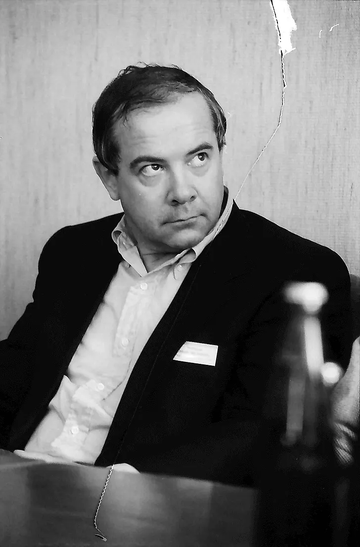
Johann Deisenhofer
born 1943 in Zusamaltheim, Germany
A farmer’s eldest son, expected to take over the farm. He chose physics instead, and became an expert at turning X-ray pictures into models of molecules.
Photo: Willy Pragher, CC BY 4.0
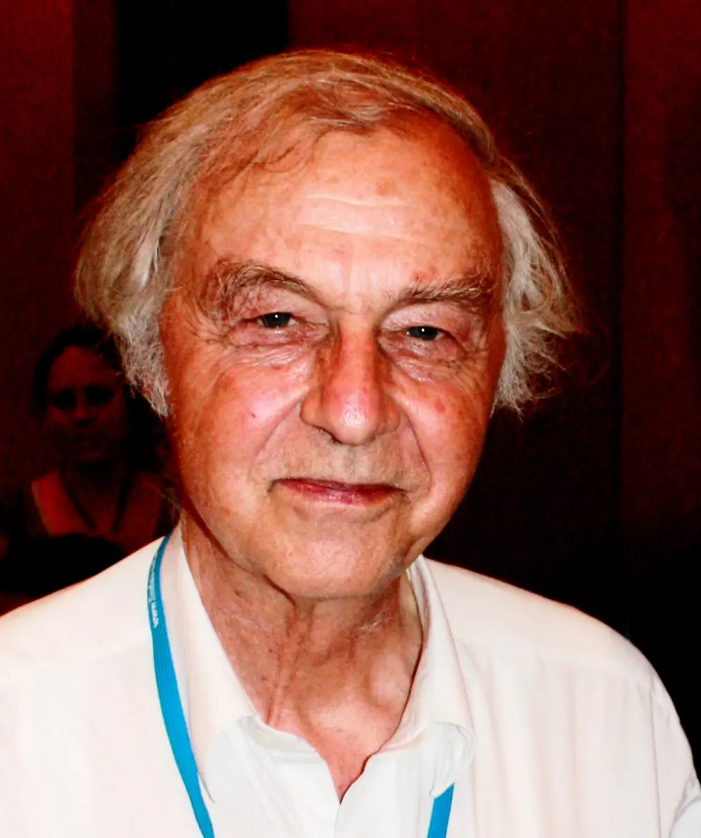
Robert Huber
born 1937 in Munich, Germany
Head of the X-ray department where the work was done. His team had built many of the methods and computer programs used to work out the shapes of proteins.
Photo: Markus Pössel ( Mapos ), CC BY-SA 3.0

Hartmut Michel
born 1948 in Ludwigsburg, Germany
As a boy he read two to four library books a week. In his early thirties he grew crystals that experts said could not be grown.
Photo: Shauchungshin, CC BY-SA 4.0
Every grain of rice in your dal bhat, every leaf a buffalo eats and every breath of oxygen you take comes from photosynthesis: green plants using the energy of sunlight to turn carbon dioxide and water into food. It is perhaps the most important chemical reaction on Earth.
The very first step, catching the light, is done by a tiny machine made of proteins. In 1988 the Nobel Prize in Chemistry went to Johann Deisenhofer, Robert Huber and Hartmut Michel, three scientists in Germany, for the first map, atom by atom, of such a machine. To get it, they had to do something most experts thought impossible.
The problem: proteins that hide in oil
Every cell is wrapped in a thin skin, the cell membrane Grade 8: The covering of the cell. The membrane is a film of fat-like molecules, only about 4 millionths of a millimeter thick. It behaves like a thin layer of oil: water and most things dissolved in water cannot get through it.
But a cell must still take in food, push out waste and pass on signals. That work is done by proteins that sit in the membrane, poking out on both sides like posts through a fence. They are called membrane proteins. In a leaf, the machines of photosynthesis are membrane proteins too, packed into the membranes inside the chloroplasts Grade 8: Plastids.
A membrane protein has two kinds of surface. The parts that stick out into the watery inside and outside of the cell are water-loving. The middle part, which sits in the oily membrane, is oil-loving: it carries an oily belt. This makes membrane proteins very hard to study:
- Take one out of its membrane into water, and its oily belts stick together. The proteins clump into a useless lump and often fall apart.
- Leave it in the membrane, and it is surrounded by a jumble of fat molecules.
By the early 1980s, scientists had worked out the exact shapes of many proteins that dissolve in water, such as hemoglobin from blood. For membrane proteins they had only blurry pictures, taken with electron microscopes. Nobody knew how a protein sits in a membrane, atom by atom.
Why do membrane proteins clump together when they are taken out of the membrane into water?
Pick an answer to see why.
How you see the atoms in a protein
Atoms are far too small to see with any light microscope. To find where they are, scientists use X-ray crystallography.
- Grow a crystal. Millions of millions of copies of the same protein must line up in perfect, repeating rows, like soldiers on parade. A protein crystal is often smaller than a grain of salt.
- Shine X-rays through it. X-rays have a wavelength about as small as the gaps between atoms Grade 9: X-rays. As they pass through the crystal, they bounce off the electrons in the atoms and come out as a pattern of thousands of dots on film.
- Work backwards. The positions and brightness of the dots depend on where the atoms are. With a great deal of mathematics, a computer turns the dots into a 3D map of where the electrons are thickest, and so where the atoms sit.
- Build the model. Scientists fit a chain of atoms into the map, piece by piece, until every atom has its place.
The key is step 1. A crystal is only useful if its copies of the protein are lined up almost perfectly. A messy crystal gives a blurry pattern, or none at all.
Why it was hard
- The detergent problem. To get a membrane protein out of its membrane, you dissolve the membrane with a detergent, a soap-like substance. Like soap Grade 10: How soap cleans, each detergent molecule has a water-loving head and an oil-loving tail. The tails cover the protein’s oily belt, and the heads face the water, so the protein dissolves. But the detergent wraps each protein in a big, soft collar. Proteins in big, soft collars cannot pack tightly into neat rows.
- Expert opinion. Most protein crystallographers believed that membrane proteins simply could not be crystallized.
- The size. Even with a crystal, the protein Michel chose was huge, with more than 10,000 atoms. In the early 1980s, working out a structure that big was a task of years.
What they did, step by step
Michel: crystals nobody thought possible
Hartmut Michel studied biochemistry and then worked in the laboratory of Dieter Oesterhelt, a German scientist who studied a purple protein, bacteriorhodopsin, from bacteria that live in very salty water.
- 1977, a hint in the freezer. Michel stored some of the purple protein in a freezer. When he took it out, it had formed solid, glass-like lumps. If the protein could pack together like that, he thought, perhaps it could also form crystals. Four weeks later he had flat, one-layer crystals of it.
- 1979, the first real crystals. Michel and Oesterhelt grew the first 3D crystals of bacteriorhodopsin. But they were too small and too disordered for X-rays to give a clear picture.
- Smaller collars. Michel guessed that the detergent collars were too big. He looked through the catalogs of the big chemical companies and ordered nearly everything that was water-loving at one end and oil-loving at the other. He also made about 20 such molecules himself. The best was a small molecule called heptane-1,2,3-triol. It was too small to form collars of its own, so it mixed into the detergent collars and made them smaller.
- 1981, a new protein. Frustrated with bacteriorhodopsin, Michel tried other membrane proteins. One was the photosynthetic reaction center of a purple bacterium called Rhodopseudomonas viridis (now named Blastochloris viridis). He found a new way to purify it, used a detergent with a smaller head, added his helper molecule, and at the end of July 1981 he had crystals, up to 2 mm long. In September they were tested with X-rays: they were excellent. A week after the first crystals, his daughter was born. He later called 1981 “the happiest and most successful year of my life”. (The Nobel committee’s summary gives 1982 for the crystals, the year Michel published them.)
Deisenhofer and Huber: from dots to atoms
Robert Huber led the X-ray department of the same institute, the Max Planck Institute for Biochemistry near Munich. His team had developed many of the methods and computer programs used to work out protein structures. In 1982 Michel asked for help, and Huber agreed that Johann Deisenhofer, one of his scientists, should join the project. Deisenhofer and Michel became best friends.
- 1982–1983, collecting the dots. Each crystal was turned in the X-ray beam a fraction of a degree at a time, with a photograph for each step. One complete set of photographs took three to four months.
- Heavy atoms as landmarks. The dots alone do not give the full answer; a vital piece of information is lost. To recover it, Michel soaked crystals in solutions of heavy atoms such as mercury and gold, which stick to a few places on the protein. Comparing the patterns with and without them, the team worked it out, using five different heavy-atom soaks.
- 1983, the first map. First the colored molecules that catch the light showed up in the map, then the protein chains wrapped round them. By the end of 1983 success was in sight. Deisenhofer built the model on a computer screen, and later wrote that it was “hard to describe the excitement” of seeing it grow.
- 1984–1985, every atom. To be sure of each chain, Michel and colleagues worked out the order of the building blocks in the proteins from their genes. The first structure was published in 1984 and the full model in 1985: four proteins and about 10,000 atoms (not counting hydrogen), each in its place.
How the light-catching machine works
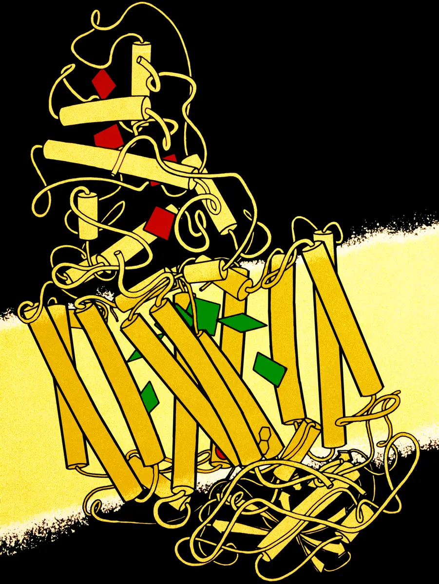
The reaction center is about 13 millionths of a millimeter long, and it crosses the membrane from one side to the other. It is made of four proteins. Two of them, called L and M, are nearly the same shape, and each crosses the membrane five times as a long, stiff spiral, a helix. A third protein, H, adds one more helix and a cap on the inside of the cell. The fourth, a cytochrome, sits on the outside, holding four iron-containing groups.
Held inside the proteins, like jewels in a setting, are the colored molecules that do the work: bacteriochlorophyll (the bacteria’s version of the green chlorophyll in leaves), related molecules called pheophytins, two quinones, and an iron atom.
When light strikes, its energy reaches a pair of bacteriochlorophylls in the middle, called the special pair. Then an electron is passed along a chain, like a bucket passed along a line of people:
- The special pair is excited by the light and throws out one electron. In about 3 picoseconds (3 millionths of a millionth of a second) it reaches a pheophytin.
- In about 200 picoseconds it moves on to the first quinone. It has now crossed most of the membrane.
- In about a ten-thousandth of a second it reaches the second quinone. This quinone collects two electrons, picks up two hydrogen ions, and leaves to deliver them.
- Meanwhile, the cytochrome on the outside passes an electron to the special pair, refilling the gap.
The result is that the energy of light has pushed electric charge across the membrane. The cell uses this to pump hydrogen ions and to make ATP, the molecule that stores energy in every living cell (Catching the energy: ATP).
The map held a surprise. The colored molecules form two branches that are almost mirror images, one held by L and one by M. Yet the electron goes down only one branch, the one held by protein L. The structure showed the question clearly for the first time; scientists are still working out all the reasons.
In the reaction center, what does the energy of light actually do?
Pick an answer to see why.
Bacteria, plants and you
Purple bacteria are much simpler than plants. Their photosynthesis does not split water and does not give off oxygen. So why does the bacteria’s machine matter for a rice field?
Because the reaction center in plants, called photosystem II, turned out to be built the same way. Its two central proteins match the bacteria’s L and M closely. Plant photosynthesis has to work this way to give the oxygen we breathe, and the bacterial structure became the guide to it.
It also explained something farmers already knew. Some weedkillers work by plugging the pocket where the second quinone sits in a plant’s photosystem II, so the electrons stop and the weed starves. Deisenhofer and Michel soaked crystals in one such weedkiller and saw it sitting in the same pocket of the bacteria’s machine Grade 10: Pesticides.
Why did Michel add a small helper molecule, heptane-1,2,3-triol, to his protein?
Pick an answer to see why.
In your life today
- Your food. All the energy in your food was first caught by reaction centers in plants and algae (Food chain; The Sun, the ultimate source of energy). Knowing the machine’s shape helps scientists understand how crops use light.
- Medicines. Many medicines work by fitting into membrane proteins: the heart medicines called beta-blockers, for example, act on a protein in the membranes of heart cells (Medicine 1988). Michel’s way of crystallizing membrane proteins opened the door to seeing their shapes.
- Weedkillers. The structure showed how weedkillers that block photosystem II do their work, and why some weeds, with one small change in the pocket, resist them.
What came next
- More membrane proteins. After 1988, other teams used Michel’s methods. Roderick MacKinnon saw the shape of a channel that lets potassium through a membrane (Chemistry 2003), and Brian Kobilka the shape of a receptor that beta-blockers also block (Chemistry 2012). Today thousands of membrane protein structures are known, many of them now found with electron microscopes (Chemistry 2017).
- Photosystem II itself. The plant machine is much bigger and was harder to crystallize. Its first X-ray structure came in 2001, and a sharp one, showing the cluster of manganese atoms that splits water, in 2011.
- Artificial photosynthesis. The Nobel committee hoped this work might one day lead to new ways of turning sunlight into fuel. Chemists are trying to copy the reaction center’s trick of moving an electron quickly and far enough that it cannot fall back. An open question: can a man-made device do this as cheaply and as long as a leaf?
In your science books
- Grade 8: the covering of the cell, plastids and mitochondria; bacteria.
- Grade 9: the food chain, the Sun as the ultimate source of energy, and X-rays.
- Grade 10: how soap cleans, detergents and pesticides.
- Going further: proteins as chains that fold, ATP, and the living things that first made oxygen.
Learn more
- The Swedish Academy’s press release, the presentation speech and a short speed read (nobelprize.org).
- The Nobel lectures: Deisenhofer and Michel, who tell the story of the crystals, and Huber.
The official reason
The prize was given “for the determination of the three-dimensional structure of a photosynthetic reaction centre.”