
Direct methods: finding atoms in X-ray spots
Nobel Prize in Chemistry, 1985: Herbert A. Hauptman and Jerome Karle
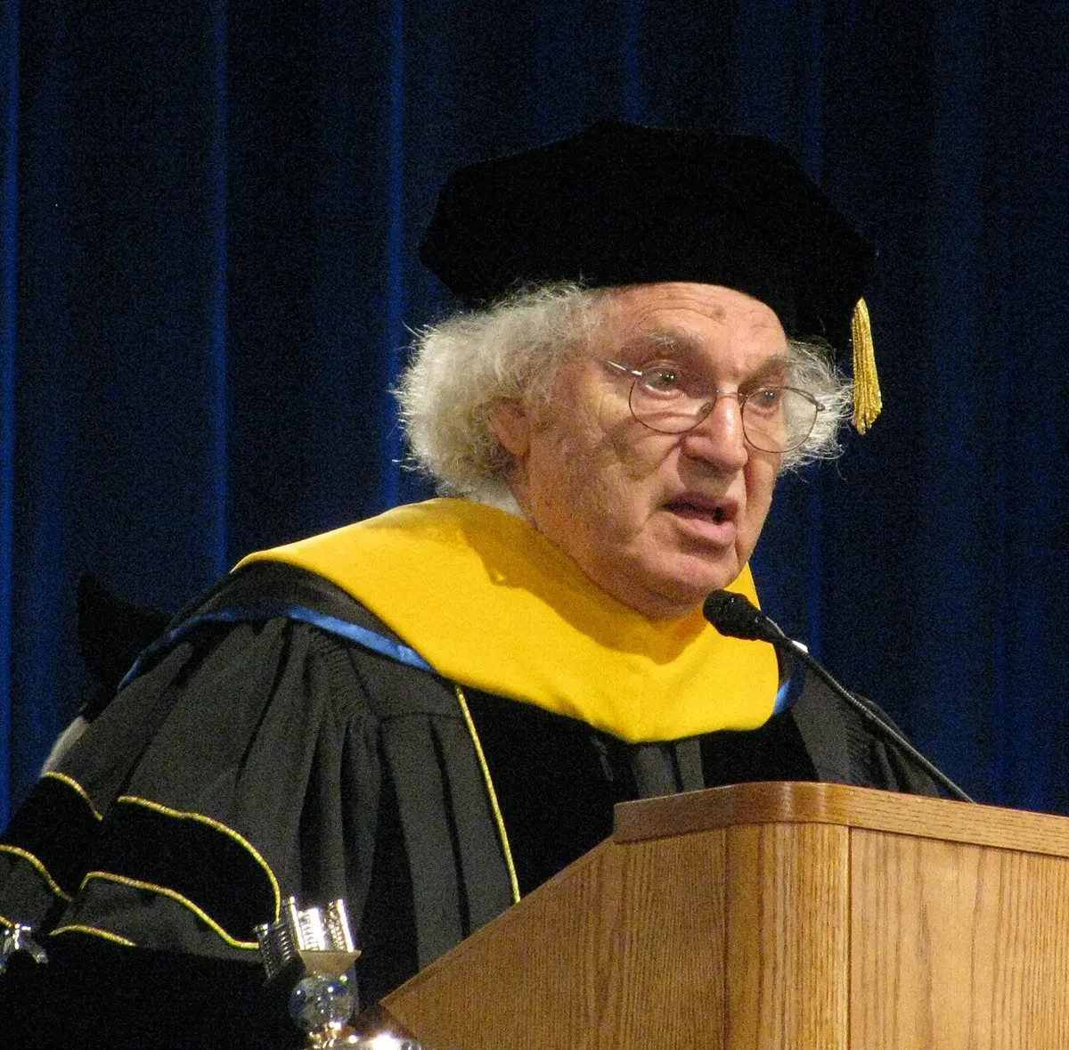
Herbert A. Hauptman
1917–2011, born in the United States
A mathematician who loved numbers from the time he could read. He won the Nobel Prize in Chemistry without ever training as a chemist.
Photo: Dave Pape, Public domain
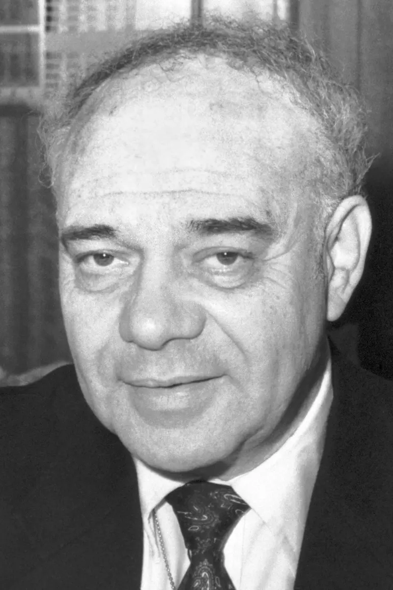
Jerome Karle
1918–2013, born in the United States
A chemist who started college at 15. He and his wife Isabella worked side by side at the same laboratory for more than sixty years.
Photo: United States Office of Naval Research, Public domain
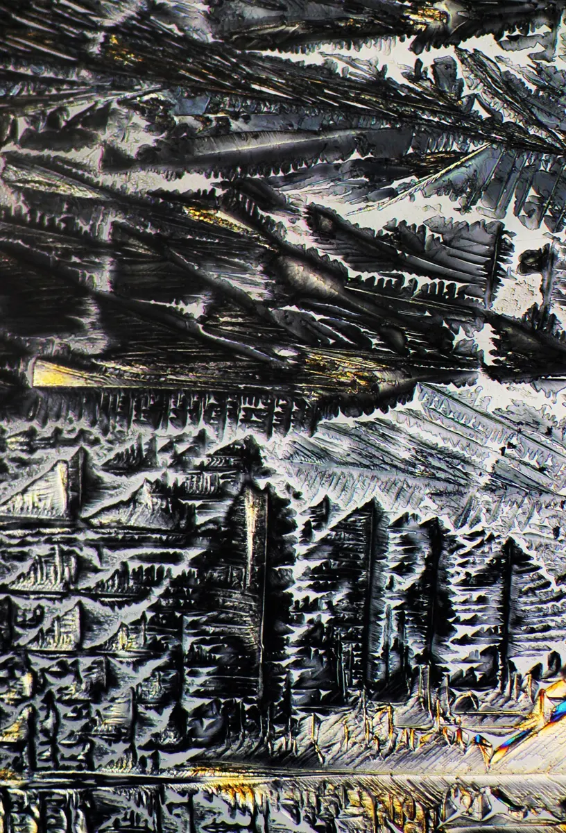
Pour a little sugar into your hand and look closely. Every grain is a tiny crystal, with flat faces and sharp edges. The flat faces are a clue: inside, the sugar molecules are stacked in perfectly regular rows, like bricks in a wall, repeated millions of times in every direction.
That regular stacking lets scientists find out the exact shape of a molecule: where every atom sits, and how far apart they are. They shine X-rays through the crystal and study the pattern of spots that comes out. But for forty years there was a catch. The spots held only half of the information needed. The other half, called the phase, was lost.
In 1985 the Nobel Prize in Chemistry went to two Americans, the mathematician Herbert Hauptman and the chemist Jerome Karle, who showed how to get the missing half back, by mathematics alone.
The problem: a picture with half the information missing
You can’t see a molecule, even with the best microscope that uses light. Atoms are about a ten-millionth of a millimeter apart, far smaller than the wavelength of light. To “see” them you need a wave whose wavelength is about as small as the gap between atoms. X-rays are such waves Grade 9: X-rays.
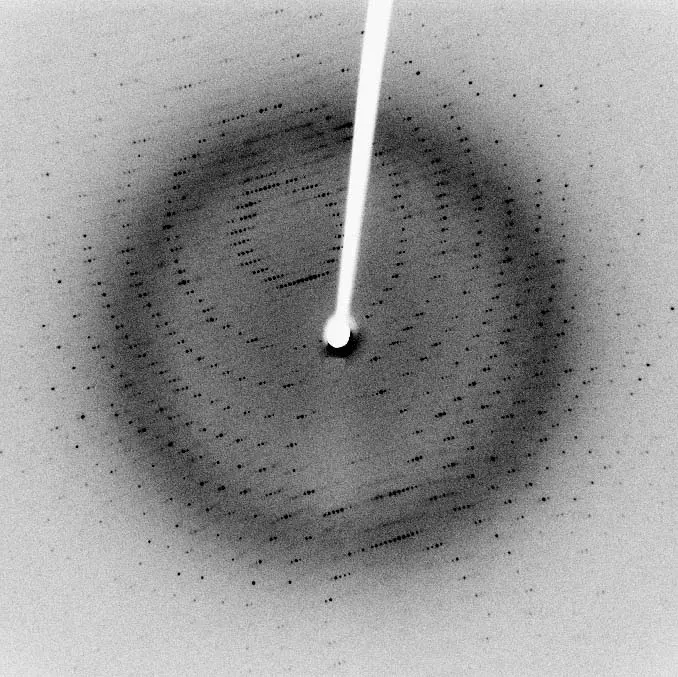
When a narrow beam of X-rays goes into a crystal, the electrons in the atoms scatter it. Because the atoms are in regular rows, the scattered waves add up only in certain directions and cancel out everywhere else (Waves that cancel). So the crystal sends out many thin beams, which make a pattern of spots on a film or detector. This is called X-ray diffraction.
In 1912 Max von Laue showed that crystals do this. Soon after, William Henry Bragg and his son William Lawrence Bragg showed how to work backwards from the spots to the positions of the atoms in simple crystals such as common salt. Both discoveries won Nobel Prizes in Physics, in 1914 and 1915.
How the spots are turned back into atoms
Each spot is made by one wave coming out of the crystal. To rebuild the picture of the atoms, you must add all these waves together again. Two things about each wave matter:
- its height (scientists say its amplitude), which decides how bright the spot is;
- its phase: where its crests and troughs sit, compared with the other waves. Two waves of the same height can be shifted against each other by a quarter of a wave, half a wave, or anything in between.
The detector measures brightness, so it tells you every wave’s height. But it can’t tell where a wave’s crests were. A wave shifted by half a wave makes exactly the same spot (Figure 2). This is the phase problem.
Without the phases, adding the waves back together gives the wrong picture, or nothing that makes sense. It is like being told how loud each instrument in a band was playing, but not when each one started: you can’t rebuild the song.
What does the brightness of an X-ray spot tell you?
Pick an answer to see why.
Why it was so hard
Before 1950, crystallographers got round the phase problem with tricks and good guesses.
- Guess and check. Build a model of the molecule from what chemists already knew, work out the spots it would make, and compare them with the real ones. Then move the atoms and try again, and again. For a molecule of a few dozen atoms this could take months or years.
- A heavy atom. An atom with many electrons, such as mercury or iodine, scatters X-rays far more strongly than light atoms such as carbon. If the molecule has one, its position can be found first, and it gives rough phases for the rest. Dorothy Hodgkin used methods like these to find the shapes of penicillin and vitamin B12, and won the Nobel Prize in Chemistry in 1964 (the story of her prize).
But most molecules of living things, and most medicines, are made only of light atoms: carbon, hydrogen, oxygen and nitrogen. For these, the tricks hardly worked. Many scientists believed that the phases were simply gone, and that no amount of mathematics could bring them back.
What they did, step by step
Hauptman and Karle were both born in New York City, a year apart, and both studied at the City College of New York, a university that charged no fees. They met at the US Naval Research Laboratory in Washington, D.C., where Karle already worked. Hauptman joined him in 1947.
Hauptman and Karle: two simple facts
- The first fact: never less than nothing. The picture you are trying to rebuild is a map of where the electrons are, called the electron density. At the center of an atom there are many electrons; between atoms there are very few. But nowhere can there be fewer than zero electrons. Hauptman and Karle saw that this simple fact is a strong rule. If you choose the wrong phases, the waves add up to a map with places of “less than no electrons”, which can’t be real (Figure 3).
- The second fact: far more spots than atoms. A crystal of a small molecule gives thousands of spots, but the molecule has only a few dozen atoms. So there are many more measurements than unknowns, and the measurements must agree with each other in many ways. That makes it possible to use probability: to work out which phases are most likely, given all the spot brightnesses together.
- 1950–1956: the papers. In a series of papers, and a short book in 1953, they turned these two facts into equations. The equations link the phases of different spots to each other through the measured brightnesses. They also showed how to start: a few phases can be chosen freely, which fixes where the picture begins, and the rest follow from them.
The rule of three
One of the most useful results is easy to say. Take two strong spots. Their positions point to a third spot. If all three are strong, then the phase of the third is very probably the phase of the first plus the phase of the second. This is only “probably”, never “certainly”, so Hauptman and Karle used many such links at once, each spot checked by many others, until the phases agreed with each other.
Other scientists had found parts of the idea before them: David Harker and John Kasper in the United States, who found one such rule in 1948, and David Sayre, who found an equation in 1952 that pointed the same way. The Nobel committee named them too. Hauptman and Karle’s work joined these pieces into one complete method.
Isabella Karle: making it work
For years, many crystallographers did not believe the method. In Jerome Karle’s words, it was a time “when a large number of fellow-scientists did not believe a word we said”.
The person who proved it worked was Isabella Karle, Jerome’s wife and a crystallographer at the same laboratory. She turned the theory into a practical recipe that she called symbolic addition, and used it to solve real crystals one after another. In 1964 the Karles’ laboratory published the first structure of a molecule made only of light atoms, in a crystal with no special symmetry to help, found directly from its spots. After that, laboratories around the world took up the method.
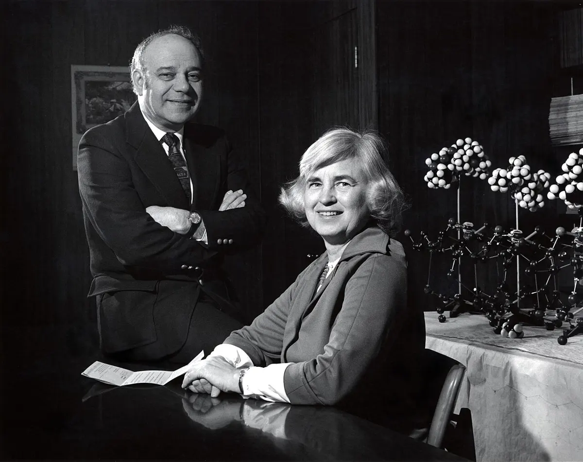
Isabella Karle was not given a share of the prize; a Nobel Prize can go to at most three people, and the committee chose two. The Royal Swedish Academy’s own announcement called her work, and that of the British crystallographer Michael Woolfson, “crucial”. Many crystallographers, Jerome Karle among them, believed she should have shared it.
Why is the rule “never less than zero electrons” so useful for finding the phases?
Pick an answer to see why.
How it works now
Direct methods needed a great deal of arithmetic: thousands of spots, each linked to many others. In the 1950s that took weeks of hand calculation. The method really took off only with fast computers, in the 1960s and 70s.
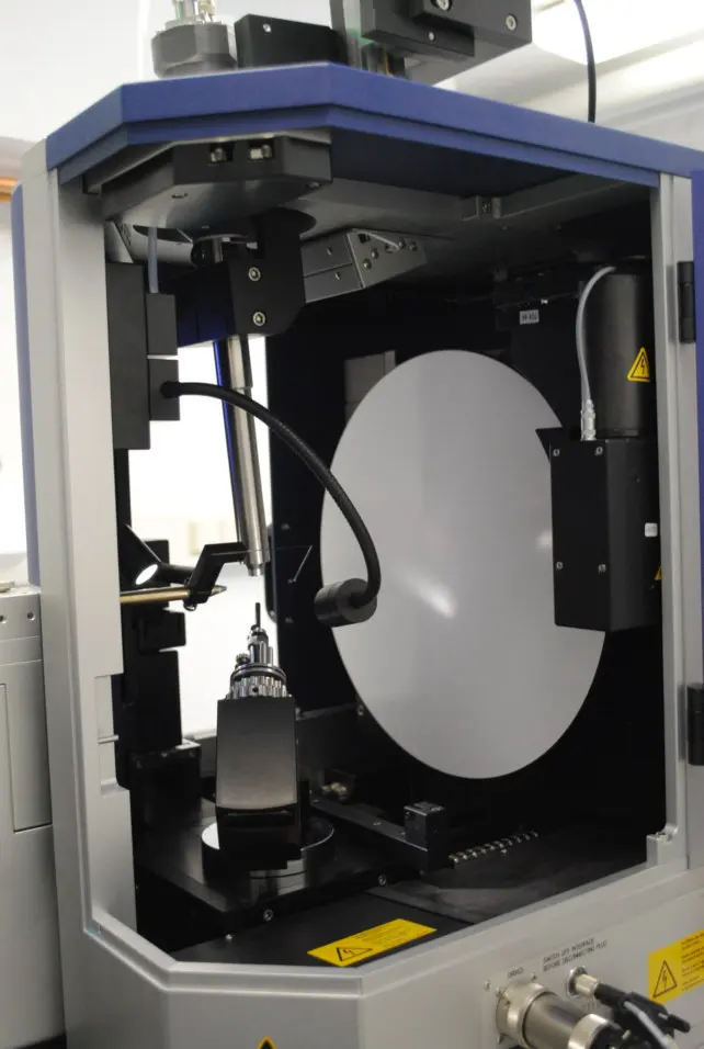
Today a chemist who has made a new compound puts a crystal smaller than a grain of salt into a machine called a diffractometer. The crystal turns slowly in the X-ray beam while a detector records tens of thousands of spots. Then a computer program finds the phases with direct methods, adds up the waves, and shows the molecule on the screen, often within minutes.
- Grow a crystal of the compound.
- Measure the brightness of every spot.
- The program finds likely phases with direct methods.
- It adds up the waves to make a map of the electrons, with peaks at the atoms.
- A chemist checks the map, names each atom and improves the positions.
At the prize ceremony, the Academy said that structures which had won the Nobel Prize in 1964 could now be solved “by a clever beginner”.
In your life today
- Medicines. Before a new medicine is made in a factory, chemists find the exact shape of its molecule, usually from a crystal and direct methods. The shape decides how the medicine fits the molecule in your body that it acts on.
- The same medicine, different crystals. Many medicines can crystallize in more than one way. Paracetamol, the tablet for fever and pain in every home in Nepal, has two main crystal forms. They are the same molecule stacked in different patterns, and they behave differently when pressed into tablets. X-ray diffraction tells a factory which one it has.
- A library of shapes. The Cambridge Structural Database, a collection of the shapes of small molecules found by crystallography, passed 1.25 million structures in 2023. Most of them were solved with direct methods.
Why do chemists need to know the exact shape of a medicine’s molecule?
Pick an answer to see why.
What came next
- Bigger molecules. Ordinary direct methods work best for molecules of up to about a hundred atoms (not counting hydrogen). Hauptman spent the rest of his life pushing them further. In the 1990s, with his colleagues in Buffalo, he wrote a program called “Shake-and-Bake” that solved structures of up to about a thousand atoms by direct methods alone, if the crystal was good enough.
- Proteins. Most protein structures are still solved in other ways: with heavy atoms, or by starting from the known shape of a similar protein. Jerome Karle worked out the theory of one of these ways too, using X-rays of several wavelengths.
- Without crystals. Some molecules refuse to crystallize. Since about 2013, cryo-electron microscopy, which photographs frozen molecules with electrons, has shown many of them (Nobel Prize in Chemistry, 2017). Electron beams also make spots from crystals too tiny for X-rays, and direct methods work on those spots too.
- An open question. Can computers now predict a molecule’s shape, and even how it will stack in a crystal, without any experiment? For proteins, programs like AlphaFold have come close (Nobel Prize in Chemistry, 2024). Predicting how a medicine will crystallize is still hard.
In your science books
- Grade 8: atoms and their structure, and molecular formulae.
- Grade 9: X-rays and the electromagnetic spectrum; covalent bonds, which hold the atoms of a molecule together.
- Going Further (9–12): waves that cancel and light bending round edges, the ideas behind diffraction; four kinds of solid.
- Going Further (6–8): molecules have shapes.
Learn more
- The Royal Swedish Academy of Sciences’ press release and the presentation speech (nobelprize.org).
- The laureates’ Nobel lectures: Hauptman and Karle.
- Dorothy Hodgkin’s prize, the shapes of penicillin and vitamin B12, found before direct methods.
The official reason
The prize was given “for their outstanding achievements in the development of direct methods for the determination of crystal structures.”