
A sunflower, a mushroom, a ladybird and a chain of tiny beads you could never see without a microscope. They look nothing alike. Yet cut any of them open, look closely enough, and you find the same thing: they are all built of cells, tiny living rooms, each one busy with the work of staying alive.
An elephant has trillions of cells. A bacterium is just one. Some living things have heads, legs and tails; plants have roots, stems, leaves and flowers; many tiny ones have no such parts at all. But every living thing is made of cells, and inside every cell the same kinds of jobs are done: taking in food, releasing energy, getting rid of wastes, and making more cells. All these cells, working together in perfect coordination, keep the body alive.
In this unit you will look inside a cell, part by part. Then you will see how living things are sorted into groups by their bodies and their cells, meet the microorganisms that live all around us (and inside us), and learn why your mother dries gundruk in the sun and keeps lemons in salt and oil.
3.1 Cell
Are all the cells in all living things the same? Do they all do the same job? No: a nerve cell, a muscle cell and a leaf cell look different and work differently. But they share a basic plan.
The cell is the basic unit of life. The body of every organism, whether microscopic or gigantic, is made of one or many cells. That makes the cell the structural unit of life: the building block.
The cell is also where life’s work happens. Digestion, respiration, reproduction, transport of materials and photosynthesis all take place inside cells, or are done by cells, through chemical reactions and through movements such as the contraction and relaxation of muscle cells. That makes the cell the functional unit of life as well. So the cell is both the structural and functional unit of life.
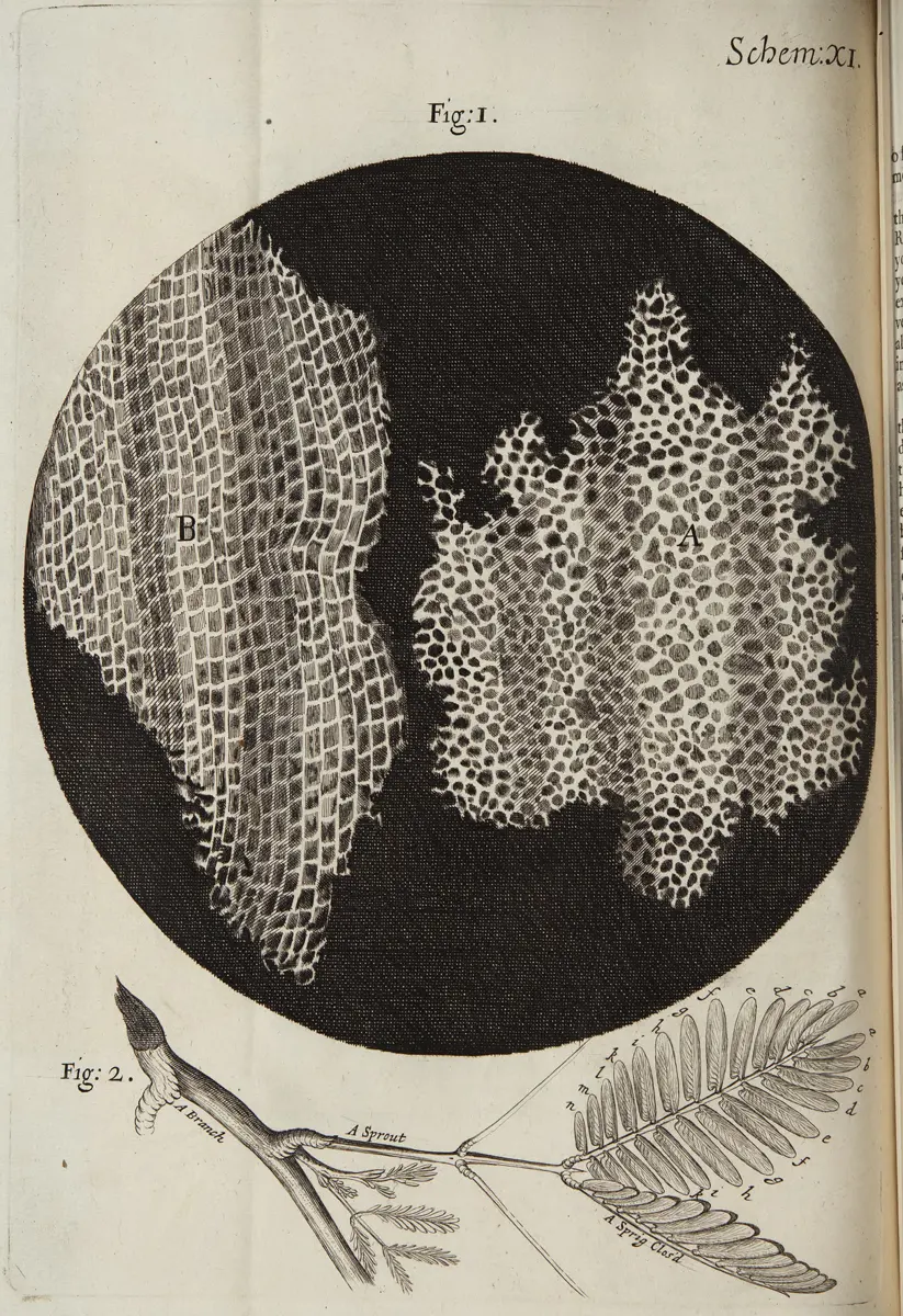
The branch of biology that studies cells is called cytology (from the Greek kytos, a hollow vessel or cell).
Why is the cell called the functional unit of life?
Pick an answer to see why.
3.1.1 Shape and size of cells
Cells come in many shapes, and a cell’s shape usually suits its job.
- Animal cells are generally irregular in shape. They may be oval, round (spherical), rod-like, long, cube-shaped or column-shaped.
- Nerve cells are thin and long, like threads, with branches at the ends: good for carrying messages over long distances. One nerve cell can run from your spine to your big toe, about a meter.
- Muscle cells are mostly spindle-shaped: long, fat in the middle and pointed at both ends. They pull by getting shorter.
- Plant cells are usually many-sided, often six-sided (hexagonal), and fit together like bricks in a wall; in an onion peel they are long and six-sided. Their stiff walls keep that shape.
- Bacteria have four main shapes: spherical (round), rod-like, filamentous (in long threads) and spiral.
Most cells are far too small to see with the naked eye. But some are big:
- The smallest known cells are those of Mycoplasma, a kind of bacterium, only about 0.2 micrometers (0.0002 mm) across.
- The biggest cell is the egg cell of an ostrich. The whole egg is about 15–18 cm long; the cell itself is its yolk (see below).
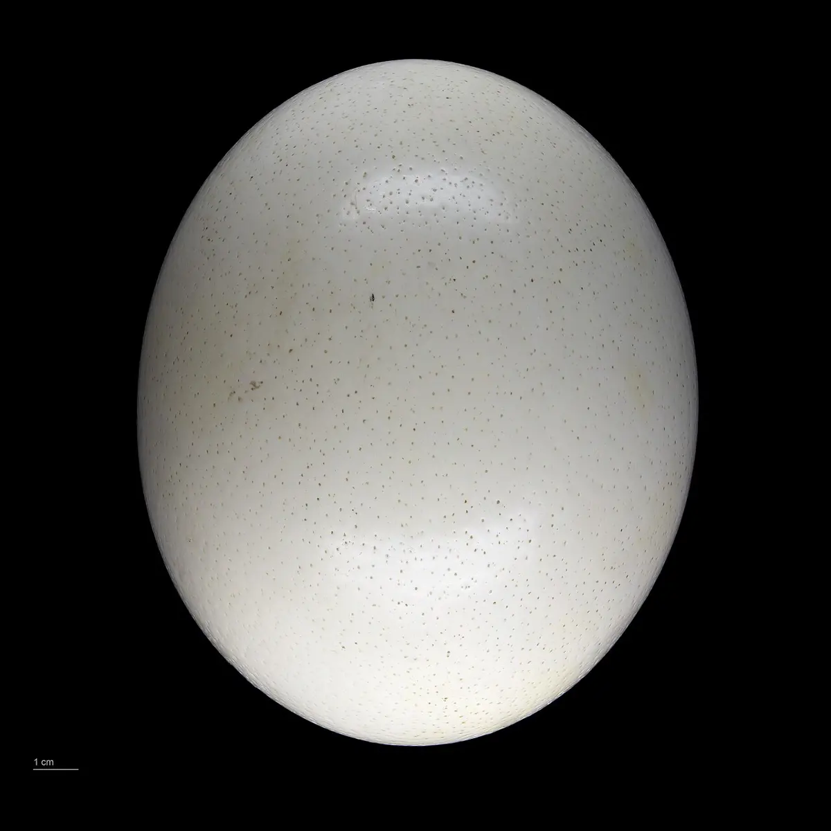
To get a feel for how small cells are, zoom in step by step.
Which of these is a single cell?
Pick an answer to see why.
Structure of a cell
A cell has several parts. To make them easy to study, we can put them into four groups:
- the cell covering (cell wall and cell membrane)
- the cytoplasm
- the cell components (organelles and inclusions)
- the nucleus
You can see real cells yourself, in an onion and in your own mouth.
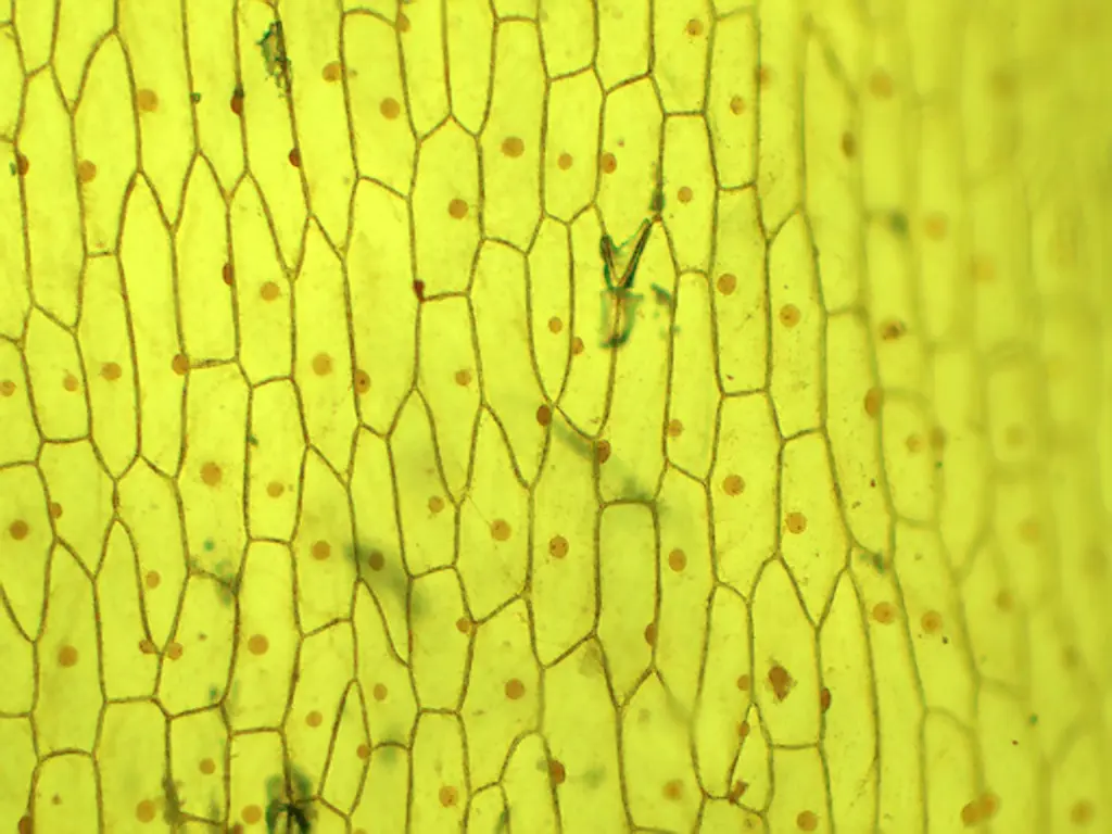
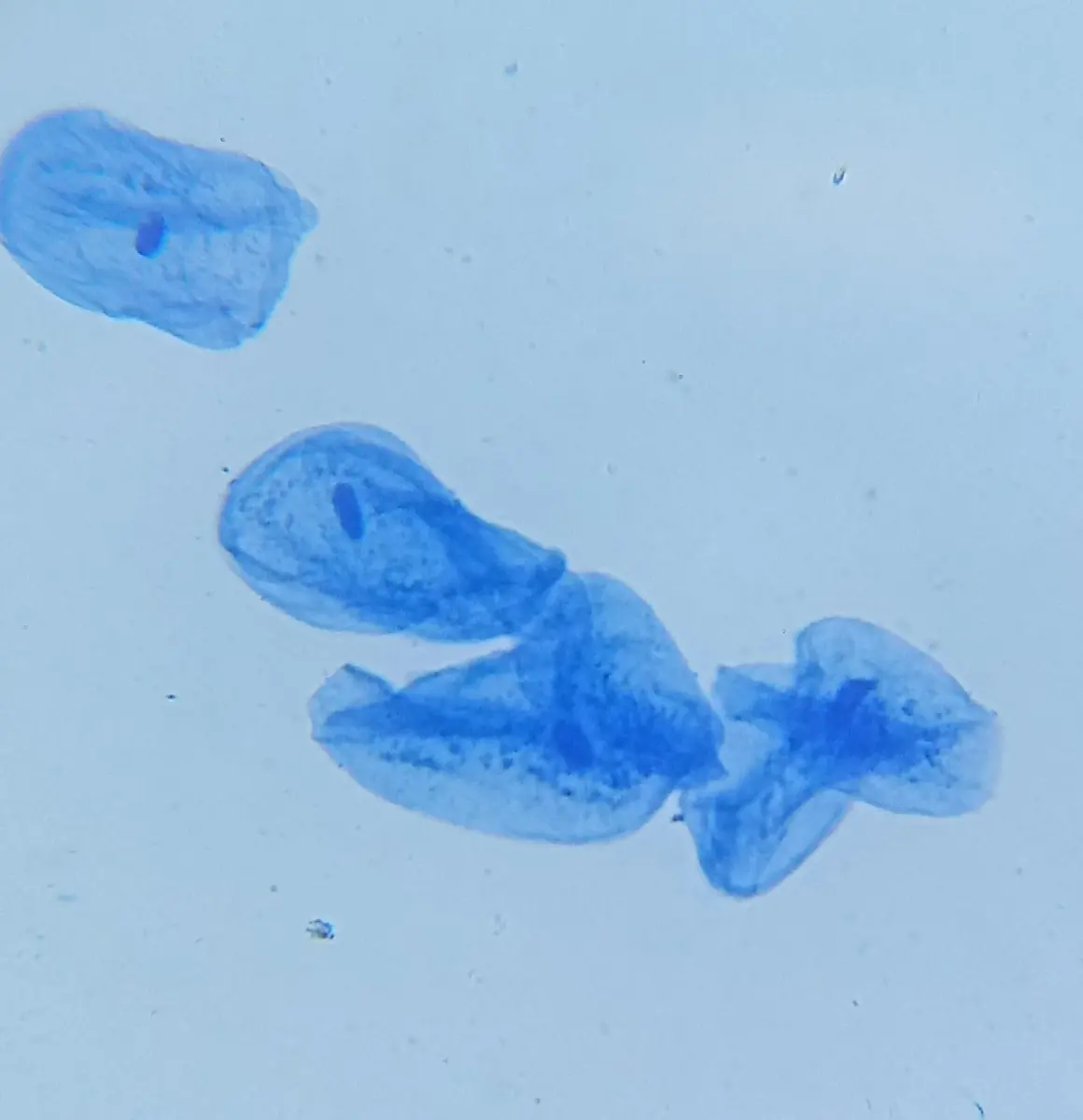
Parts of a cell
Now the four groups, one at a time.
1. The covering of the cell
The covering is the outermost layer of a cell. There are two kinds: the cell wall and the cell membrane.
The cell wall is found only in the cells of plants, fungi and bacteria. Animal cells and most protozoa (single-celled animal-like organisms) have none. It is tough, rigid, a little elastic, and non-living. In plants it is made mainly of cellulose, which is also what cotton and paper are made of.
The cell membrane, also called the plasma membrane, is found in every cell. In animals and most protozoa it is the outermost layer of the cell. In plants, fungi and bacteria it lies just inside the cell wall. It is much thinner, smoother and more elastic than the wall, and it is living.
Functions of the cell wall and the cell membrane:
- The cell wall gives support and a fixed shape to plant, bacterial and fungal cells.
- The cell wall protects the cell, for example from bursting and from attack by some germs.
- In the root hairs of plants, the cell wall lets water and dissolved minerals from the soil pass through to the membrane, which takes them in.
- The cell membrane controls what goes into and out of the cell: water, food and oxygen in, wastes out. It lets some substances through and stops others, so it is called semi-permeable (selectively permeable).
- In single-celled protozoa such as Amoeba, the cell membrane pushes out pseudopodia (“false feet”) to move and to catch food, helps get rid of wastes, and protects the cell from its surroundings.
2. Cytoplasm
The cytoplasm is a clear, sticky, jelly-like fluid that fills the cell between the cell membrane and the nucleus. It is mostly water, with dissolved salts, minerals, sugars, proteins, vitamins and more. The cell’s components, such as ribosomes, plastids, mitochondria, Golgi bodies, endoplasmic reticulum and vacuoles, float in it.
Functions of the cytoplasm:
- Most of the cell’s chemical reactions, its metabolism, take place in the cytoplasm.
- It keeps the right balance of water and minerals in the cell.
- It holds the cell components (mitochondria, Golgi bodies, plastids and others) and lets materials move between them.
3. Cell components
The working parts inside a cell are called cell components. There are two types: cell organelles and cell inclusions.
- Cell organelles (“little organs”) are the living parts. Each one does a particular job that life needs, just as the organs of your body do. Most organelles are found in both plant and animal cells; a few only in one.
- Cell inclusions are non-living substances in the cytoplasm: droplets of water, crystals of salts and minerals, stored food such as starch grains and fat droplets, pigments, and wastes. They are raw materials, stores of energy and leftovers of the organelles’ work.
Here are the main organelles, one by one.
Mitochondria (singular: mitochondrion)
A cell has many mitochondria, from a few to thousands. They are rod-shaped or thread-like, with a double membrane: a smooth outer one and a highly folded inner one. The folds give a huge surface where cellular respiration happens: glucose is broken down with oxygen, giving carbon dioxide and water and releasing energy.
Part of this energy is stored in molecules of ATP (adenosine triphosphate), which carry it to every part of the cell that needs it; the rest escapes as heat, which keeps you warm. The carbon dioxide and water are passed out of the cell. Because mitochondria release the energy that every other part of the cell runs on, they are called the power-houses of the cell. Without them, the cell can’t survive.
Functions:
- They release energy from food (glucose) by cellular respiration, and store it in ATP.
- They contain a little DNA of their own, which is passed on to the offspring (in humans, only from the mother).
Plastids
Plastids are organelles found only in plants, algae and a few protists such as Euglena. Many are colored. By their color, there are three types:
A. Chloroplasts are the green plastids. They contain the green pigment chlorophyll. They are usually round or oval, but in the alga Spirogyra they are long spiral ribbons. Each has a double membrane, and inside, stacks of disc-like membranes (grana) holding the chlorophyll, in a fluid called the stroma.
Functions of chloroplasts:
- They make plants green.
- In sunlight they carry out photosynthesis: they make carbohydrates (glucose, then starch) from carbon dioxide and water. Carbohydrates are the basic food of almost all living things, so green plants are called the producers of an ecosystem.
- They also help make fatty acids and amino acids (the building blocks of fats and proteins).
- Like mitochondria, they contain their own DNA, which is passed on to the offspring.
B. Chromoplasts are the colored plastids other than green: red, orange and yellow. They are found in the colored parts of plants: flowers, ripe fruits, some seeds, carrots and some leaves.
Functions of chromoplasts:
- They make flowers colorful, and the colors attract insects and birds, which carry pollen: pollination.
- They color ripe fruits and seeds, which attract birds and other animals that eat them and scatter the seeds: seed dispersal.
C. Leucoplasts are colorless (white) plastids, usually oval. They are found in the parts of a plant that get no light: roots, underground stems, the white flesh of fruits, and seeds (their cotyledons or endosperm).
Function of leucoplasts: they store food, such as starch (in a potato or a grain of rice), proteins and fats (oils), for later use.
A potato tuber is white inside and full of starch. Which plastids does it have most of?
Pick an answer to see why.
Ribosomes
Ribosomes are the smallest organelles: tiny grains made of RNA and protein, with no membrane around them. Some float free in the cytoplasm; many are stuck on the surface of the endoplasmic reticulum. Mitochondria and chloroplasts have small ribosomes of their own.
Functions:
- They join amino acids together to make proteins, following instructions copied from the DNA in the nucleus. So they are called the protein factories of the cell.
- This is how a cell’s genes are put to work, or expressed: a gene in the DNA is copied into a message (RNA), and the ribosome reads the message and builds the protein.
Endoplasmic reticulum (ER)
The endoplasmic reticulum, or ER for short, is a network of folded membranes forming narrow canals and flat sacs, running from the nucleus out towards the cell membrane. Where its outer surface is covered with ribosomes, it looks grainy and is called rough ER; where it has no ribosomes, it is smooth ER.
Functions:
- It forms a framework that supports the cell, so it is sometimes called the cell’s skeleton (endoskeleton).
- It contains enzymes that help in the cell’s metabolism: rough ER helps make proteins, smooth ER makes fats (lipids) and breaks down some poisons.
- It makes new membranes, which the cell needs when it grows and divides, and it helps form the Golgi body.
- It is the cell’s transport system: materials move through its canals from one part of the cell to another.
Golgi body
The Golgi body was discovered by the Italian scientist Camillo Golgi in 1898 and is named after him. It is a stack of curved, flattened, membrane-bound sacs called cisternae (like a pile of puris), with tiny bubbles called vesicles budding off its edges. It is also called the Golgi apparatus or Golgi complex.
Functions:
- It sorts, packs and sends out the substances a cell makes. Gland cells use it to secrete mucus, saliva, enzymes, hormones and, in plants, gums. So it is called the cell’s post office.
- During cell division it helps make the new cell membrane (in animal cells) and the new cell plate, which becomes the wall between the two new cells (in plant cells).
- It helps in transport inside the cell and in sorting proteins to where they are needed.
Lysosomes
A lysosome is a very small, round organelle with a single membrane, full of digestive enzymes. Lysosomes are found in most animal cells, and in large numbers in white blood cells and in the cells of the liver, kidneys, pancreas and spleen. (Plant cells do this kind of digesting mainly in their vacuole.)
Functions:
- Their enzymes digest proteins, fats and carbohydrates such as glycogen, so food taken into the cell can be used.
- They digest worn-out parts of the cell, dead cells and germs swallowed by white blood cells: they are the cell’s clean-up crew. The broken-down materials are reused.
- When a cell is badly damaged or old, its lysosomes may burst and their enzymes digest the cell itself. For this reason lysosomes are called suicide bags (or suicidal bags).
- In germinating seeds, similar digestive enzymes break down stored food to feed the growing embryo.
Vacuoles
A vacuole is not really a working organelle but a space, a sac surrounded by a membrane called the tonoplast. It is filled with a watery liquid called cell sap: water with dissolved salts, sugars, enzymes and wastes. In animal cells, vacuoles are small, or there are none. In a mature plant cell, one large vacuole fills most of the cell.
Some special vacuoles:
- Some water-dwelling bacteria have air (gas) vacuoles that help them float near the surface.
- Freshwater protozoa such as Amoeba and Paramecium have contractile vacuoles that fill with the extra water that keeps seeping in, and squirt it out, so that the cell doesn’t burst.
- Amoeba has food vacuoles, in which it stores and digests the food it has swallowed; the undigested waste is passed out.
Functions:
- Vacuoles keep the balance of water and salts in the cytoplasm, and a plant cell’s large vacuole keeps the cell firm.
- They collect wastes before these are passed out of the cell, and store water, food and pigments. (The purple of an onion’s skin and the red of a beetroot are pigments in vacuoles.)
A cell has a cell wall, chloroplasts and one large vacuole. What kind of cell is it?
Pick an answer to see why.
Now explore the two kinds of cell yourself.
4. Nucleus
The nucleus is the central part of the cell. It is usually round and is surrounded by a double membrane with pores (holes) in it. It is found in the cells of all eukaryotes: plants, animals, protozoa and fungi. (A few kinds of cell lose it as they mature, such as your red blood cells.) Bacteria are prokaryotes: they have no proper nucleus, only genetic material (DNA) lying loose in the cytoplasm, with no membrane around it.
The nucleus controls and coordinates the work of all the other parts of the cell, so it is called the brain (or control center) of the cell. It has four main parts:
- nuclear membrane
- nucleolus
- nucleoplasm
- chromatin fibers
Nuclear membrane. The nuclear membrane is the outer covering of the nucleus: two layers of fat (lipid) and protein, joined at many places to leave pores. It is elastic and protects the nucleus, and through its pores it lets substances pass into and out of the nucleus: materials from the cytoplasm come in, and the messages copied from the DNA go out to the ribosomes.
Nucleolus. The nucleolus is a dark, round body inside the nucleus, with no membrane. It is made of protein and RNA, and it makes ribosomes, which then pass out through the pores into the cytoplasm. A cell that makes a lot of protein has a big nucleolus. It disappears during cell division and appears again afterwards.
Nucleoplasm. The nucleoplasm is the liquid that fills the nucleus: water with dissolved amino acids, minerals, the building blocks of DNA and RNA, and enzymes. The nucleolus and the chromatin fibers lie in it. It provides the materials and enzymes for making new DNA and RNA, and it gets them from the cytoplasm through the pores of the nuclear membrane.
Chromatin fibers. Chromatin is a tangled network of very long, extremely thin threads, too thin to see with an ordinary microscope. It is the genetic material: the instructions that control all the cell’s work. Chromatin is made of DNA wound around proteins called histones. Each species has a fixed number of these threads: every body cell of a human has 46, in 23 pairs. Through them, the characters of the parents pass to their offspring.
When a cell is about to divide, the chromatin threads coil up tightly into short, thick, visible bodies called chromosomes. After the division they unwind and become thin, invisible chromatin again.
Functions of the nucleus:
- It controls and coordinates all the cell’s activities: its metabolism, its growth and the movement of materials in it.
- It carries the genetic characters and passes them from parents to offspring through reproduction.
- It takes part in cell division: first the nucleus divides into two, then the rest of the cell divides, so that each new cell gets a nucleus. This is how the body grows and repairs itself.
- In single-celled organisms such as Amoeba, the division of the nucleus is the start of reproduction itself: one cell becomes two new organisms.
Comparison between plant and animal cells
Which of these is found in both plant and animal cells?
Pick an answer to see why.
3.2 Interrelationship among cell, tissue and organ in human body
How is an organ like the stomach built? Do cells help it work? And how is the whole human body put together?
The human body is very complex. It has about 200 different kinds of cells, around 30 trillion cells in all, and they are organized in levels, each built from the one before:
- Cells of the same kind (or of a few kinds) group together to form a tissue: muscle tissue, nerve tissue, the lining tissue of the stomach.
- Different tissues join, at a particular place in the body, to form an organ, which does one or more particular jobs: the stomach, the heart, the lungs.
- Organs that work together, directly connected and coordinated, form an organ system, which carries out one major task: the digestive system, the respiratory system.
- All the organ systems, working together, make up the whole organism: the body.
Digestion, breathing, reproduction, transport, the building of new body material (assimilation) and the removal of wastes (excretion): every one of these begins with the work of one or many cells. It is cells that make every tissue, and that start every chemical and physical process of the body.
Take digestion. In the walls of the food pipe, stomach and intestine, the middle layer is muscle tissue, made of long muscle cells (muscle fibers). This muscle squeezes and relaxes in waves, one after another, pushing the food along: this is called peristalsis. The inner wall of the stomach and intestine is lined with glandular tissue, whose cells make digestive juices with enzymes that break down the different nutrients in food. So muscle tissue and gland tissue, each made of cells, together make the stomach work; the stomach, intestine and other organs together make the digestive system work; and the digestive system feeds every cell of the body.
So cells, tissues, organs and organ systems depend on one another to build the body and keep it working.
Which list is in the right order, from the smallest level to the largest?
Pick an answer to see why.
Exercise (3.1 and 3.2)
1. Select the correct option
a. What is the branch of science that studies cells called?
Pick an answer to see why.
b. In what form is the energy released in a cell stored before it is used by the organelles?
Pick an answer to see why.
c. What is common to the nuclear membrane and the cell membrane?
Pick an answer to see why.
d. What is marked R in this figure?
What is R?
Pick an answer to see why.
e. Which of these is done by both plant and animal cells?
Pick an answer to see why.
f. What would happen if a cell had no cell membrane?
Pick an answer to see why.
g. Which function of the nucleus is needed for the growth and development of the body?
Pick an answer to see why.
2. Differentiate between
Cytoplasm and nucleoplasm
Answer
| Cytoplasm | Nucleoplasm |
|---|---|
| the jelly-like fluid between the cell membrane and the nucleus | the liquid inside the nucleus |
| holds the organelles: mitochondria, ER, Golgi body, plastids, ribosomes | holds the nucleolus and the chromatin fibers |
| most of the cell’s metabolism happens in it | DNA and RNA are made in it |
| surrounded by the cell membrane | surrounded by the nuclear membrane |
Chloroplast and chromoplast
Answer
| Chloroplast | Chromoplast |
|---|---|
| green plastid | colored plastid other than green (red, orange, yellow) |
| contains chlorophyll | contains other pigments, such as carotene and lycopene |
| in leaves and young green stems | in flowers, ripe fruits, some roots (carrot) and seeds |
| makes food by photosynthesis | attracts insects and birds for pollination and animals for seed dispersal |
Cell wall and cell membrane
Answer
| Cell wall | Cell membrane |
|---|---|
| found only in plants, fungi and bacteria | found in all cells |
| non-living | living |
| thick, rigid, made of cellulose in plants | thin, flexible, made of lipid and protein |
| lets most substances through (fully permeable) | lets only some substances through (semi-permeable) |
| gives shape and support | controls what enters and leaves the cell |
Cell organelles and cell inclusions
Answer
| Cell organelles | Cell inclusions |
|---|---|
| living parts of the cell | non-living substances in the cell |
| each does a particular job, such as respiration or making proteins | are raw materials, stored food, pigments or wastes |
| e.g. mitochondria, chloroplasts, ribosomes, Golgi body | e.g. starch grains, fat droplets, crystals of salts, water droplets |
3. Give reasons
Ribosomes are called the protein factories of the cell.
Answer
Ribosomes join amino acids together, in the order set by the instructions from the DNA, to make all the cell’s proteins. Since they produce the cell’s proteins, as a factory produces goods, they are called its protein factories.
The cell wall gives a fixed shape to plant cells.
Answer
The cell wall is a thick, rigid layer of cellulose around the cell. It doesn’t change shape easily, and the water-filled vacuole presses the cell against it from inside. So the cell keeps the wall’s shape.
Metabolism takes place in the cytoplasm.
Answer
The cytoplasm holds the water, dissolved substances and enzymes that the cell’s chemical reactions need, and the organelles (mitochondria, ribosomes, ER and others) float in it. So most of the cell’s chemical reactions, its metabolism, take place in the cytoplasm.
The cell is called the basic unit of life.
Answer
Every living thing is made of one or more cells, so the cell is the structural unit of life. All of life’s processes (respiration, digestion, reproduction and others) happen in and by cells, so it is also the functional unit. And new cells come only from existing cells. The cell is the smallest thing that is alive by itself, so it is the basic unit of life.
Cells, tissues and organs must be properly coordinated to carry out the life processes.
Answer
No single cell or tissue can carry out a life process alone. In digestion, for example, muscle tissue in the stomach and intestine pushes food along while gland tissue makes the enzymes that digest it, and both must work at the right time and place. Cells make tissues, tissues make organs and organs make systems, each depending on the others. If they are not coordinated, the process fails.
4. Answer these questions
What is a cell?
Answer
A cell is the basic structural and functional unit of life: the smallest unit of a living thing that is itself alive. It has a cell membrane, cytoplasm and (in eukaryotes) a nucleus, and all the processes of life happen in it. Every organism is made of one or more cells.
Draw a labeled diagram of a plant cell and an animal cell.
Answer
Draw the two cells side by side, as in Figure 3.4.
Plant cell: a box-like shape with a thick cell wall, the cell membrane just inside it, cytoplasm, a large central vacuole, a nucleus pushed to one side, chloroplasts, mitochondria and a Golgi body.
Animal cell: a rounded, irregular shape with only a cell membrane outside, cytoplasm, a central nucleus, mitochondria, a Golgi body, lysosomes and small vacuoles.
Use a sharp pencil, label with straight lines that don’t cross, and give each drawing a title.
Where are mitochondria found in a cell? Write their functions.
Answer
Mitochondria are found in the cytoplasm of the cells of all eukaryotes, plants and animals alike: many of them, especially in busy cells such as muscle cells.
Functions:
- They carry out cellular respiration: they break down glucose with oxygen and release energy.
- They store this energy in ATP, which supplies the rest of the cell, so they are called the power-houses of the cell.
- They have a little DNA of their own, which is passed to the offspring.
Which organelle of the cell digests itself, and why?
Answer
The lysosome. It is a bag full of strong digestive enzymes. When a cell is old or badly damaged, its lysosomes burst, and their enzymes digest the cell itself. This clears away dead and useless cells so their materials can be reused. Because of this self-digestion, lysosomes are called suicide bags.
Make a list of the cell organelles and cell inclusions found in a cell.
Answer
Cell organelles: mitochondria, plastids (chloroplasts, chromoplasts, leucoplasts), ribosomes, endoplasmic reticulum, Golgi body, lysosomes (and the nucleus, the largest organelle). Vacuoles are often listed with them.
Cell inclusions: droplets of water, crystals of salts and minerals, starch grains, fat droplets, pigments, and waste products.
Mention the functions of plastids in a plant cell.
Answer
- Chloroplasts make the plant green and make its food by photosynthesis.
- Chromoplasts color flowers, which attract insects and birds for pollination, and ripe fruits, which attract animals that disperse the seeds.
- Leucoplasts store food such as starch, proteins and oils in roots, tubers and seeds.
Complete the Venn diagram comparing plant and animal cells.
Answer
Draw two overlapping circles: Plant on the left, Animal on the right, and Both where they overlap.
Plant only: cell wall, chloroplasts, one large central vacuole, a regular shape, stores starch.
Both: cell membrane, cytoplasm, nucleus, mitochondria, Golgi body, ER, ribosomes.
Animal only: no cell wall, lysosomes, centrioles, small vacuoles (or none), an irregular shape, stores glycogen.
Mention the roles of the Golgi body and the vacuole in a cell.
Answer
Golgi body: sorts, packs and sends out the cell’s products (mucus, saliva, enzymes, hormones, gums); helps make the new cell membrane or cell plate during cell division; helps in transport inside the cell.
Vacuole: stores cell sap (water, salts, sugars, pigments); keeps the balance of water and salts, and keeps a plant cell firm; collects wastes before they are passed out of the cell.
Study the figure and answer:
- i.Copy the figure and label A, B, C and D.
- ii.What is the function of B?
- iii.What happens if a cell has no nucleus?
Answer
- i.The figure shows a nucleus. A: nucleolus. B: nuclear membrane. C: nuclear pore. D: chromatin fibers (lying in the nucleoplasm).
- ii.The nuclear membrane protects the nucleus and separates it from the cytoplasm, and through its pores it controls what passes into and out of the nucleus.
- iii.Without a nucleus, a cell can’t control its activities, can’t make new proteins for long, and can’t divide, so it soon dies. (Mature red blood cells in humans have no nucleus, and that is why they live only about 120 days and can’t divide.)
In your opinion, which organelle of the cell is the most important, and why?
Answer
There is no single correct answer; a good answer gives a reason. For example:
The nucleus, because it holds the DNA, the instructions for everything the cell does; it controls all the other parts and is needed for the cell to divide. Without it, the cell soon dies.
Or the mitochondria, because every activity of the cell needs energy, and without them most cells can’t release enough energy to live.
What would happen to a plant’s life processes if it had no chloroplasts?
Answer
Without chloroplasts the plant couldn’t carry out photosynthesis, so it couldn’t make its own food. Its respiration, growth and reproduction would soon stop for lack of food and energy, and the plant would die once its stored food ran out. Animals that eat plants would lose their food too.
Why is the nucleus called the brain of the cell? Explain.
Answer
Just as the brain controls the body, the nucleus controls the cell. It holds the DNA, the cell’s instructions, and decides which proteins are made, and so controls the cell’s metabolism, growth and division. It also passes on the characters to the offspring. So it is called the brain, or control center, of the cell.
If no metabolism happened in a cell, what would be the effect on the organism’s life processes?
Answer
Metabolism is all the chemical reactions in a cell: releasing energy from food, making new materials and breaking down wastes. Without it, no energy would be released, so the cell couldn’t move, grow, take in materials or divide; no new substances would be made; and wastes would not be dealt with. The cell would die, and if this happened in all the cells, the organism’s life processes (digestion, respiration, excretion, growth, reproduction) would stop and the organism would die.
3.3 Classification of living beings
There are millions of kinds of living things on Earth. Each kind has its own features that make it different from the others. They differ in their habitat (where they live), their mode of nutrition (how they get food), how they reproduce, and their body structure.
But look carefully and you also find likenesses. A bat flies and a rabbit runs, yet both give birth to live young and feed them on milk until they are old enough to eat solid food. Both have hair. So bats and rabbits are put in the same group, the mammals (class Mammalia). In the same way, all known organisms are sorted into groups and smaller groups.
Sorting living things into groups by their likenesses and differences is called classification. Its aims:
- It makes the study of millions of organisms easy and systematic: study one member of a group and you know a lot about all of them.
- It shows how organisms are related, and so gives an idea of how life has evolved on Earth.
Living things can be classified in different ways, but there are two main systems: the two-kingdom system and the five-kingdom system.
3.3.1 Two-kingdom system of classification
The two-kingdom system was proposed by the Swedish biologist Carl Linnaeus in the 1700s (in his books Systema Naturae, 1735, and Species Plantarum, 1753). He sorted all living things into just two kingdoms: Plantae (plants) and Animalia (animals).
Basis of the two-kingdom system. Linnaeus looked at nutrition, movement, growth and reproduction:
- Organisms that move about, take in ready-made food (heterotrophic nutrition) and stop growing after a certain age were put in the animal kingdom.
- Organisms that don’t move about, make their own food (autotrophic nutrition) and keep growing all their lives were put in the plant kingdom.
Features of the two-kingdom system:
- All living things are divided into two kingdoms, plants and animals.
- It was the first scientific system of classification.
- It is simple: plants and animals are easy to tell apart.
Drawbacks of the two-kingdom system:
- Organisms that can’t make their own food were put in the plant kingdom with those that can: fungi, which have no chlorophyll, were put with algae, which do photosynthesis.
- Prokaryotes (bacteria, with no proper nucleus) and eukaryotes (such as algae, with a nucleus) were put in the same kingdom.
- It doesn’t separate unicellular (one-celled) from multicellular organisms.
- Some organisms fit in neither kingdom. A lichen is a fungus and an alga living together as one; Euglena makes food like a plant but swims and has no cell wall, like an animal.
Five-kingdom system of classification
To remove these drawbacks, the American ecologist Robert H. Whittaker proposed the five-kingdom system in 1969, more than 200 years after Linnaeus. His five kingdoms are Monera, Protista, Fungi (also called Mycota), Plantae and Animalia.
Basis of the five-kingdom system. Whittaker sorted organisms by:
- type of cell: prokaryotic (no proper nucleus) or eukaryotic (with a nucleus);
- body structure: unicellular or multicellular;
- mode of nutrition: autotrophic (making their own food) or heterotrophic (taking food from others);
- role in the ecosystem: producer, consumer or decomposer;
- phylogenetic relationship: the line of evolution, from the simplest to the most developed.
So:
| Kingdom | Who is in it | Examples |
|---|---|---|
| Monera | all unicellular prokaryotes | bacteria, blue-green algae (cyanobacteria), mycoplasma, archaea |
| Protista | unicellular eukaryotes (except fungi such as yeast) | Amoeba, Paramecium, Euglena, Plasmodium |
| Fungi (Mycota) | eukaryotes with a cell wall and no chlorophyll that absorb food from dead matter (saprophytes) or from living hosts (parasites), digesting it outside their bodies | mushroom, yeast, Mucor, yarsagumba |
| Plantae | multicellular green autotrophs: producers | moss, fern, pine, mustard |
| Animalia | multicellular eukaryotic consumers with no cell wall | earthworm, tortoise, bat, human |
Features of the five-kingdom system:
- Unicellular and multicellular organisms are put in separate kingdoms.
- Prokaryotes and eukaryotes are separated.
- Autotrophs (Plantae) are separated from heterotrophs (Animalia, which take in food and digest it inside, and Fungi, which absorb food).
- The kingdoms are arranged in order from the simplest to the most developed, which shows the path of evolution.
Mushrooms were put in the plant kingdom by Linnaeus. Why are they in a separate kingdom, Fungi, today?
Pick an answer to see why.
In which kingdom is Amoeba placed in the five-kingdom system?
Pick an answer to see why.
Try sorting here first.
3.4 Microorganism
There are many kinds of living things around us that are too small to see with the naked eye. They are found in air, water and soil, in food left uncovered, in dirt, and on and inside the bodies of living, dead and decaying organisms. These are called microorganisms (micro, small). They affect our daily lives in many ways, directly and indirectly. The rotting of the broken egg is their work.
Microorganisms include bacteria, some fungi (yeast, molds), some algae (Chlamydomonas, Chlorella), protozoa (Amoeba, Paramecium, Plasmodium, Vorticella), and viruses (which are not quite living, as you will see). Some small worms, like the eggs and young of the blood fluke Schistosoma, are microscopic too. Microorganisms are commonly called germs or microbes. The branch of biology that studies them is microbiology.
(A) Virus
Viruses are the smallest germs. They can infect plants, animals, bacteria, fungi, and every other kind of organism. The word virus is Latin for poison.
A virus is not a cell: it is acellular. Its body is only some genetic material, DNA or RNA, wrapped in a coat of protein called a capsid. Some viruses, such as the coronavirus and the flu virus, have an extra, fatty outer covering called an envelope. Viruses come in many shapes and sizes.
Viruses show features of both living and non-living things, so they are called a link (or bridge) between the living and the non-living:
- Outside a living cell, a virus is like a non-living particle. It doesn’t eat, drink, breathe or grow, and has no metabolism of its own. Some can even be crystallized like salt.
- Inside a living host cell, it takes over the cell and uses the cell’s own materials and machinery to make hundreds of copies of itself, which usually damages or kills the cell. Because it can multiply only inside a living host, a virus is called an obligate parasite.
Because a virus is not a cell and has no metabolism, it is not counted as an organism, and it belongs to none of the five kingdoms. Viruses are found everywhere: in air, water and soil, and in every kind of living thing.
Examples: rhinovirus (the common cold), coronaviruses (including SARS-CoV-2, which causes COVID-19), the tobacco mosaic virus (a disease of tobacco and tomato leaves), bacteriophages (viruses that infect bacteria) and even virophages, viruses that infect other viruses.
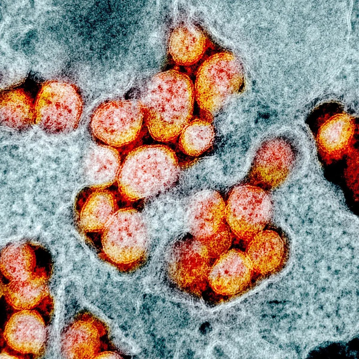
Why is a virus called a link between living and non-living things?
Pick an answer to see why.
(B) Bacteria
Bacteria (singular: bacterium) are unicellular prokaryotes: single cells with no proper nucleus, so they belong to the kingdom Monera. Their cells may be round, rod-shaped, thread-like (filamentous) or spiral (Figure 3.2). They live everywhere: in air, water and soil, and on and inside other organisms. Some live in boiling hot springs, in ice, or in very salty water.
Almost every bacterium has a cell wall (tiny Mycoplasma is one of the few without), and many have a slimy capsule outside it, which protects them. Some can turn into tough spores that survive heat, cold and dryness for years, waiting for better conditions.
Some bacteria make their own food (they are autotrophic), like the green cyanobacteria. But most are saprophytic: they feed on dead matter, which makes them the great decomposers of every ecosystem. Some are parasitic and cause diseases in plants and animals.
Examples: cyanobacteria (blue-green algae), Rhizobium, Azotobacter, Salmonella, Pseudomonas, Lactobacillus.
(C) Amoeba
Amoeba is a unicellular eukaryote (a protozoan), in the kingdom Protista. It lives in water, damp soil and decaying matter, and some kinds live inside other animals.
It has no fixed shape: it can push out pseudopodia (“false feet”) from any point of its cell membrane. With these temporary bulges it moves, and it catches food by flowing around it and enclosing it in a food vacuole. Its single cell does everything: digestion, excretion, reproduction (by splitting into two) and the rest.
There are many kinds of amoebae:
- Entamoeba histolytica lives in water and food contaminated with feces. If it gets into the intestine, it causes amoebic dysentery (amoebiasis): stomach pain and diarrhea with blood and mucus. Boiling or filtering drinking water, and washing hands, prevents it.
- Naegleria fowleri lives freely in warm fresh water, such as warm ponds and poorly cleaned pools. Drinking such water is not what harms you: it is dangerous only if water goes up the nose, for example while diving or swimming. From there it can reach the brain and cause a rare but usually deadly brain infection.
(D) Fungi
Fungi (singular: fungus) are eukaryotes, unicellular or multicellular, that have a cell wall but no chlorophyll. They form the kingdom Fungi (Mycota). Most grow on dead and decaying matter and absorb their food from it, so they are called saprophytes. Some are parasites: they infect plants and animals and cause fungal diseases, such as rust in wheat and ringworm and athlete’s foot in people.
A fungus’s body is usually made of very thin threads called hyphae; the whole network of threads is called the mycelium (plural mycelia). Yeast is an exception: it is a single cell.
Examples: Mucor (bread mold), mushrooms, yeast, yarsagumba (Cordyceps, which grows out of a caterpillar), Penicillium, and Puccinia, the fungus that causes rust disease in wheat.
3.4.1 Effects of microorganisms
Microorganisms are often thought of only as “germs”, but most of them are harmless, and many are essential to us.
Positive effects
- Amoebae and other protozoa feed on bacteria. In soil, water and the tanks that clean sewage, they keep bacteria in check and help clean the water.
- Most bacteria are decomposers. They break down the excreta and dead bodies of organisms, turning complex organic substances back into simple minerals, which makes the soil fertile again. Without them, the world would be piled high with the dead. Bacteria such as Clostridium decompose dead matter; others, such as Nitrosomonas and Nitrobacter, then turn the ammonia that is released into nitrates, which plants can use. In this way bacteria keep the ecosystem in balance.
- Rhizobium bacteria live in the root nodules of leguminous plants (beans, peas, lentils, gram). They take nitrogen from the air and turn it into compounds the plant can use, which makes the soil more fertile and increases crop yields.
- Lactobacillus and Lactococcus bacteria turn milk into curd (dahi) and yoghurt; Lactococcus lactis (also called Streptococcus lactis) is used to make cheese from milk.
- Bacteria and fungi are used to make medicines: antibiotics (penicillin comes from the fungus Penicillium), vaccines, vitamins, and even human insulin, which is now made by bacteria and yeast given the human insulin gene.
- Yeast, a fungus, turns the sugar in foods into alcohol and carbon dioxide. This is fermentation. Factories use yeast to make alcohol and, with the help of other bacteria, vinegar (acetic acid). The carbon dioxide it makes is what makes bread and sel roti dough rise.
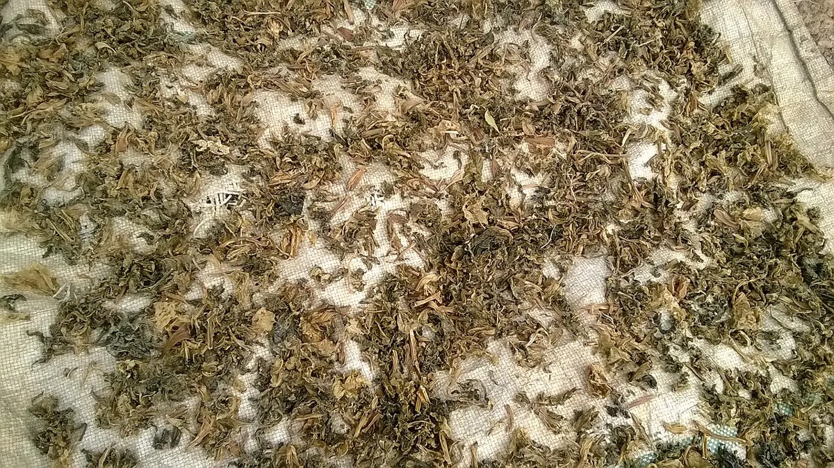
- In Nepal, microbes have been used at home for centuries: yeast and molds in marcha to make jaand and raksi, and lactic acid bacteria to ferment gundruk, sinki and pickles.
- Bacteriophages kill bacteria. They are used in some countries to treat bacterial infections that antibiotics can no longer cure (such as some kinds of dysentery and diarrhea), and in cleaning water and protecting food.
- In genetic engineering, harmless viruses are used to carry genes into cells: to add, replace or repair genes, to develop high-yielding plants and animals, and to treat some diseases.
- Viruses lie on the border between living and non-living, so studying them helps us understand how life may have begun and how it has evolved.
Negative effects
- Microorganisms cause most communicable diseases (diseases that spread from one to another) in people, animals and plants. Diseases lower the quality and amount of production in every kind of farming: crops, fish farming, silkworm farming (sericulture), bee-keeping (apiculture), fruit growing (horticulture), animal husbandry and nurseries. Human diseases include tuberculosis (bacteria), AIDS (a virus, HIV), pneumonia (bacteria or viruses), typhoid (bacteria, Salmonella Typhi) and COVID-19 (a virus). Plant diseases include tobacco mosaic (a virus), blight and root rot (fungi and bacteria).
- Fruit, vegetables and cooked food left uncovered for hours or days start to smell and rot. Microbes such as Clostridium get into the food from air, water, hands or flies, multiply, and make poisons (toxins). Eating such food causes food poisoning: diarrhea, vomiting, fever, loss of water from the body (dehydration), a dry mouth (xerostomia) and headache. Food poisoning can be caused by Clostridium, Salmonella, norovirus and Giardia; molds such as Mucor spoil food, and some molds make poisons of their own.
- Pimples, boils and pus in wounds are caused by infection with harmful microbes. If the germs or their poisons get into the blood, they can cause sepsis, in which organs fail one after another, and it can kill. To stay safe, keep wounds and pimples clean with gauze or cotton soaked in a suitable antiseptic (such as Dettol, Betadine or Savlon), dress wounds on time, take antibiotics only as a doctor prescribes, and keep yourself clean.
- Microbes spoil stored grain, vegetables and other food, and farmers and shopkeepers lose a great deal.
- At rubbish dumps, microbes acting on organic waste give off foul smells that pollute the air around.
Which of these is a useful effect of bacteria?
Pick an answer to see why.
3.4.2 Methods of food preservation
We keep food for a long time by preserving it. Preserving does two things: it keeps the food’s nutrients from breaking down, and it keeps microorganisms from spoiling it. To do this, the food is given physical and chemical conditions in which microbes can’t grow and the food doesn’t decay: dryness, cold, salt, sugar, acid, or no air.
Protecting food from microorganisms while keeping its nutritional value is called food preservation.
Microbes need four things to grow: food, water, warmth and (most of them) air. Every method of preserving food takes away at least one of these, or adds something they can’t stand, like strong salt or acid.
1. Dry preservation
Drying food until very little moisture is left, before storing it, is called dry preservation. Without water, microbes can’t grow, and with only a little moisture left the food keeps its nutrients. It can then be stored at room temperature for months or even years. It is very easy and cheap.
In Nepal it has been used for centuries for grains, dried meat (sukuti), dried fish, dried fruits, nuts, dried vegetables and fermented vegetables (gundruk, sinki). Food is dried for some days in moderate warmth (about 20–30 °C), in the sun or in an electric dryer, until it is completely dry outside and has very little moisture inside.
2. Wet preservation
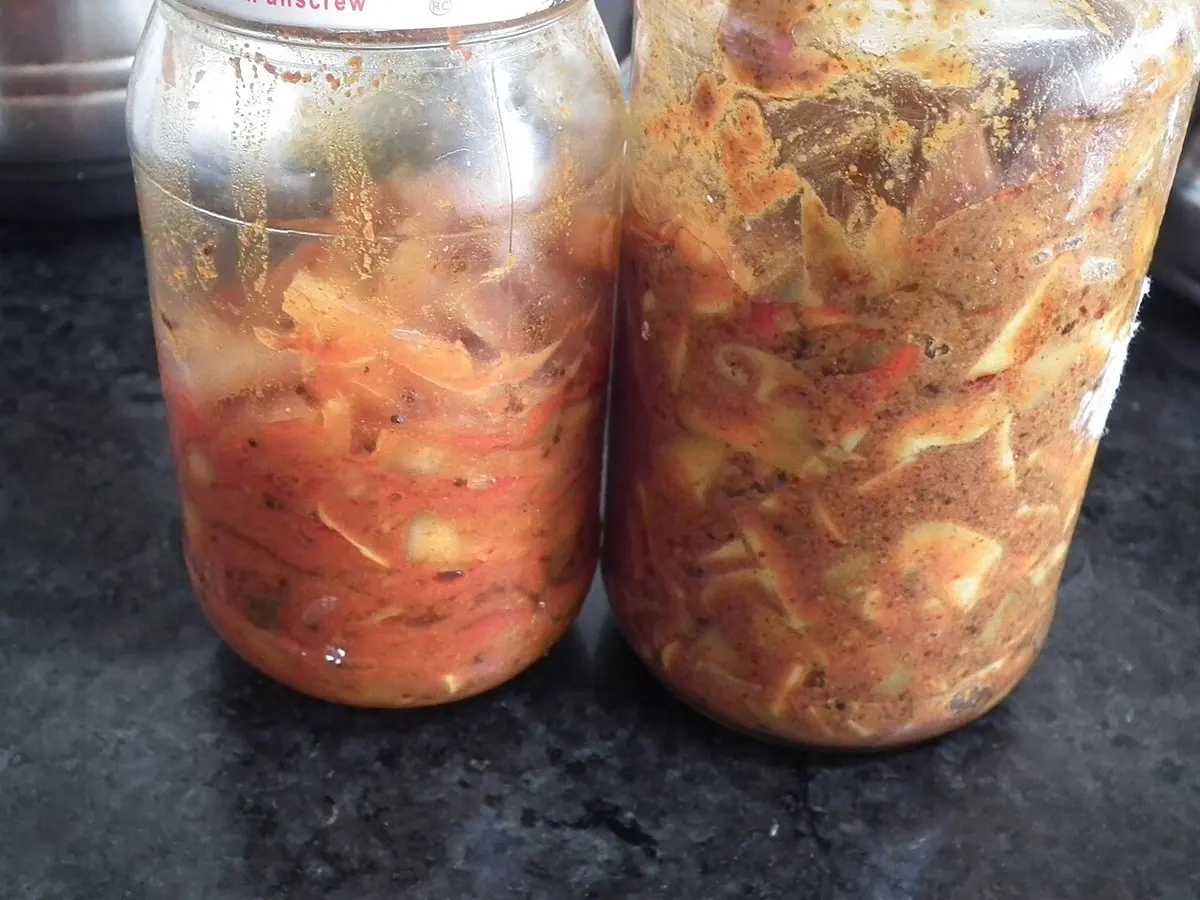
Keeping food dipped in a liquid in which microbes can’t grow is called wet preservation. The common liquids are a strong salt solution (brine), citric acid (the juice of lemons and other citrus fruits), vinegar, and mustard oil or olive oil.
The food must be free of germs to start with, so it is washed or boiled and then dried well before it goes into the liquid. The jar must be closed airtight, so that no germs or mold spores get in from the air. In Nepal, pickles (achar) of lemon, chilli, ginger, garlic, fenugreek (methi), radish and bamboo shoot (tama) have been made this way since ancient times.
3. Cold preservation
Keeping food in closed rooms or boxes at a low temperature, usually between about −18 °C and 4 °C, is called cold preservation. At these temperatures most microorganisms become inactive: they grow very slowly or not at all. Examples: keeping raw or cooked food in a refrigerator (about 4 °C) or freezer (about −18 °C), and storing potatoes, fruits and vegetables in cold stores for months.
An old way of keeping farm produce cool is still used in Nepal: potatoes, ginger, turmeric, squash (iskus) and fresh paddy are kept in a dry, well-covered pit in the ground, where it stays cool all year.
See how much difference the place you keep food makes.
Exercise (3.3 and 3.4)
1. Select the best answer
a. Who proposed the two-kingdom system of classification?
Pick an answer to see why.
b. To which kingdom do protozoa belong in the five-kingdom system?
Pick an answer to see why.
c. Why are bacteria called decomposers?
Pick an answer to see why.
d. Which virus causes COVID-19?
Pick an answer to see why.
e. What is the main thing to aim for when preserving food?
Pick an answer to see why.
f. Which of these is a feature of the five-kingdom system?
Pick an answer to see why.
g. What should be done to preserve fresh, wet gundruk?
Pick an answer to see why.
2. Differentiate between
Bacteria and viruses
Answer
| Bacteria | Viruses |
|---|---|
| are cells (unicellular prokaryotes) | are not cells (acellular) |
| have a cell wall, cytoplasm, ribosomes and DNA | have only DNA or RNA in a protein coat |
| carry out metabolism; can grow and multiply on their own | have no metabolism; multiply only inside a living host cell |
| can be seen with a light microscope | can be seen only with an electron microscope |
| belong to the kingdom Monera | belong to no kingdom |
| killed by antibiotics | not affected by antibiotics |
| e.g. Rhizobium, Lactobacillus, Salmonella | e.g. coronavirus, rhinovirus, bacteriophage |
The two-kingdom system and the five-kingdom system
Answer
| Two-kingdom system | Five-kingdom system |
|---|---|
| proposed by Carl Linnaeus (1700s) | proposed by R. H. Whittaker (1969) |
| two kingdoms: Plantae and Animalia | five kingdoms: Monera, Protista, Fungi, Plantae, Animalia |
| based on nutrition, movement, growth and reproduction | based on cell type, body structure, nutrition, role in the ecosystem and evolution |
| prokaryotes and eukaryotes are not separated | prokaryotes and eukaryotes are separated |
| unicellular and multicellular organisms are not separated | unicellular and multicellular organisms are separated |
| fungi are put with plants | fungi have their own kingdom |
3. Give reasons
A virus is called a very small infectious particle, not an organism.
Answer
A virus has no cell, no cytoplasm and no metabolism: it is only genetic material (DNA or RNA) in a protein coat. It can’t feed, grow or multiply by itself, only inside a living host cell, where it causes infection. Since it is not made of cells and can’t live on its own, it is called an infectious particle rather than an organism.
We should not eat stale and smelly food.
Answer
A bad smell shows that microorganisms have grown in the food in large numbers and are breaking it down. Many of them make poisons (toxins). Eating such food can cause food poisoning, with vomiting, diarrhea, fever and dehydration. (Food can be unsafe even before it smells, so leftovers should be kept cold and eaten soon.)
The five-kingdom system gives a proper place to almost all organisms.
Answer
It sorts organisms by many features together: whether the cell has a nucleus, whether the body has one cell or many, how the organism gets food, and its role in the ecosystem. So prokaryotes (Monera), unicellular eukaryotes (Protista) and fungi, which didn’t fit the two-kingdom system, each get their own kingdom, and organisms like Euglena and bacteria are no longer forced into “plants” or “animals”.
Fruits don’t spoil if they are preserved after drying well.
Answer
Microorganisms need water to grow and multiply. Well-dried fruit has very little water left, so bacteria and molds can’t grow on it, and it doesn’t rot. That is why dried fruits, such as raisins, dates and dried apricots, keep for months.
4. Answer these questions
What is a microorganism? Give an example of each kind.
Answer
Living things too small to be seen with the naked eye, which can be seen only with a microscope, are called microorganisms (microbes or germs).
Examples: bacteria, Lactobacillus; fungi, yeast; algae, Chlamydomonas; protozoa, Amoeba; viruses (not true organisms), the coronavirus.
What are the methods of food preservation? Describe any one of them, and mention why food preservation is important.
Answer
The main methods are dry preservation, wet preservation and cold preservation.
Dry preservation: food is dried in the sun or in a dryer, at a moderate warmth (about 20–30 °C), until very little moisture is left. Microbes can’t grow without water, so the food keeps for months at room temperature. Grain, sukuti, dried fish, gundruk and dried fruit are preserved this way.
Importance: preservation keeps food safe from microbes and food poisoning, keeps its nutrients, lets us store food for times when it isn’t grown (for winter, or for a bad harvest), reduces waste and loss to farmers, and lets food be carried to faraway places.
What is food poisoning? Mention its causes and how to be safe from it.
Answer
Illness caused by eating food or drinking water contaminated with harmful microorganisms or the poisons they make is called food poisoning. Its signs are vomiting, diarrhea, stomach pain, fever, dehydration, a dry mouth and headache.
Causes: microbes such as Clostridium, Salmonella, norovirus and Giardia, which get into food left uncovered, kept warm for hours, cooked with dirty hands or utensils, or made with dirty water.
To be safe: wash your hands before cooking and eating; keep food covered from flies and dust; eat food fresh and hot, or keep leftovers in a fridge; don’t eat stale, smelly or moldy food; drink boiled or filtered water; wash fruit and vegetables well.
Why is the five-kingdom system more advanced than the two-kingdom system?
Answer
The five-kingdom system uses more and deeper features: the type of cell, the number of cells, the mode of nutrition, the role in the ecosystem and evolutionary relationships. So it separates prokaryotes from eukaryotes and unicellular from multicellular organisms, gives fungi their own kingdom apart from green plants, and arranges the kingdoms in the order of evolution. Organisms that had no proper place in the two-kingdom system now have one.
Make a list of two diseases caused by each kind of microorganism.
Answer
| Microorganism | Diseases |
|---|---|
| virus | COVID-19, the common cold (also flu, measles, AIDS) |
| bacteria | typhoid, tuberculosis (also cholera, pneumonia) |
| protozoa | malaria (Plasmodium), amoebic dysentery (Entamoeba) |
| fungi | ringworm, athlete’s foot (in plants, rust of wheat) |
What are the common words for microorganisms?
Answer
Germs and microbes.
What is classification? How does classification make the study of organisms easier?
Answer
Sorting living things into groups and smaller groups according to their likenesses and differences is called classification.
It makes study easier because instead of studying millions of organisms one by one, we study groups: the features of a group belong to all its members, so knowing one member tells us a lot about the others. It puts organisms in order, makes them easy to identify and name, and shows how they are related and how they evolved.
Write any four advantages of microorganisms.
Answer
- Bacteria and fungi decompose dead bodies and wastes and return minerals to the soil, keeping it fertile.
- Rhizobium in the root nodules of legumes fixes nitrogen from the air, increasing crop yields.
- Lactobacillus turns milk into curd; yeast makes bread rise and is used to make alcohol; lactic acid bacteria ferment gundruk.
- Antibiotics, vaccines, vitamins and insulin are made with the help of bacteria and fungi.
Mention the merits and drawbacks of the two-kingdom system.
Answer
Merits: it was the first scientific system of classification; it is simple; it divides living things into two clear groups, plants and animals, which are easy to tell apart.
Drawbacks: it puts prokaryotes (bacteria) with eukaryotes; it doesn’t separate unicellular from multicellular organisms; it puts fungi, which have no chlorophyll, with green plants; and organisms such as Euglena and lichens fit in neither kingdom.
Mention the bases and features of the five-kingdom system.
Answer
Bases: the type of cell (prokaryotic or eukaryotic), the body structure (unicellular or multicellular), the mode of nutrition (autotrophic or heterotrophic), the role in the ecosystem (producer, consumer or decomposer), and evolutionary relationships.
Features: unicellular and multicellular organisms are separated; prokaryotes and eukaryotes are separated; autotrophs (Plantae) are separated from heterotrophs (Animalia and Fungi); and the kingdoms are arranged from the simplest to the most developed, showing the path of evolution.
Study the figure and answer:
- i.What is shown in the figure?
- ii.Name a disease it causes, and two ways to prevent that disease.
Answer
- i.A coronavirus (such as SARS-CoV-2): a round virus with an envelope covered in spike proteins, and RNA inside.
- ii.It causes COVID-19. Prevention: get vaccinated; wash hands often with soap and water; wear a mask in crowded places when there is an outbreak; keep sick people apart and cover your mouth and nose when coughing or sneezing.
The two-kingdom system didn’t separate prokaryotes and eukaryotes. How does the five-kingdom system correct this? Describe briefly.
Answer
The five-kingdom system first asks whether the cell has a proper nucleus. All prokaryotes (bacteria, cyanobacteria, mycoplasma, archaea), which have no nuclear membrane, are put in a kingdom of their own, Monera. All eukaryotes go into the other four kingdoms: Protista (unicellular), Fungi, Plantae and Animalia (multicellular). So blue-green algae, which are prokaryotes, are no longer kept with the true algae and plants.
How are amoebae useful to people?
Answer
Amoebae and other protozoa feed on bacteria. In soil and water, and in the tanks of sewage treatment plants, they eat large numbers of bacteria, which helps clean the water and keeps bacteria in check. Free-living amoebae are also studied in laboratories to learn how cells move, feed and divide.
Fungus sometimes grows in pickles kept in a bottle or jar. Why does it happen? How can pickles be protected from it?
Answer
Why: mold spores get in when the jar is opened with a wet or dirty spoon, or when it isn’t closed airtight, or if the vegetables weren’t dried well, or there isn’t enough salt, oil or acid to cover them. Where there is moisture and air, the spores grow.
Protection: wash and dry the vegetables (and the jar) well before making the pickle; use enough salt, acid and oil, with the pickle covered by oil; close the jar airtight; always take pickle out with a clean, dry spoon; keep the jar in the sun from time to time, or in a cool place.
Worksheet: Living Beings and Their Structure: the cell (PDF, 4 pages)
Worksheet: Living Beings and Their Structure: classification and microorganisms (PDF, 4 pages)
Practice every question in the book, and keep track of the ones you miss →