
How cells switch proteins on and off
Nobel Prize in Physiology or Medicine, 1992: Edmond H. Fischer and Edwin G. Krebs
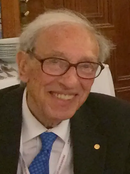
Edmond H. Fischer
1920–2021, born in China
Born in Shanghai to a Swiss-French family, he was sent to school in Switzerland at seven. He studied piano at the Geneva Conservatory and wondered about becoming a professional pianist, but kept music for pleasure.
Photo: Betsythedevine, CC BY-SA 4.0
-
Edwin G. Krebs
1918–2009, born in the United States
A minister’s son from a small town who trained as a doctor and served as a navy doctor. He kept his license to practice medicine in case research did not work out. It did.
A street dog suddenly barks and runs at you. Before you have even decided what to do, your heart is pounding and your legs are ready to run. In those few seconds your muscles need fuel, fast, and they get it from a store of sugar kept inside them.
How does a muscle cell know, so quickly, that it must open its sugar store? And how does it close the store again afterwards, so the sugar isn’t wasted? In the 1950s two scientists in Seattle, in the United States, found the answer while trying to solve a quite different puzzle. It turned out to be one of the most important switches in all of life. In 1992 Edmond Fischer and Edwin Krebs shared the Nobel Prize in Physiology or Medicine for it.
The problem: a store of sugar with a lock
Your body stores sugar as glycogen, a huge molecule made of thousands of glucose units joined in branching chains. There is glycogen in your liver, which releases sugar into the blood, and in your muscles, which keep it for their own work Grade 9: Skeletal muscle.
To use the store, glucose units must be cut off the glycogen one at a time. The cutting is done by an enzyme, a protein that speeds up one particular reaction without being used up: a catalyst made by a living cell Grade 10: Catalyst. This enzyme is called phosphorylase. Carl and Gerty Cori had discovered it, and won the 1947 Nobel Prize for their work on how the body uses glycogen.
The Coris had also found that phosphorylase comes in two forms. They called them phosphorylase b, which hardly works unless a small helper molecule called AMP is added, and phosphorylase a, which works on its own. A muscle at rest has mostly the b form; a working muscle has more of the a form. But what turned one form into the other? Nobody knew.
Where does a muscle get the sugar it needs for a sudden sprint?
Pick an answer to see why.
How proteins work, as far as we need
Almost every job in a cell is done by proteins: they carry things, build things, and as enzymes they run thousands of chemical reactions (Proteins do the work). A protein is a long chain that folds up into a particular shape, and its shape decides what it can do. An enzyme has a pocket that fits the molecule it works on, the way a lock fits a key.
So if a cell wants to switch an enzyme off, it needs a way to change its shape a little, so the pocket no longer works. And to switch it back on, it must be able to undo that change. This is what Fischer and Krebs found.
Why it was hard
In the early 1950s, scientists already knew that some proteins carry phosphate, a small group made of one phosphorus atom and four oxygen atoms, with a negative charge. But they thought of these as food proteins, such as the casein in milk and the proteins in egg yolk, where the phosphate is just part of the nutrition. Nobody imagined that a cell might stick phosphate onto an enzyme and take it off again, over and over, to control it.
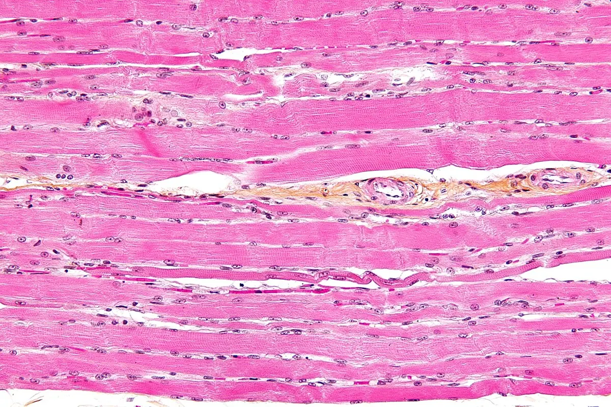
The experiments were also hard to do. Enzymes had to be pulled out of ground-up muscle and purified step by step, and a small change in the recipe could change the result, as Fischer and Krebs found out by accident.
What they did, step by step
Krebs had learned about phosphorylase in the Coris’ laboratory in St. Louis. Fischer had studied the same enzyme in potatoes in Geneva, where it works without any AMP. When Fischer came to the University of Washington in the autumn of 1953, the two decided, as a side project, to find out why the muscle enzyme needed AMP and the potato one didn’t. They never answered that question. Instead, they found something much bigger.
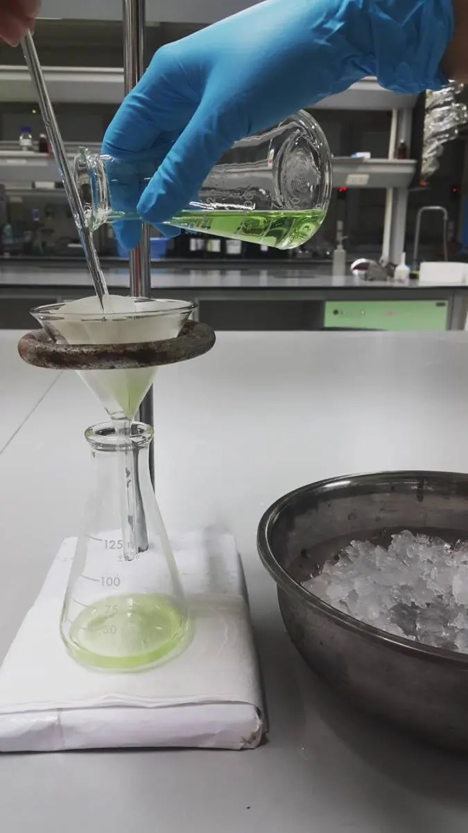
- 1953–54, a failed recipe. They tried to make pure phosphorylase a from rabbit muscle, following the Coris’ method exactly, except for one small change: to clear the muscle juice, they spun it in a centrifuge instead of pouring it through filter paper. They got only the b form, and it would not form crystals.
- The filter paper. They put the filtering step back, and this time they got phosphorylase a, which crystallized as it should. Something in the paper was switching the enzyme on. When they washed the paper well before use, the effect disappeared. The missing ingredient from the paper turned out to be calcium.
- The second ingredient. If the muscle juice was left to stand before filtering, nothing happened. Something in the fresh juice had run out. It was ATP, the molecule cells use to carry energy (Catching the energy: ATP).
- 1955–56, the switch. They showed that ATP hands its end phosphate to the enzyme itself: phosphorylase b + ATP → phosphorylase a + ADP. The job is done by another enzyme, which they called phosphorylase kinase. A kinase is an enzyme that adds a phosphate group to something. The phosphate goes onto one particular building block of the protein, an amino acid called serine.
- Late 1950s, the way back. A second enzyme takes the phosphate off again, turning phosphorylase a back into b. An enzyme that removes a phosphate group is called a phosphatase.
| What they did | What they got |
|---|---|
| spun the muscle juice in a centrifuge | phosphorylase b (off) |
| poured it through filter paper | phosphorylase a (on) |
| used filter paper washed first | phosphorylase b (off) |
| let the juice stand, then filtered it | phosphorylase b (off) |
So the enzyme could be switched on by adding a phosphate, and off by removing it, again and again. This is reversible protein phosphorylation, the words in their prize.
What does a protein kinase do?
Pick an answer to see why.
How it works now
The switch
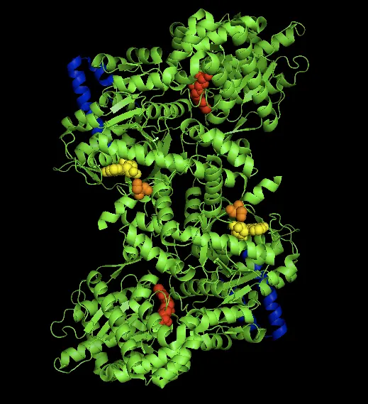
The phosphate group is small, but it carries negative charges. Stuck onto the protein, it pulls and pushes on the charged parts nearby, and the protein’s shape shifts. In phosphorylase, the pieces move so that the working pocket opens: the enzyme switches on (Figure 2). Take the phosphate off, and the protein relaxes back into its old shape: switched off.
The Nobel presentation compared the phosphate to a pair of ballet shoes: very small, but they change completely how the wearer moves, and they can be put on and taken off again and again.
A chain of switches that multiplies
Fischer, Krebs and others then found that the switches come in chains. Phosphorylase kinase is itself switched on by a phosphate, put on by another kinase. In 1968 Krebs’s team found that kinase too: protein kinase A, which is switched on by a messenger molecule inside the cell, cyclic AMP (discovered by Earl Sutherland, Nobel Prize 1971).
Now follow the barking dog. Your adrenal glands release the hormone adrenaline into the blood Grade 9: Adrenal glands. When it reaches a muscle or liver cell, the cell makes cyclic AMP, which switches on protein kinase A. That tags and switches on phosphorylase kinase, which tags and switches on phosphorylase, which cuts glucose off the glycogen. At the same time protein kinase A puts a tag on the enzyme that builds glycogen and switches it off, so the cell doesn’t store sugar while it is trying to release it. In a working muscle, calcium, released inside the cell each time the muscle contracts, switches on phosphorylase kinase directly. That is the calcium Fischer and Krebs found in their filter paper.
Each switched-on enzyme switches on many copies of the next one, so the signal grows at every step (Figure 3). The Nobel presenter compared this to the brakes of a car: a gentle push on the pedal can stop a heavy bus. Textbooks estimate that one molecule of adrenaline can free somewhere between ten thousand and a hundred million glucose units; the exact number depends on the cell and on how it is counted.
When the danger is over, adrenaline falls, and the phosphatases take over, removing the tags so that the whole chain switches off again.
Why is a chain of kinases better for the body than a single switch?
Pick an answer to see why.
In your life today
- Almost everything in your cells. The switch Fischer and Krebs found in muscle is used everywhere: to control growth and cell division, memory in the brain, the beating of the heart, the release of hormones and the body’s defenses. There are about 500 different protein kinases in a human, roughly 2% of all our genes, counted when the human genome was read in the early 2000s. (In 1992 the Nobel committee had guessed about 1%.) Up to a third of all our proteins may carry phosphate tags.
- Medicines for cancer. In some cancers a kinase gets stuck “on”, telling cells to divide again and again. In chronic myeloid leukemia, a blood cancer, a faulty kinase called BCR-ABL does this. The medicine imatinib, approved in 2001, fits into its pocket and blocks it. In the United States, the share of patients still alive five years after diagnosis rose from about 31% in 1993 to 59% in 2009, thanks in large part to imatinib and the similar medicines that followed. Imatinib is on the World Health Organization’s list of essential medicines. By 2025 more than 80 medicines that block kinases had been approved in the United States.
- Transplants. The medicine cyclosporin, which stops the body from rejecting a transplanted kidney or liver, works by blocking a phosphatase called calcineurin that the immune cells need.
- Your next fright. Every time adrenaline gets you ready to run, the chain of switches in Figure 3 runs in your muscles and liver.
What came next
- Phosphatases matter too. Fischer later turned to the enzymes that take phosphate tags off, and in 1988 his team purified the first tyrosine phosphatase, which removes phosphate from the amino acid tyrosine. It became clear that a cell needs both kinds of enzyme, in balance, and that too much or too little of either can cause disease.
- New medicines. Drug companies are now searching for medicines that block one kinase without touching the other 500, for cancers, arthritis and other diseases.
- An open problem. Cancers often become resistant to kinase-blocking medicines: a small change in the kinase’s pocket stops the medicine from fitting. Designing medicines that keep working, and finding which of the thousands of phosphate tags in a cell matter for a disease, are questions being worked on now.
In your science books
- Grade 9: skeletal muscle, endocrine glands, the adrenal glands (adrenaline), the pancreas (insulin and glucagon), and nerves and hormones working together.
- Grade 10: catalysts, with the enzymes of living things.
- Going further: proteins do the work, ATP, your muscles in a sprint, and feedback loops.
Learn more
- The Nobel Prize’s press release for 1992, with pictures of the switch and the chain.
- The Nobel lectures: Krebs, Protein phosphorylation and cellular regulation I, with the filter paper story, and Fischer, Protein phosphorylation and cellular regulation II.
- Their own life stories: Fischer and Krebs (nobelprize.org).
The official reason
The prize was given “for their discoveries concerning reversible protein phosphorylation as a biological regulatory mechanism.”