
Split genes: the gene that comes in pieces
Nobel Prize in Physiology or Medicine, 1993: Richard Roberts and Phillip Sharp

Richard Roberts
born 1943 in England
A motor mechanic’s son who loved puzzles and a chemistry set, and became an expert at cutting DNA into pieces.
Photo: paloma.cl, CC BY-SA 2.0
Phillip Sharp
born 1944 in the United States
A farm boy from Kentucky who paid for college by raising cattle and growing tobacco.
Imagine a recipe for sel roti copied out by hand, but with long passages of nonsense written between the steps. “Mix the rice flour with sugar”, then two pages of random letters, then “add milk and ghee”, then more nonsense. Before anyone could cook from it, someone would have to cut out the nonsense and tape the useful parts together in the right order.
In 1977, two teams found that most of the genes in our own cells are written exactly like that. In 1993 Richard Roberts and Phillip Sharp won the Nobel Prize in Physiology or Medicine for the discovery of these split genes.
The problem: what a gene was supposed to look like
By the 1970s, scientists knew how a gene works Grade 10: DNA.
- A gene is a stretch of DNA that holds the recipe for one protein. DNA is written in four letters, A, T, G and C.
- When the cell needs the protein, it copies the gene into a single strand of messenger RNA, DNA’s cousin Grade 10: RNA. In RNA the letter T is replaced by U.
- The messenger RNA travels to a ribosome, which reads it three letters at a time and joins the matching amino acids into a protein (how a gene makes a protein).
Most of this was learned from bacteria and from the viruses that infect them, because they are simple and grow fast. In bacteria, a gene is one unbroken stretch of DNA, and its messenger RNA is an exact copy of it, letter for letter. Everyone assumed the genes of plants, animals and people were the same.
But there were puzzles. In the cells of animals, the nucleus, where DNA is kept, was full of RNA molecules much longer than the messenger RNAs that came out of it into the rest of the cell. Much of that RNA never left the nucleus. Where was it going?
In 1975, what did nearly every scientist believe about a gene in a human cell?
Pick an answer to see why.
A virus as a model
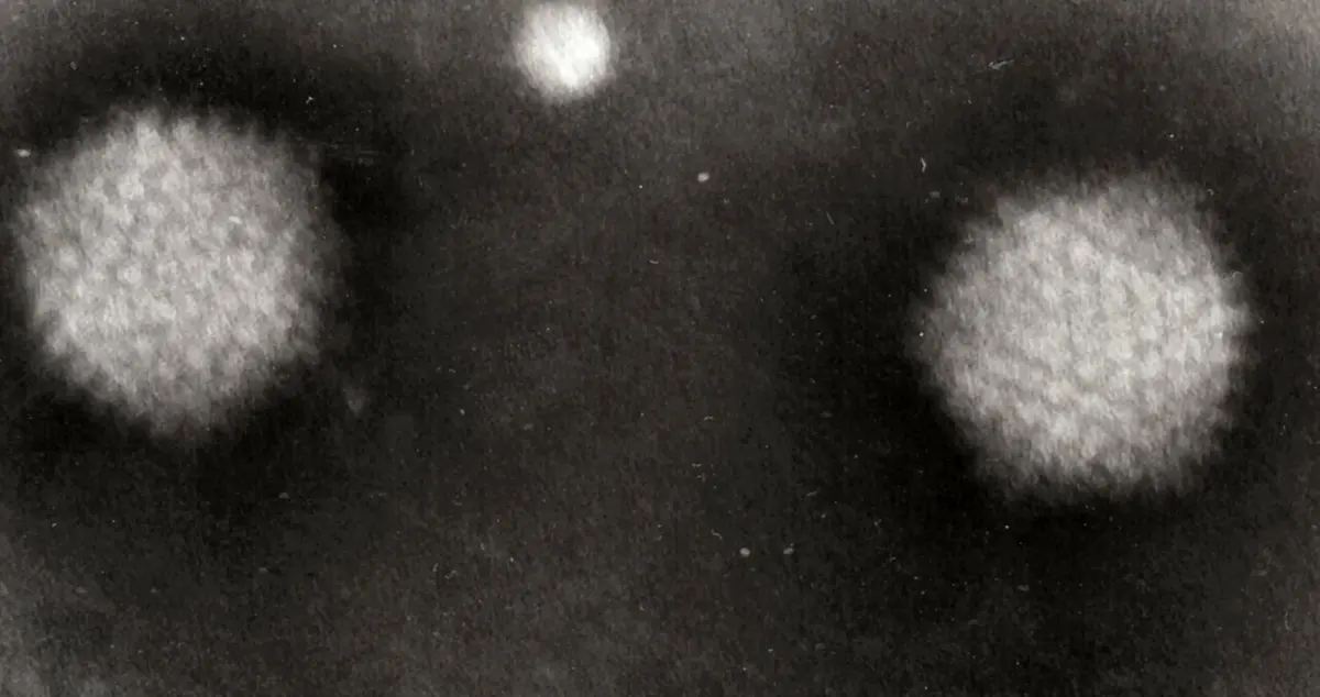
Human DNA is enormous: about 3 billion letter pairs. In the 1970s nobody could study one human gene on its own. So both teams studied a virus instead: the adenovirus, one of the viruses that cause the common cold Grade 8: viruses.
A virus is a good model because it cannot make anything by itself. It gets inside a cell and uses the cell’s own machinery to copy its genes and make its proteins. So a virus that infects human cells has its genes read exactly the way the cell reads its own. And the adenovirus has only about 35,000 letter pairs of DNA, so its genes could be mapped one by one.
How you can see where a message comes from
Both teams used the same trick, and it depends on the pairing rule of DNA. The letters of RNA pair with the letters of a DNA strand: A with T (or U), G with C. A messenger RNA is a copy of one strand of its gene, so if the two are mixed in the right conditions, the RNA sticks to the strand it was copied from, letter by letter, as a zip closes.
Under an electron microscope, which uses a beam of electrons instead of light and can show single molecules, DNA and RNA look like very thin threads. Where the RNA has stuck to the DNA the thread looks thicker; where single DNA has nothing to pair with, it looks thinner.
If a gene were one unbroken stretch, the RNA would cover it from one end to the other.
Why it was hard
- Everyone “knew” the answer. Bacteria had shown how genes looked, and there seemed no reason for animals to be different. A strange result was more likely to be a mistake in the experiment than a new rule.
- The pieces were far apart. The parts of the message came from places thousands of letters apart on the DNA. Ordinary chemical tests could find that something odd was going on, but not show what.
- The microscope work was delicate. Getting RNA and DNA to pair, spreading them out flat without tangling and photographing them took great skill.
What they did, step by step
The two teams worked separately, in different cities, and found the same thing in the same year.
Roberts’ team: a beginning that didn’t belong
- 1972, Cold Spring Harbor. Richard Roberts, a chemist from England, joined the Cold Spring Harbor Laboratory near New York. His team collected restriction enzymes, enzymes from bacteria that cut DNA at particular sequences of letters. Three quarters of the first ones known were found or studied there. With them, the adenovirus DNA could be cut into pieces and mapped.
- 1974–1976, a puzzle. With Richard Gelinas, Roberts looked at the very start of the virus’s messenger RNAs. They found something strange: many different messages, made late in an infection, all began with the same short stretch of letters. And that stretch was not written in the DNA next to the rest of each gene.
- March 1977, seeing it. Roberts designed the experiment to prove it, and the electron microscope experts Louise Chow and Tom Broker carried it out. They let a messenger RNA pair with pieces of the virus’s DNA, and could see it: the beginning of the message paired with DNA far away from the rest.

Sharp’s team: loops in the thread
- 1974, MIT. Phillip Sharp, who grew up on a farm in Kentucky, set up his laboratory at the Massachusetts Institute of Technology (MIT). With Jane Flint he found that cells infected with adenovirus held long RNAs in the nucleus that never left it.
- 1977, the experiment. With Susan Berget and Claire Moore, Sharp paired the messenger RNA for one of the virus’s coat proteins with a single strand of its DNA, and looked at it in the electron microscope.
- The loops. The main part of the message paired neatly with its gene. But its first part paired with three short stretches far away, and the DNA between them had nothing to pair with. Those spare stretches of DNA bulged out as loops. The message had come from four separate pieces of DNA.
So a gene in an animal cell can be split. The cell copies the whole thing, pieces and gaps together, into a long RNA. Then it must cut out the gaps and join the pieces. This was what the long RNAs in the nucleus had been: unfinished messages.
How split genes work
In 1978 the American scientist Walter Gilbert gave the pieces their names, which are still used.
- Exons are the pieces that are kept and expressed: they make it into the finished message.
- Introns are the stretches in-between, which are cut out.
The cutting and joining is called splicing, the word for joining the ends of two ropes or two strips of film. It is done in the nucleus by a large machine made of RNA and proteins, called the spliceosome. It finds the ends of each intron by short signal letters, cuts it out, and joins the exons together, accurate to the single letter. One letter out of place would make every three-letter word after it wrong.
Most human genes are split. An average human gene has about 8 introns, and the introns are usually much longer than the exons. Together the exons of all our genes make up only a small part of our DNA.
A gene has exons 1, 2 and 3, with introns between them. What does the finished messenger RNA contain?
Pick an answer to see why.
One gene, many proteins
Splicing turned out to do more than tidy up. A cell can choose which exons to keep. In one kind of cell it may join exons 1, 2 and 3; in another, 1 and 3 only. The same gene then makes two different proteins. This is called alternative splicing, and the adenovirus was already doing it in 1977: its long RNA was spliced in different ways to make different proteins.
It is thought that about 95% of human genes that have more than one exon are spliced in more than one way, although scientists still argue about how many of these versions really matter. It helps to explain how about 20,000 genes can make the many more kinds of protein our bodies use.
In your life today
- Thalassemia. Hemoglobin, the protein that carries oxygen in red blood cells Grade 10: red blood cells, is made of two kinds of chain. In beta-thalassemia, an inherited disease common around the Mediterranean and in South Asia, one of those chains is made badly or not at all. In many families the cause is a single wrong letter that upsets splicing: an intron is not cut out properly, or a new, false exon is made. The red blood cells die early, and many children with the disease need blood transfusions every few weeks.
- Medicines that fix splicing. Spinal muscular atrophy is an inherited disease that weakens the muscles of babies. They lack a working copy of one gene, but have a near-copy that is usually spliced wrongly. In 2016 the first medicine for it, nusinersen, was approved in the United States: it sticks to the RNA and makes the cell splice it correctly, so the protein is made.
- Some cancers. In one blood cancer, chronic myeloid leukemia, two chromosomes break and swap ends. Exons from two different genes are then spliced into one message, which makes a harmful mixed protein Grade 10: chromosomes.
- Making medicines in bacteria. Bacteria cannot splice. That is why, to make human insulin in bacteria, scientists give them a copy of the finished message, not the gene (insulin from bacteria).
What came next
- Shuffling exons in evolution. Often one exon makes one working part of a protein. If exons from different genes get swapped or copied over millions of years, new proteins can be built from old parts, like building something new from the same Lego bricks. This may be one way complex living things evolved new proteins quickly Grade 9: variation and mutation.
- RNA that cuts itself. In 1982 Thomas Cech found an intron, in a single-celled pond creature, that cuts itself out of its RNA with no help from proteins. It showed that RNA can act as an enzyme, and won Cech a share of the 1989 Nobel Prize in Chemistry.
- Why introns at all? Bacteria manage without them. Did the first living things have introns that bacteria later lost, or did introns arrive later, in the cells of plants and animals? Scientists are still debating it.
In your science books
- Grade 8: viruses.
- Grade 9: variation and mutation.
- Grade 10: DNA, RNA, chromosomes and red blood cells.
- Going further: how a gene makes a protein, from copying the gene to reading the message.
Learn more
- The Nobel Prize’s press release, with drawings of the loops and of splicing.
- The laureates’ own stories: Richard Roberts and Phillip Sharp.
The official reason
The prize was given “for their discoveries of split genes.”