
Seeing molecules with X-rays
Nobel Prize in Chemistry, 1964: Dorothy Crowfoot Hodgkin
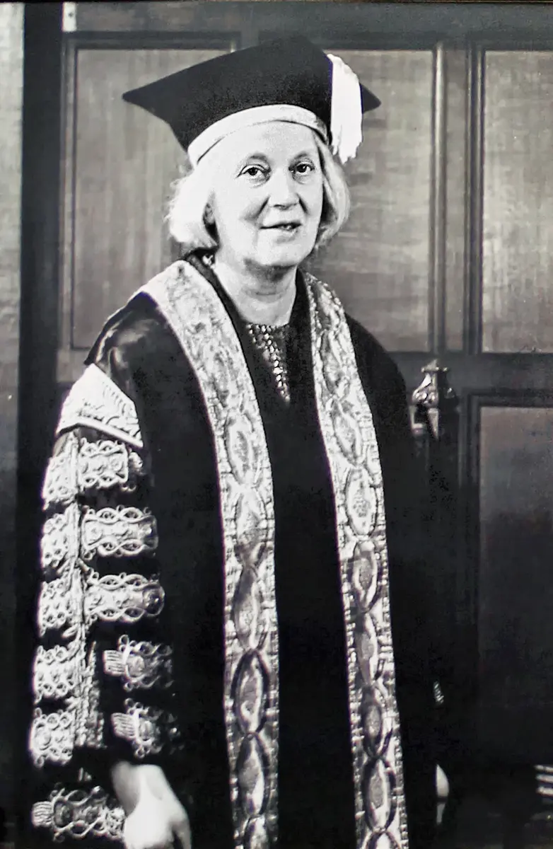
Dorothy Crowfoot Hodgkin
1910–1994, born in Cairo, Egypt
Fell in love with crystals at the age of ten, and spent 35 years on one molecule, insulin, until she had solved it.
Photo: University of Bristol, CC BY-SA 2.0
Take a grain of sugar or salt and look at it closely with a magnifying glass. It has flat faces, straight edges and sharp corners. It is a crystal. Its neat shape comes from something you can never see directly: its atoms and molecules are stacked in a perfectly regular pattern, the same arrangement repeated millions of times in every direction.
Dorothy Hodgkin found a way to use that pattern to see the molecules themselves. By shining X-rays through crystals, she worked out exactly where every atom sits in some of the most important molecules in medicine: penicillin, vitamin B12 and later insulin. For this she received the Nobel Prize in Chemistry in 1964.
The problem: molecules too small to see
Every medicine works because of its shape. A molecule of a drug fits into a molecule in your body, or in a germ, a little like a key in a lock. So if chemists know a medicine’s shape, they can understand how it works, make it in a factory, and design better ones.
But molecules are far too small to see. Even the best light microscope can’t show anything smaller than about a five-thousandth of a millimeter, and atoms are about a thousand times smaller still. Light simply can’t show something so much smaller than its own wavelength.
Before the 1940s, chemists worked out a molecule’s structure in a slow, roundabout way. They broke the molecule into smaller pieces whose structures they already knew, then guessed how the pieces fitted together, and checked by trying to build the molecule from scratch. For small molecules this worked. For the big, complicated molecules of living things, it often failed.
How X-rays can show atoms
X-rays are electromagnetic waves, like light, but with a much shorter wavelength: about the same as the distance between two atoms Grade 9: Electromagnetic spectrum. You know them from hospital X-ray pictures of bones Grade 9: X-rays.
When waves pass through a pattern of small, regularly spaced objects, they spread out and add up in some directions and cancel in others. This is called diffraction. You can see it if you look at a street light through a fine cloth or an umbrella: you see a pattern of bright spots, not one light. The spacing of the spots tells you about the spacing of the threads.
In 1912 the German physicist Max von Laue showed that X-rays do the same thing when they pass through a crystal: the rows of atoms act like the threads of the cloth. A beam of X-rays going into a crystal comes out as many thin beams, which make a pattern of spots on a photographic film. Soon after, the British father and son William Henry Bragg and William Lawrence Bragg showed how to work backwards from the spots to the positions of the atoms. Both discoveries won Nobel Prizes in Physics, in 1914 and 1915.
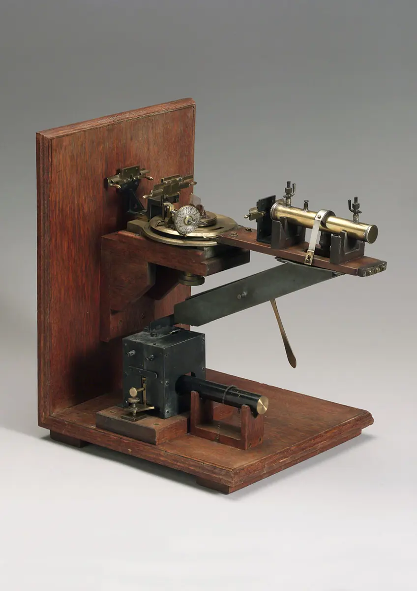
Why can X-rays show the arrangement of atoms in a crystal when ordinary light can’t?
Pick an answer to see why.
Why it was so hard
The spots don’t show the atoms directly. A film shows how bright each spot is, but not a second piece of information, called the phase, which is needed to turn the spots back into a picture of the atoms. Crystallographers had to find the phases by clever tricks and by guessing a part of the structure, then checking it against the spots and improving it, again and again.
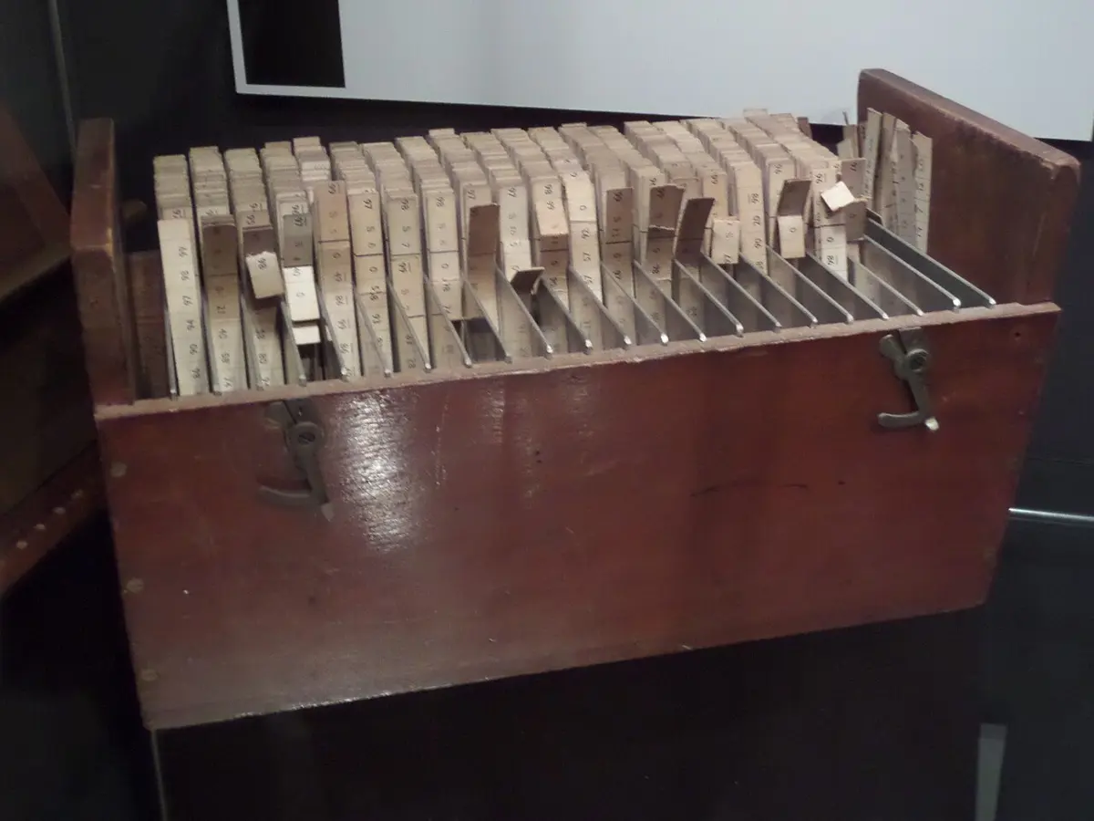
And the work was enormous. A big molecule gives thousands of spots. For each one, the brightness had to be measured by eye from the film, and then long sums had to be done for every point in the crystal, mostly by hand. For one picture of a molecule, crystallographers could spend weeks adding up columns of numbers, with the help of printed cardboard strips (below).
What she did, step by step
Growing up with crystals (1920s–1934)
Dorothy Crowfoot was born in Cairo, Egypt, in 1910, where her father worked; she grew up mostly in England. At about ten, she grew crystals from a children’s book of experiments and was fascinated. At her school, she and one other girl were allowed to join the boys’ chemistry lessons. She studied chemistry at Oxford and then worked at Cambridge with the crystallographer J. D. Bernal.
- 1934: the first X-ray photograph of a protein. With Bernal, she took the first X-ray photograph of a crystal of a protein, the digestive enzyme pepsin. It showed that even huge molecules from living things are regular enough to form crystals, and that X-rays could one day reveal them.
- 1934: a hard year. At 24, she developed rheumatoid arthritis, a painful disease of the joints. It slowly bent her hands and feet. She kept working for the rest of her life, building models and adjusting delicate equipment with swollen fingers.
She went back to Oxford in 1934, raised money for her own X-ray equipment, and stayed there for the rest of her career. In 1937 she married Thomas Hodgkin, a historian; they had three children.
Penicillin (1942–1945)
During the Second World War, penicillin had just been shown to cure infections (the story of penicillin). Factories needed to make much more of it, and chemists wanted to know its structure so they could make it, or better versions of it, in the laboratory. But they couldn’t agree on its shape.
Hodgkin and her student Barbara Low began the X-ray work in 1942, at the same time as a team at a chemical company. By 1945 they had the answer. Penicillin’s atoms include a small, tightly bent ring of just four atoms, called the β-lactam ring (beta-lactam). Many chemists had thought such a ring was too strained to be stable. The X-rays showed it was there.
Vitamin B12 (1948–1956)
Vitamin B12 is needed to make red blood cells and keep nerves healthy. Without it, people can develop pernicious anemia, which was deadly until the 1920s, when doctors found that eating raw liver cures it; B12 is the substance in liver that does it. B12 was first purified in 1948, and Hodgkin began work on it the same year.
It is a much bigger molecule than penicillin: 181 atoms, against penicillin’s 41, with a cobalt atom at its center. The sums were too great to do by hand. Hodgkin’s team took the X-ray photographs and measured the spots in Oxford; Kenneth Trueblood in California ran the calculations on SWAC, one of the first electronic computers, and posted the results back by ordinary mail. Piece by piece, the picture grew. By 1956, after eight years, the whole structure was known: the largest molecule ever solved at that time.
Insulin (1934–1969)
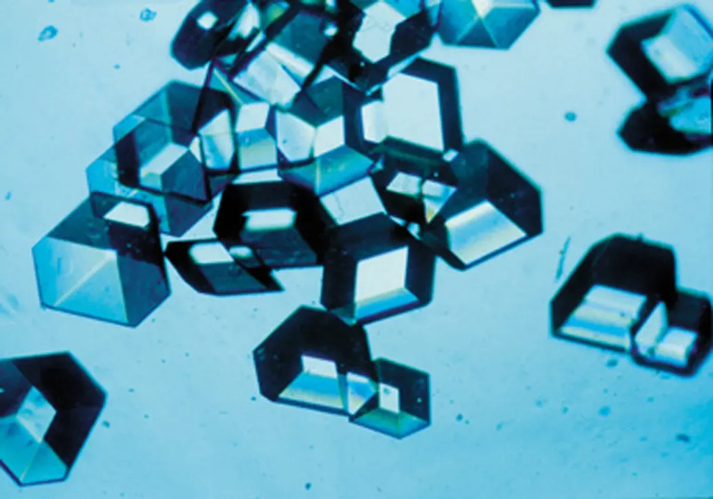
The prize came in 1964, but Hodgkin’s longest project wasn’t finished. In 1934 she had been given a tiny sample of crystals of insulin, the hormone that controls the sugar in your blood Grade 10: Diabetes. An insulin molecule has about 800 atoms. With better X-ray machines, better computers and a larger team, she finally solved its structure in 1969, 35 years after her first photograph.
How it works now
X-ray crystallography is still one of the main ways scientists find the shapes of molecules:
- A pure sample of the molecule is made to form a crystal, often only a fraction of a millimeter across. For a protein this can take months of trying.
- The crystal is placed in a strong beam of X-rays, often at a synchrotron, a huge ring-shaped machine that makes X-rays millions of times brighter than a hospital X-ray tube.
- A detector records the spots while the crystal slowly turns.
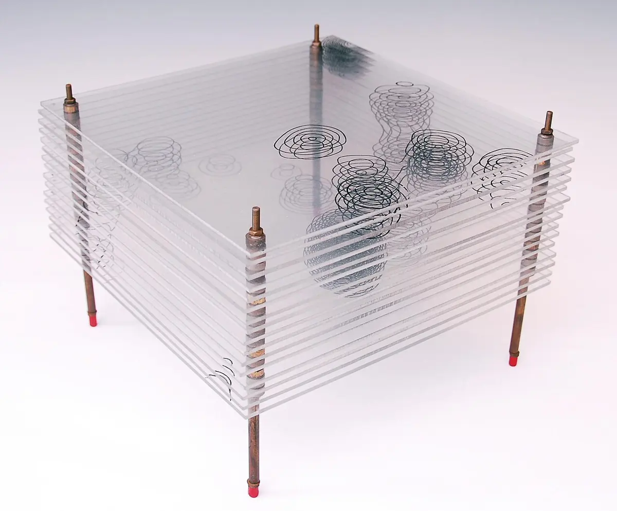
- A computer works out a map of the electrons, and the scientists fit the atoms into it, as Hodgkin did with her glass sheets.
What took Hodgkin years can now often be done in days.
In Hodgkin’s glass-sheet map of penicillin, what do the blobs made by the contour lines show?
Pick an answer to see why.
In your life today
- New medicines. Knowing penicillin’s shape helped chemists make new penicillins, such as amoxicillin, which many people in Nepal take for ear and throat infections. Today, most new medicines are designed with the help of the shape of the protein they are meant to fit.
- Insulin. Millions of people with diabetes inject insulin every day. Knowing its structure helped scientists make insulins that act faster or last longer.
- Vitamin B12. Only bacteria can make B12. People get it from milk, curd, eggs, fish and meat, so those who eat few animal foods can run short of it, and a doctor may suggest B12 tablets.
- A library of shapes. The structures of more than 200,000 proteins and other large molecules, most of them found by X-ray crystallography, are freely available to anyone in an online library, the Protein Data Bank.
What came next
- Bigger and bigger molecules. In the 1950s, X-ray photographs of DNA by Rosalind Franklin helped James Watson and Francis Crick to work out its double helix. Others solved the first protein structures, and later huge machines of the cell such as the ribosome, which makes proteins (How a gene makes a protein).
- Without crystals. Some molecules refuse to form crystals. Since about 2013, cryo-electron microscopy, which freezes molecules and photographs them with electrons, has shown many of them (Nobel Prize in Chemistry, 2017).
- The open question: in 2024 the Nobel Prize in Chemistry went partly to scientists who taught computers to predict a protein’s shape from its recipe alone. Will computers one day replace experiments like Hodgkin’s? So far, the predictions are checked against shapes measured by experiments, and some molecules, and the way they move, are still hard to predict.
In your science books
- Grade 8: what an atom is, and molecular formulas.
- Grade 9: X-rays and the electromagnetic spectrum; X-ray imaging in hospitals; the crystals of diamond and graphite.
- Grade 10: insulin and diabetes.
- Going further: What does a molecule look like? and its four ways to show a molecule; waves that bend round edges and cancel; proteins.
Learn more
- The prize at nobelprize.org: The Nobel Prize in Chemistry 1964, with the presentation speech.
- Hodgkin’s own Nobel lecture, “The X-ray analysis of complicated molecules”, and her biography, written at the time of the prize.
The official reason
The prize was given “for her determinations by X-ray techniques of the structures of important biochemical substances.”