
Fill a thin glass tube with hydrogen and pass a spark through it, and it glows pink. Look at that glow through a prism or a diffraction grating, and the pink comes apart into just four bright lines, red, blue-green, blue and violet, with darkness between. Not a rainbow, four colors. Why should hydrogen, the simplest atom of all, give out light of only a few exact wavelengths?
The answer turned physics over. Inside an atom, energy can’t take any value it likes: it comes in steps, quantized. This unit follows that idea from Bohr’s model of the hydrogen atom, its orbits, energy levels and spectral lines, to the surprising discovery that electrons behave as waves, the limit on what can ever be measured about them, and then to X-rays: how they are made, what they are used for, and how crystals reveal their wavelength.
23.1 Bohr’s atomic model
Why don’t the electrons in an atom spiral into the nucleus?
The German physicist Max Planck had shown in 1900 that light is given out and absorbed in packets, quanta, each of energy , where is the frequency and Planck’s constant, J s (the full story). Bohr applied the same idea, that energy comes in steps, to the atom.
In 1911 Rutherford’s nuclear model had shown that an atom is a tiny, dense, positive nucleus with electrons going round it. But the model met two problems it couldn’t solve:
- The stability of atoms. An electron going round the nucleus is always changing direction, so it is accelerating, and by classical electromagnetism an accelerating charge radiates energy. The electron should lose energy continuously and spiral into the nucleus, and the atom should collapse (Figure 23.2). Yet atoms are stable.
- The line spectrum of hydrogen. A spiraling electron would go round faster and faster, giving out light of every frequency: a continuous spectrum. But hot hydrogen gives out light of only certain exact wavelengths, separate bright lines.
In 1913 Niels Bohr kept Rutherford’s nucleus but added three rules, his postulates:
Postulates of Bohr’s atomic model
i. Stationary orbits. An electron goes round the nucleus only in certain allowed circular orbits, each with a definite radius and a definite energy. In these orbits it does not radiate energy, however long it stays. They are called stationary orbits (or stationary states).
ii. Quantization of angular momentum. The allowed orbits are those in which the electron’s angular momentum is a whole-number multiple of . If an electron of mass moves with speed in an orbit of radius ,
where is the principal quantum number. Equation (23.1) is Bohr’s quantization condition. ( is often written , “h-bar”, the reduced Planck constant.)
iii. Frequency condition. An atom gives out energy only when an electron jumps from an orbit of higher energy to one of lower energy, and takes in energy only when it jumps from a lower to a higher one. The energy goes out or comes in as a single photon. If the electron jumps from the orbit of energy to the orbit of energy , the frequency of the light is given by
Equation (23.2) is Bohr’s frequency condition.
In Bohr’s model, an electron in a stationary orbit:
Pick an answer to see why.
23.2 Bohr’s theory of the hydrogen atom
How big is a hydrogen atom, and how much energy does its electron have?
A hydrogen atom is a nucleus (a proton) of charge and one electron of charge going round it in a circle of radius . The electric force between them is
where is the permittivity of free space; N m² C⁻².
Radius of the nth orbit
For the electron to go round in a circle, it needs a centripetal force , and the electric attraction provides it:
From Bohr’s quantization condition, , so
Putting this into equation (23.3) and multiplying both sides by :
Multiplying both sides by and dividing by :
With J s, C² N⁻¹ m⁻², C and kg,
So : the radii of the stationary orbits are in the ratio 1 : 4 : 9 : 16 … The smallest, Å, is the Bohr radius, the size of a hydrogen atom in its normal state. (1 ångström, Å, is m.)
For a nucleus of charge with one electron (He⁺, Li²⁺ …), the force is , and the same steps give
Speed of the electron in the nth orbit
Putting from (23.5) into (23.4):
With the values of , and , this is m/s, or : in the first orbit the electron goes at about 1/137 of the speed of light. So : the farther out the orbit, the slower the electron.
Energy of the electron in the nth orbit
The electron’s total energy is its kinetic energy (from its motion) plus its potential energy (from the attraction of the nucleus):
Kinetic energy. From equation (23.3), , so
Potential energy. The potential of the nucleus at distance is , and the electron’s charge is , so
Total energy.
Putting in from (23.5):
With the values of the constants, J, and since 1 eV = J,
The wavelengths of the lines: Rydberg’s formula
Let the electron jump from an orbit with quantum number to a lower one with (). From (23.7) their energies are
By Bohr’s frequency condition, the photon given out has
so
For light, , so , and
, the number of waves in one meter, is called the wave number. The constant in front, , is written and called Rydberg’s constant; its measured value is m⁻¹. So
This is the Rydberg formula. The Swedish physicist Johannes Rydberg had found it in 1888 by fitting the measured wavelengths, with no idea why it worked. Bohr’s theory gave its reason, and the value of worked out from , , and agrees with the measured one: that was the triumph of the theory.
In Bohr’s model of hydrogen, the radius of the electron’s orbit for n = 3 is:
Pick an answer to see why.
As the electron moves to orbits farther out (larger n), its speed and its total energy:
Pick an answer to see why.
23.3 Limitations of Bohr’s atomic model
Bohr’s model was a great step, but it has limits:
- It works only for hydrogen and for ions with a single electron (He⁺, Li²⁺); it can’t describe atoms with more than one electron accurately.
- It can’t explain the fine structure of spectral lines (a line seen with a better spectrometer is really two or more close lines), or the splitting of lines in a magnetic field (the Zeeman effect).
- It doesn’t take into account the wave nature of electrons, or quantum mechanics as it was later worked out.
- It can’t explain the chemical behavior of atoms or how they bond into molecules.
- It gives the electron an exact orbit, an exact position and an exact momentum at once, which the uncertainty principle (Section 23.11) says is impossible.
In modern quantum mechanics, the electron has no orbit at all, only a cloud of where it is likely to be found. But Bohr’s energies, eV, come out exactly the same.
23.4 Spectral series of the hydrogen atom
Why do hydrogen’s lines come in families?
When an electron in a hydrogen atom drops from a higher level to a lower one, the energy difference goes out as light of one particular wavelength: a spectral line. Different pairs of levels give different lines. All the lines given by jumps that end on the same lower level form a spectral series (Figure 23.3). Each comes from the Rydberg formula, , with a different .
1. Lyman series. Jumps from to :
These are big jumps, so the photons have large energies: the lines lie in the ultraviolet (121.6 nm down to 91.2 nm).
2. Balmer series. Jumps from to :
Its first lines are in the visible region: 656 nm (red), 486 nm (blue-green), 434 nm (blue) and 410 nm (violet), the four lines of the opening. The rest crowd together towards 365 nm, in the ultraviolet.
3. Paschen series. Jumps from to :
in the infrared (1875 nm down to 820 nm).
4. Brackett series. Jumps from to :
also in the infrared.
5. Pfund series. Jumps from to :
in the far infrared.
In each series, the first line () has the longest wavelength, and the lines get closer together towards the series limit (), the shortest wavelength, .
23.5 Energy level diagram of the hydrogen atom
How can all of hydrogen’s lines be shown in one picture?
From equation (23.7), the energy of the electron in the nth orbit is
so
- eV (), the ground state of the hydrogen atom: its normal state, of lowest energy;
- eV, the first excited state;
- eV, the second excited state;
- eV, eV, eV, and so on;
- for .
As n increases, the energy becomes less negative and approaches zero. For large n the levels are so close together that they almost form a continuous band. At the electron is no longer bound to the nucleus: it is free, and above zero it can have any energy at all.
Drawing each energy as a horizontal line at its height, with the jumps as arrows, gives the energy level diagram of the hydrogen atom (Figure 23.4). Each arrow is one spectral line; its length is the photon’s energy.
23.6 Excitation and ionization potentials
How much energy does it take to lift the electron, or to take it out of the atom?
Excitation energy and potential
Excitation energy is the energy needed to raise an electron from the ground state to a higher level, an excited state. For hydrogen, the electron in the ground state has eV, and in the first excited state eV, so the energy needed to raise it from the ground state to the first excited state is
This, 10.2 eV, is the first excitation energy of hydrogen.
An electron accelerated through a potential difference of 1 V gains 1 eV of energy. So an electron accelerated through 10.2 V has just enough energy to raise a hydrogen atom it hits from the ground state to the first excited state. The accelerating voltage that gives an electron the excitation energy is the excitation potential: 10.2 V is the first excitation potential of hydrogen.
Similarly, from to :
the second excitation energy; the second excitation potential is 12.09 V.
Ionization energy and potential
Ionization energy is the energy needed to remove the electron completely from the atom in its ground state, to an infinite distance from the nucleus, where it is free with no kinetic energy. For hydrogen, that is a jump from to :
It equals the size of the ground state energy, as a positive number, because energy must be supplied. The accelerating potential that gives an electron this energy, 13.6 V, is the ionization potential of hydrogen. What is left is a positive ion, for hydrogen a bare proton: hence the name.
23.7 Emission and absorption spectra
Why does a spectrum sometimes show bright lines, and sometimes dark ones?
Emission spectra
An emission spectrum is the spectrum of the light given out by a substance when its atoms or molecules lose energy, their electrons dropping from higher to lower levels. There are three kinds (Figure 23.5):
- Continuous spectrum. All wavelengths, with no gaps, one color merging into the next. It is given out by hot solids, liquids and dense gases, where the atoms are so close together that their levels merge. Examples: the filament of an old light bulb, a red-hot iron rod, and (nearly) the Sun.
- Line spectrum. Separate bright lines of particular wavelengths on a dark background. It is given out by atoms of a gas (spread far apart, so each behaves on its own), when they are excited by heat or an electric discharge. Each line is one jump between two levels. Example: the hydrogen spectrum, sodium’s yellow street lamps. Every element has its own lines, like a fingerprint, so a line spectrum identifies the element.
- Band spectrum. Bands, each made of many lines so close that they look like a smudge with one sharp edge. It is given out by molecules (such as N₂, O₂ or CO₂), whose energy changes when they vibrate and rotate as well as when their electrons jump, so each electron jump comes with many slightly different extra energies.
Absorption spectra
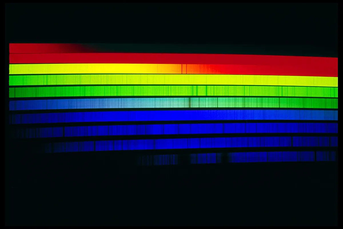
When white light, with all wavelengths, passes through a cooler gas, the atoms of the gas absorb exactly those wavelengths whose photons can lift their electrons to higher levels, and let the rest pass. The spectrum of the light that comes through is continuous, but crossed by dark lines at the absorbed wavelengths. This is an absorption spectrum. Its dark lines are at exactly the same wavelengths as the bright lines in the same gas’s emission spectrum, since the same pairs of levels are involved.
23.8 Dual nature of radiation
Is light a wave or a stream of particles?
Both, depending on what we do with it. Interference, diffraction and polarization of light can be explained only if light is an electromagnetic wave. But the photoelectric effect and the Compton effect (X-rays bouncing off electrons as if they were billiard balls) can be explained only if light comes as particles, photons, each of energy and momentum . Radiation has a dual nature: it shows wave properties in some experiments and particle properties in others.
23.9 de Broglie’s dual hypothesis
If light can behave like particles, can particles behave like waves?
In 1924 the French physicist Louis de Broglie (pronounced “duh BROY”) argued from the symmetry of nature: the universe is made of radiation and matter, and if radiation has a dual nature, matter should too. A moving particle, an electron for example, should behave as a particle when it is given out or absorbed, but as a wave while it travels.
It was soon confirmed. Electrons passed through a thin crystal (or a pair of slits) make interference and diffraction patterns, exactly as light and X-rays do. The electron microscope, which magnifies up to millions of times, works because electrons are waves of very short wavelength. Protons, neutrons, atoms and even large molecules have since been seen to diffract.
de Broglie hypothesis
A moving material particle behaves sometimes as a wave and sometimes as a particle. The wave linked with a moving particle is called a matter wave (or de Broglie wave), and its wavelength, the de Broglie wavelength, is
where and are the particle’s mass and speed, and is Planck’s constant.
Where the formula comes from
By Planck’s quantum theory, a photon of light of frequency and wavelength has energy
By the theory of relativity, anything that travels at the speed of light with energy carries momentum
So a photon has momentum
de Broglie assumed that equation (23.10) holds for material particles as well as for photons. A particle of mass moving with speed has momentum , so the wavelength of its wave is
This is the de Broglie wave equation for a material particle.
23.10 de Broglie wavelength of an electron
What is the wavelength of an electron fired by a voltage?
An electron of mass and charge , starting from rest, is accelerated through a potential difference . It gains kinetic energy equal to the work done on it:
Its de Broglie wavelength is then
Putting in J s, kg and C:
An electron accelerated through 100 V has a wavelength of 1.23 Å, about the spacing of atoms in a crystal, which is why crystals diffract electrons. At the 100 000 V or so of an electron microscope, the wavelength is a few hundredths of an ångström, a hundred thousand times shorter than light’s: that is why it can show such small things.
A proton and an electron are moving at the same speed. Which has the longer de Broglie wavelength?
Pick an answer to see why.
23.11 Uncertainty principle
Can we know exactly where an electron is and how fast it moves?
In classical physics, a moving particle has at every moment an exact position, an exact momentum and an exact energy, and all of them can, in principle, be measured at once as accurately as we like. Pairs of quantities such as position and momentum, or energy and time, are called complementary (or conjugate) variables.
Quantum theory says no. It is impossible to know both the exact position and the exact momentum of a particle at the same time. The more precisely its position is pinned down (the smaller the uncertainty ), the less precisely its momentum can be known (the larger ), and the other way round. In the same way, the more precisely an energy is known, the longer the time over which it must be measured. This isn’t a fault of our instruments: it is how nature is, because a particle is also a wave, and a wave squeezed into a small space has no single wavelength, so no single momentum.
Heisenberg’s uncertainty principle
In any simultaneous measurement of a pair of complementary variables of a particle, the product of their uncertainties is at least about . For position and momentum,
and for energy and time,
where are the uncertainties in position, momentum, energy and time. The German physicist Werner Heisenberg stated it in 1927 (the full story).
Applications of the uncertainty principle
1. Electrons can’t be inside the nucleus. If an electron were inside a nucleus, its position would be known to within the nucleus’s size, about m. Then its momentum would be uncertain by at least
so its momentum would be at least this large. Dividing by the electron’s mass, as Newton’s mechanics would, gives a speed of m/s, nineteen times the speed of light: impossible. Worked out properly with relativity, an electron with this momentum would have an energy of at least J, about 10 MeV. That is far more than the energies of the electrons that nuclei give out in beta decay, mostly under 1 MeV. So electrons can’t exist inside the nucleus. (The electrons of beta decay are created at the moment they are emitted.)
2. The stability of atoms. An electron that fell into the nucleus would have its position known almost exactly, so its momentum, and with it its kinetic energy, would be enormous. The size of an atom is the balance: squeezing the electron closer lowers its potential energy but raises its kinetic energy. The smallest total energy comes at about 0.5 Å, the size of the hydrogen atom. So the uncertainty principle explains why atoms don’t collapse.
3. Energy levels in atoms. An electron held in the small space of an atom behaves as a wave confined to that space, and only certain waves fit, just as only certain notes fit on a guitar string. That is why its energy can have only certain values.
4. The width of spectral lines. An atom stays in an excited state only for a short time, about s, so the energy of that state is uncertain by . The photons given out then have slightly different energies, and every spectral line has a small natural width instead of being perfectly sharp.
23.12 Discovery of X-rays
How did a covered photographic plate get fogged?
X-rays were discovered by accident in 1895 by the German physicist Wilhelm Conrad Röntgen. He was studying the discharge of electricity through a gas at very low pressure in a glass tube (a cathode ray tube). He noticed that a screen coated with a fluorescent chemical, lying near the tube, glowed, even though the tube was completely covered in thick black paper. Photographic plates kept near the tube, wrapped in black paper, were found fogged. When he put objects between the tube and the screen, their shadows appeared on it, and the shadow of his own hand showed the bones inside.
He concluded that some unknown radiation was coming from the tube, one that could pass through things opaque to ordinary light. Since nothing was known about it, he called it X-rays, X for the unknown. They are also called Röntgen rays. He won the first Nobel Prize in Physics, in 1901 (the full story).

23.13 Production of X-rays
How can X-rays be made?
X-rays are produced when fast electrons strike a target made of a heavy element and are suddenly stopped. A device that does this is an X-ray tube. It needs three things:
- i.a source of electrons;
- ii.a way to accelerate them to high speed (a high voltage);
- iii.a metal target of high atomic number for them to strike.
Röntgen’s early tubes were gas-filled tubes, which got their electrons from the gas; they were hard to control and are now obsolete. The tube used today in hospitals and industry was designed by the American physicist William Coolidge.
23.14 Coolidge X-ray tube
How does a modern X-ray tube work?
The modern X-ray tube was invented by W. D. Coolidge in 1913. It works in a high vacuum, and is called the Coolidge tube.
Construction
The Coolidge tube (Figure 23.11) is a highly evacuated glass bulb, containing:
- a filament of tungsten, the cathode C, heated by a low-voltage supply (a few volts) so that it gives out electrons (thermionic emission);
- a focusing cup M, a metal shield round the filament at the cathode’s negative potential, which pushes the electrons into a narrow beam;
- a target T, the anode (also called the anticathode), a block of tungsten set in a thick copper rod, its face sloped at about 45° to the beam so the X-rays come out sideways;
- a high voltage (from about 10 000 V to over 100 000 V) between the cathode and the target, which accelerates the electrons;
- cooling: the copper rod carries away the heat through fins in air, or with circulating water or oil.
The tube sits in a box lined with lead about 5 mm thick, with a small window for the X-rays to come out.
The target must have:
- i.a high atomic number (heavy atoms), since the X-rays produced increase with it;
- ii.a high melting point, to stand the heat;
- iii.a high thermal conductivity, to carry the heat away;
- iv.inner energy levels far enough apart that the jumps between them give out X-rays.
Platinum, tungsten and tantalum all have these properties, but tungsten (atomic number 74, melting point 3422 °C) is the most suitable.
Working
When a current is passed through the filament, it gives out electrons, which leave it with almost no speed. The high potential difference between the filament and the target accelerates them. Each electron reaches the target with kinetic energy
In the target, the electron is slowed down and stopped by collisions with the atoms. In most collisions it just gives energy to the atoms, which heats the target. But sometimes, as it swerves past a nucleus and is sharply slowed down, it gives out an X-ray photon carrying part or all of its kinetic energy (this X-ray light, of every wavelength above a smallest one, is called braking radiation, Bremsstrahlung in German). And sometimes it knocks an electron out of an inner shell of a target atom; an outer electron then drops into the gap, giving out an X-ray photon of a particular energy, characteristic of the metal. So the X-rays have a continuous spectrum with a few sharp characteristic lines on top. For a tungsten target the innermost (K) electrons are held so tightly that the tube needs more than 69.5 kV to knock them out; below that, tungsten’s strongest characteristic lines are missing.
Only about 1 % of the electrons’ energy (often less) becomes X-rays; the rest becomes heat in the target.
Control of the quality and the intensity
In a Coolidge tube, the quality and the intensity of the X-rays can be controlled separately:
i. Quality (penetrating power). The energy of each electron is . A higher voltage gives faster electrons and so more energetic X-ray photons, of shorter wavelength, which go through materials more easily. X-rays of high energy (from high voltages) are called hard X-rays, those of low energy soft X-rays. Hard X-rays penetrate more. So the voltage controls the quality.
ii. Intensity. The intensity of the X-rays (how many photons come out each second) depends on how many electrons strike the target each second, that is, on the tube current, which depends on the filament’s temperature. So the filament current controls the intensity.
iii. The highest frequency and shortest wavelength. The most energetic X-ray photon is made when an electron gives up all its kinetic energy in one go:
and since , the shortest wavelength is
So : the higher the voltage, the shorter the shortest wavelength. This is the Duane–Hunt law. With V m, Å when V is in volts.
23.15 Properties of X-rays
X-rays have the following properties:
- i.They are electromagnetic waves of very short wavelength, from about 0.06 Å to 100 Å.
- ii.They travel through a vacuum at the speed of light, m/s.
- iii.They carry no charge, so they are not deflected by electric or magnetic fields.
- iv.They affect photographic plates strongly.
- v.They ionize the gas they pass through.
- vi.They make some substances fluoresce (glow), such as zinc sulphide, barium platinocyanide and calcium tungstate.
- vii.Like light, they cause the photoelectric effect.
- viii.They travel in straight lines, and cast shadows of objects in their path.
- ix.They penetrate materials opaque to visible and ultraviolet light: paper, wood, thin metal sheets and flesh, but not dense materials such as bone and heavy metals.
- x.They are harmful to living things: they ionize atoms in cells and can damage them and their DNA.
- xi.When they fall on a metal, they knock electrons out and make secondary X-rays characteristic of that metal.
23.16 Applications of X-rays
X-rays are used in many fields:
- i.Medical diagnosis: to find fractures, kidney and gall stones, swallowed objects and bullets, and diseases of the lungs (tuberculosis, pneumonia), and to follow how a broken bone heals. A CT scanner takes X-ray pictures from all round the body and a computer builds them into slices.
- ii.Radiotherapy: carefully aimed doses kill cancer cells. Softer X-rays are used for growths near the surface, harder ones for deep tumors.
- iii.Engineering: to find cracks, flaws and gas bubbles in metal castings and welds, in bridges and pipelines, and in the engines of cars and airplanes.
- iv.Security and customs: to scan luggage at airports, find explosives and smuggled goods (including gold hidden in the body), and tell real gems from fakes.
- v.Industry: to test the uniformity of insulating materials and timber, find defects in tyres and balls, and find pearls inside oysters.
- vi.Scientific research: to find the structure of crystals and molecules (Section 23.17); the double helix of DNA was found from an X-ray photograph.
- vii.Art: to see the earlier paintings under an old painting, and changes the painter made.
23.17 X-ray diffraction
How do we know X-rays are waves?
In 1912 the German physicist Max von Laue and his colleagues sent a narrow beam of X-rays through a crystal of zinc sulphide, with a photographic plate behind it (Figure 23.12). After several hours, the developed plate showed a bright central spot surrounded by many faint spots in a regular pattern, the Laue pattern. It was made by the diffraction of X-rays.
The experiment showed two things.
First, X-rays are waves, since they diffract. But why hadn’t anyone diffracted them before? A slit or grating diffracts light noticeably only when its spacing is comparable to the wavelength. An ordinary diffraction grating has about 6000 lines a centimeter, so its lines are about m apart, suited to visible light. X-ray wavelengths are about m, thousands of times shorter: a grating for them would need lines thousands of times closer, which no one can rule.
Second, the regular pattern showed that the atoms in a crystal are arranged in a regular three-dimensional pattern, about m apart. Laue realized that a crystal is a natural three-dimensional diffraction grating with just the right spacing for X-rays. W. L. Bragg soon explained the Laue spots in a much simpler way.
23.18 Determination of the wavelength of X-rays: Bragg’s law
How can a crystal measure an X-ray’s wavelength?
Visible light (400–700 nm) is diffracted by slits about its own size, but X-rays (about 0.1 nm) need “slits” the size of atoms, which no grating can have. A crystal, whose atoms are regularly spaced about 0.1 nm apart, does the job. In 1912–13 W. L. Bragg (then 22) and his father W. H. Bragg explained X-ray diffraction by thinking of the crystal as stacks of parallel layers of atoms, the Bragg planes, a distance apart, and built the first X-ray spectrometer. They shared the 1915 Nobel Prize in Physics (the full story).
Bragg’s law
A beam of X-rays of wavelength falls on a crystal at a glancing angle (the angle between the ray and the planes, not the normal). Each plane of atoms reflects a little of the beam, at an equal angle (Figure 23.13). The ray reflected from one plane interferes with the ray reflected from the plane below. The two rays travel almost the same path, but the lower one goes a little farther. They reinforce each other, giving a strong reflected beam, only when this path difference is a whole number of wavelengths:
To find the path difference, drop perpendiculars PL and PM from the point P, where the upper ray is reflected, to the lower ray before and after its reflection at Q. Up to L, and from M on, the two rays have gone equal distances, so the extra path of the lower ray is . In the right-angled triangles PLQ and PMQ, the hypotenuse is and the angle at P is , so
and the total path difference is . For a strong reflection,
This is Bragg’s law. For X-rays of one wavelength, strong reflection happens only at certain glancing angles , for , the first, second, third orders of reflection. At these angles the rays from all the planes add up and the crystal reflects strongly (obeying the law of reflection); at other angles the reflections from the many planes cancel, and almost nothing is reflected. For the first order,
So knowing the spacing of the crystal’s planes and measuring the glancing angle of the strong reflection gives the wavelength of the X-rays. The other way round, X-rays of known wavelength give the spacing of the atoms: that is how the structures of crystals, and of molecules such as DNA and proteins, are found.
In Bragg’s law, , θ is:
Pick an answer to see why.
Exercise
Choose the correct option
- The reciprocal of wavelength is:
Pick an answer to see why.
- The angular momentum of the electron in the second orbit of a hydrogen atom is:
Pick an answer to see why.
- How many electrons strike the target each second in an X-ray tube carrying a current of 0.02 A?
Pick an answer to see why.
- Compared with the binding energy of a nucleus, the ionization energy of an atom is:
Pick an answer to see why.
- The ionization energy of a hydrogen atom is 13.6 eV. The energy needed to remove the electron from a singly ionized helium ion, He⁺, in its ground state is:
Pick an answer to see why.
- The energy of a hydrogen atom in its ground state is −13.6 eV. The energy of the level n = 5 is:
Pick an answer to see why.
- The ionization energy of hydrogen is 13.6 eV. The energy needed to excite the electron of He⁺ from its first orbit to its second is:
Pick an answer to see why.
- An electron in the n = 1 orbit of hydrogen is bound by 13.6 eV. How much energy is needed to ionize a hydrogen atom in the n = 3 state?
Pick an answer to see why.
- Which spectral series of hydrogen contains the line of wavelength 4860 Å?
Pick an answer to see why.
- In a sodium ion with only one electron left (Z = 11), the electron’s speed in the second orbit is v. Its speed in the fifth orbit would be:
Pick an answer to see why.
- If the potential energy of a hydrogen atom in its ground state is taken as zero, its potential energy in the first excited state will be:
Pick an answer to see why.
- The internal structure of a crystal can be studied with:
Pick an answer to see why.
- The voltage across an X-ray tube is 20 kV. The shortest wavelength of its X-ray photons is:
Pick an answer to see why.
- An X-ray tube works at 42 000 V. The maximum frequency of its X-rays is about:
Pick an answer to see why.
- In radiotherapy, X-rays are used to:
Pick an answer to see why.
- Electrons of energy 10 keV strike a tungsten target in a vacuum. The radiation given out by the target is:
Pick an answer to see why.
- X-rays of wavelength 1 Å give a first-order reflection from the principal planes of a sodium chloride crystal at a glancing angle of 10°. The spacing of the planes is about:
Pick an answer to see why.
Answer these questions
- a.State the postulates of Bohr’s theory of the hydrogen atom, especially his idea of stationary orbits.
- b.Derive expressions for the radius of the nth orbit, the speed of the electron in the nth orbit, and the total energy of the electron in the nth orbit of a hydrogen atom.
Hint
Answer
Discuss the postulates of Bohr’s theory of atomic structure. How does it explain the spectrum of hydrogen?
Answer
What wavelengths will be given out by a sample of hydrogen gas (in the ground state) if electrons of energy 12.2 eV collide with its atoms?
Hint
Answer
- a.Explain the terms excitation potential and ionization potential.
- b.Show that the ionization potential of hydrogen is 13.6 V.
- c.Explain emission spectra.
- d.When white light is passed through hydrogen gas at room temperature, absorption lines are seen only in the Lyman series. Explain.
Answer
- a.What are X-rays? How are they produced?
- b.What is meant by the penetrating power and the intensity of X-rays? How are (i) the penetrating power and (ii) the intensity of the X-rays from a Coolidge tube controlled?
- c.Can aluminium be used as the target in an X-ray tube?
Answer
- a.Explain Bragg’s law of diffraction of X-rays.
- b.X-rays fall on a zinc sulphide crystal whose planes are cm apart. The first-order reflection is at a glancing angle of 12°. Calculate the wavelength of the X-rays.
Answer
- a.A proton and an electron have the same kinetic energy. Which has the longer de Broglie wavelength?
- b.Using Bohr’s theory of the hydrogen atom, calculate the circumference of the first orbit and the momentum of the electron in it. Find the de Broglie wavelength of this electron, and say how the two are related.
Hint
Answer
- a.When a Coolidge tube is used for a long time, it becomes hot. Where does the heat come from?
- b.An X-ray tube operated at a d.c. potential difference of 10 kV produces heat at a rate of 720 W. If 0.5 % of the energy of the electrons is converted into X-rays, calculate the tube current and the speed of the electrons striking the target. (e/m = C/kg)
Answer
The diagram shows three of the energy levels of an isolated hydrogen atom. The lowest is the ground state.
- a.Explain what happens to an electron in the ground state when it absorbs the energy of a photon of energy J.
- b.i. Explain why a photon is given out when an electron makes a transition from the level n = 3 to n = 2.
- ii.Calculate the wavelength of the radiation given out in this transition.
Answer
- a.State what is meant by wave number.
- b.The lowest energy levels of an isolated hydrogen atom are shown below.
- i.An electron is at first in the level −0.85 eV. State how many different wavelengths may be given out as it loses energy.
- ii.Photons from the electron’s fall from the −0.85 eV level strike a sample of platinum, whose work function is 5.6 eV. Find: 1. the maximum kinetic energy, in eV, of a photoelectron from the platinum; 2. the wavelength of the photon that gives this photoelectron.
Answer
- a.State one effect that is evidence for (i) the wave nature of a particle, and (ii) the particle nature of electromagnetic radiation.
- b.Four electron energy levels of an isolated atom are shown below. For the emission spectrum from these levels:
- i.name the transition that gives the shortest wavelength (in the exam, mark it with an arrow on the diagram);
- ii.show that the wavelength of this transition is m.
- c.i. State what is meant by the de Broglie wavelength.
- ii.Calculate the speed of an electron whose de Broglie wavelength equals the wavelength in (b)(ii).
Answer
Printable PDFs
- The unit as a book (A5) 35 pages Answers as an A4 booklet, 9 sheets
- Study notes 3 pages
- Worksheet 4 pages Answer key
- Harder practice questions 2 pages Answer key
Quiz yourself: Matter, Atoms and Reactions, Grades 5–12 →
Practice every question in the book, and keep track of the ones you miss →
A companion to your textbook, not a replacement for it: study from the book your school uses, and where the two differ, follow your teacher. Found a mistake? Please tell us.