
X-rays: the rays that see through skin
Nobel Prize in Physics, 1901: Wilhelm Conrad Röntgen
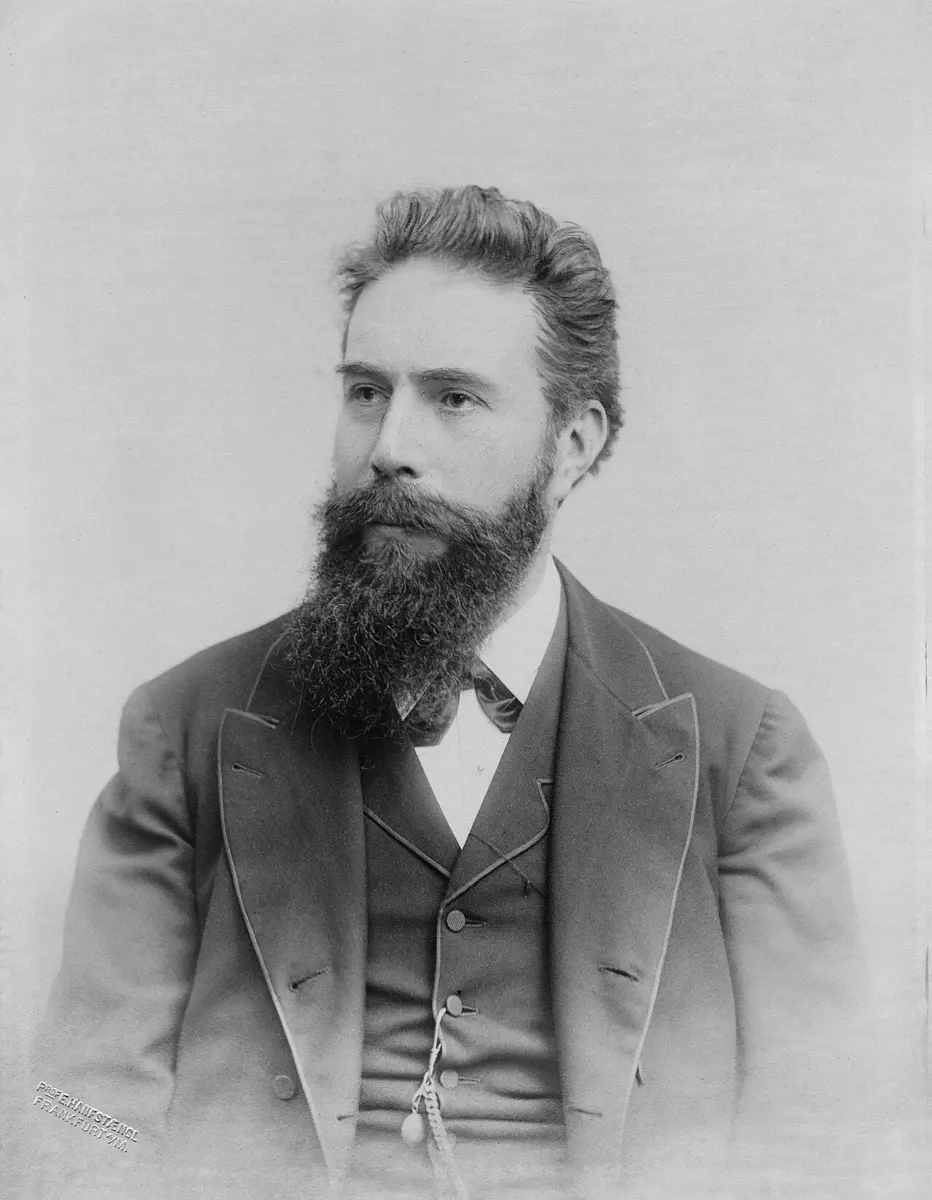
Wilhelm Conrad Röntgen
1845–1923, born in Germany
A shy professor who liked to work alone and built much of his own apparatus. He gave his discovery to the world without a patent.
Photo: Erwin Hanfstaengl, Public domain
Have you ever broken a bone, or known someone who has? At the hospital, the doctor sends you for an X-ray. You hold your arm still for a second, and a few minutes later the doctor holds up a gray picture in which your bones show white, and the crack in one of them is plain to see. Nobody cut you open, and you felt nothing.
Before 1895, that was impossible. Then, one evening in November, a physics professor in Germany noticed a faint glow in his dark laboratory. Within weeks, doctors all over the world were looking inside living people. In 1901, at the very first Nobel Prize ceremony, the first Nobel Prize in Physics went to that professor, Wilhelm Conrad Röntgen.
The problem: nobody could see inside a living body
For all of history, a doctor could only learn what was inside you from the outside. They could feel a swelling, tap your chest and listen, or look at your skin and eyes. Was a bone broken, or only bruised? Where exactly was the bullet in a soldier’s leg, or the needle a child had stepped on? To find out, a surgeon had to cut and search, often for a long time, without painkillers as good as today’s and with a real danger of infection.
What doctors needed was a way to see through skin and muscle. Nobody thought it could be done. Light doesn’t pass through your hand: shine a torch behind your fingers and you see only a red glow at the edges.
Glowing tubes and cathode rays
Röntgen’s discovery came out of an experiment many physicists were doing in the 1890s, with a discharge tube. This is a glass tube with a metal plate (an electrode) sealed in at each end and most of the air pumped out. Join the plates to a very high voltage, and something invisible streams out of the negative plate, the cathode. Where it hits the far end of the glass, the glass glows green.
Nobody yet knew what this stream was. It was called cathode rays. Scientists such as William Crookes in England and Philipp Lenard in Germany had found that cathode rays travel in straight lines, that a magnet bends them, and that in ordinary air they travel only a few centimeters before they are stopped. (In 1897, two years after Röntgen’s discovery, J. J. Thomson showed that cathode rays are a stream of tiny charged particles, the electrons that are part of every atom: see Electron.)
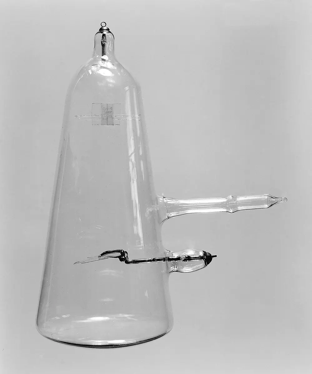
In a discharge tube, what makes the end of the glass glow green?
Pick an answer to see why.
What Röntgen did, step by step
Röntgen was a professor of physics at the University of Würzburg, in Germany. He was 50 years old, careful and quiet, and he liked to work alone.
1. 8 November 1895: a glow in the dark
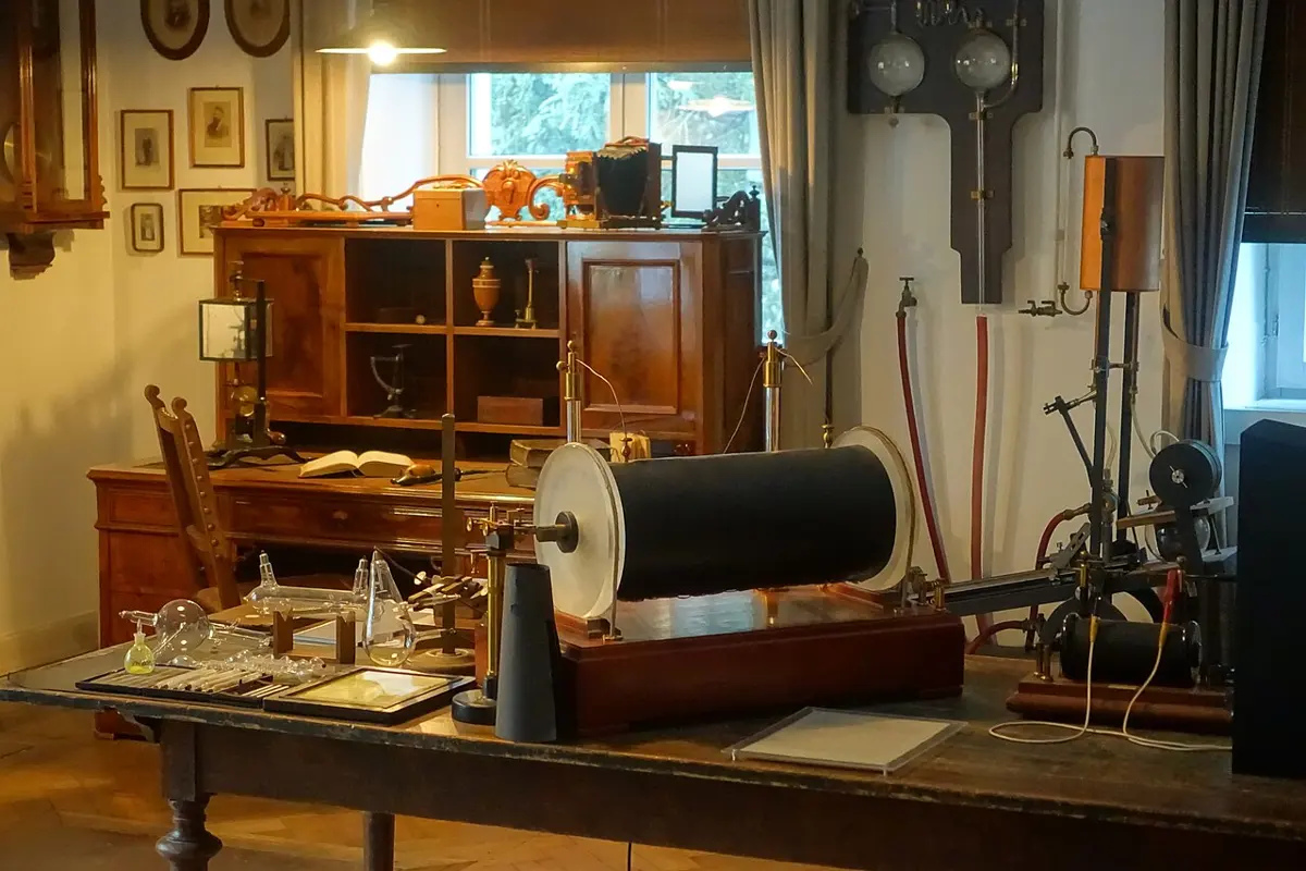
- Röntgen wanted to study the cathode rays without the glow of the tube getting in the way. He drove the tube with an induction coil, which turns the current from a battery into many thousands of volts. He covered the tube completely with black cardboard, and darkened the room to make sure that no light could leak out.
- When he switched the tube on, he noticed a faint shimmer a little way off. It came from a sheet of paper painted with barium platinocyanide, a chemical that glows when certain rays fall on it (it is fluorescent). He had it on his bench for another experiment.
- Nothing he knew could explain it. No light could get through the cardboard, and cathode rays are stopped by a few centimeters of air. Yet the screen glowed whenever the tube was on, and went dark when it was off. It glowed even when it was as far as two meters from the tube.
Something new and invisible was coming out of the tube, passing through the cardboard and through the air, and making the screen glow.
2. Seven weeks alone in the laboratory
For the next seven weeks Röntgen hardly left his laboratory. He ate and slept there, and told almost nobody what he was doing. He tested the new rays one property at a time.
- What do they pass through? He put things between the tube and the screen. Behind a bound book of about a thousand pages, the screen still glowed brightly. Wood, packs of playing cards and thin sheets of aluminium let the rays through. Lead stopped them almost completely.
- His own bones. While he was holding a small piece of lead in the beam, he saw on the screen the shadow of the lead, and around it the shadow of the bones of his own hand, with only a faint outline of the flesh.
- Photographs. The rays darkened photographic plates, the glass plates that cameras used before film, even inside their light-proof wrappers. So he could record the shadows as pictures.
- Where they come from. The rays started from the place where the cathode rays hit the glass of the tube.
- Not cathode rays. A magnet bends cathode rays, but it did not bend the new rays at all. They went in straight lines and cast sharp shadows, like light. But they could not be seen, and glass lenses did not bend them.
He still did not know what the rays were. In mathematics, x stands for an unknown, so he called them X-rays.
Why did Röntgen decide that the new rays were not cathode rays?
Pick an answer to see why.
3. 22 December 1895: a hand with a ring
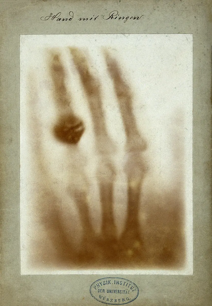
Röntgen asked his wife, Anna Bertha Röntgen, to hold her hand still on a photographic plate in the path of the rays. The picture showed the bones of her fingers, the ring on one of them as a dark lump, and the softer flesh around them as a pale shadow, because the flesh let more rays through. She had to keep her hand still for many minutes; the sources don’t agree on exactly how long. She is said to have cried out, “I have seen my death!”
4. New Year 1896: the news goes round the world
- Over Christmas, Röntgen wrote a short paper of ten pages, On a new kind of rays. The Physical-Medical Society of Würzburg accepted it on 28 December 1895.
- On 1 January 1896 he posted copies to about 90 physicists all over Europe. To a few of them he sent X-ray photographs too, including the picture of his wife’s hand.
- One of the photographs reached a newspaper editor in Vienna. On 5 January 1896 his paper, Die Presse, put the story on its front page (with Röntgen’s name spelled wrong). Within days, newspapers around the world had it.
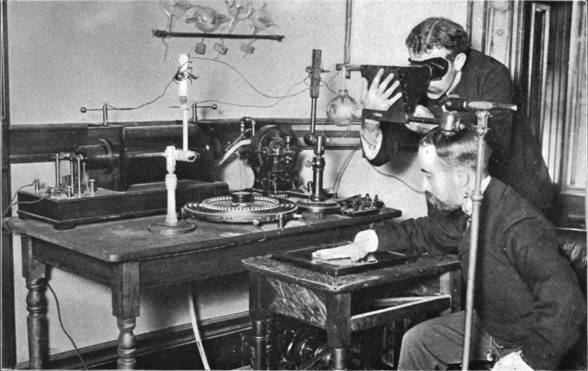
- Within weeks, doctors were using X-rays to find bullets and needles and to look at broken bones.
5. A gift to the world
Röntgen could have patented the X-ray tube, so that anyone making one would have to pay him. He refused: he believed that discoveries belong to the world. Anyone could build an X-ray machine, and by the end of 1896 more than a thousand books and papers about X-rays had been published. He wrote only two more papers about them himself. When he received the Nobel Prize in Stockholm on 10 December 1901, he didn’t give the usual Nobel lecture, and he is said to have given the prize money to his university in Würzburg for science.
How an X-ray picture is made today
In 1912, the German physicist Max von Laue showed what X-rays are: they are electromagnetic waves, like light, radio waves and ultraviolet rays, but with a far shorter wavelength, thousands of times shorter than that of visible light. A shorter wavelength means more energy, which is why X-rays can pass through things that stop light Grade 9: Electromagnetic spectrum.
A modern X-ray tube works much like Röntgen’s, but it is built to make X-rays well.
- A thin wire, the filament, is heated until it glows, and electrons boil off it.
- A very high voltage, tens of thousands of volts, pulls the electrons across the empty tube at enormous speed.
- They crash into a block of tungsten, a heavy metal that melts only at about 3,400 °C. As the electrons are suddenly stopped, a little of their energy comes out as X-rays. Only about 1% does: the rest becomes heat, so the tungsten has to be cooled, and in many tubes it spins so that the beam never hits the same spot for long.
- The X-rays leave through a window. Everywhere else the tube is wrapped in lead, so that X-rays go only where the doctor aims them.
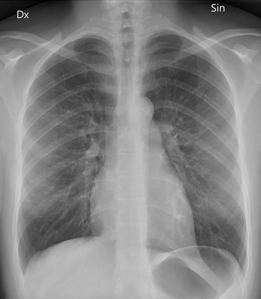
The beam passes through the patient to a detector (in older machines a sheet of film) on the other side. Different parts of the body stop different amounts of X-rays. Bones contain a lot of calcium, which is good at stopping X-rays, so few rays get through them. Muscle, fat and skin stop fewer, and air in the lungs hardly any. The picture is made so that wherever few X-rays arrive it is light, so bones look white, flesh gray and air black.
On an X-ray picture of your chest, your ribs look white and your lungs look dark. Why?
Pick an answer to see why.
In your life today
- Hospitals and health posts. X-rays show broken bones, the lungs of a patient with pneumonia or tuberculosis, and the roots of your teeth at the dentist’s Grade 9: X-ray imaging. A CT scan takes hundreds of X-ray pictures from all round the body, and a computer builds them into slices Grade 9: CT scan.
- Airports. At Tribhuvan International Airport, as at every airport, your bags go through an X-ray scanner before a flight.
- Treating cancer. Strong, carefully aimed beams of X-rays are used to destroy cancer cells.
- The shapes of molecules. X-rays shone through a crystal are scattered into a pattern of spots, and from the pattern scientists can work out where every atom sits. This is how the shape of DNA was found, and the shapes of thousands of medicines.
What came next
- Radioactivity (1896). Hearing about X-rays, the French scientist Henri Becquerel tested whether glowing uranium salts give out X-rays too. He found something else: uranium gives out rays all by itself. Becquerel and Pierre and Marie Curie shared the 1903 Nobel Prize in Physics for this radioactivity Grade 9: Radioactive elements.
- The electron (1897). J. J. Thomson found that cathode rays, the rays that make X-rays, are electrons. He won the Nobel Prize in Physics in 1906.
- More prizes. X-rays led to many more Nobel Prizes: Max von Laue (1914) and William and Lawrence Bragg, father and son (1915), for looking at the atoms inside crystals, and Allan Cormack and Godfrey Hounsfield (Physiology or Medicine, 1979) for the CT scanner.
- The X-ray sky. Hot gas around black holes and exploding stars gives out X-rays. Our air stops them, luckily for us, so X-ray telescopes have to fly in space.
An open question: X-rays in large doses cause cancer, but nobody knows for sure whether the tiny doses of a single X-ray picture do any harm at all. It is too small to measure, so doctors play safe: they take an X-ray only when it is needed, and use the smallest dose that gives a clear picture.
In your science books
- Grade 8: the electron, the particle that makes up cathode rays.
- Grade 9: electromagnetic waves, the electromagnetic spectrum, X-rays and their uses, X-ray imaging, CT scans and radioactivity.
- Going Further: Is phone radiation dangerous?, on why X-rays can harm cells and radio waves can’t.
Learn more
- The Nobel Prize’s own story of the discovery and the news: An illuminating accident (nobelprize.org). Röntgen gave no Nobel lecture.
- The presentation speech from the first Nobel Prize ceremony, 10 December 1901.
The official reason
The prize was given “in recognition of the extraordinary services he has rendered by the discovery of the remarkable rays subsequently named after him.”