
How your cells recycle themselves
Nobel Prize in Physiology or Medicine, 2016: Yoshinori Ohsumi
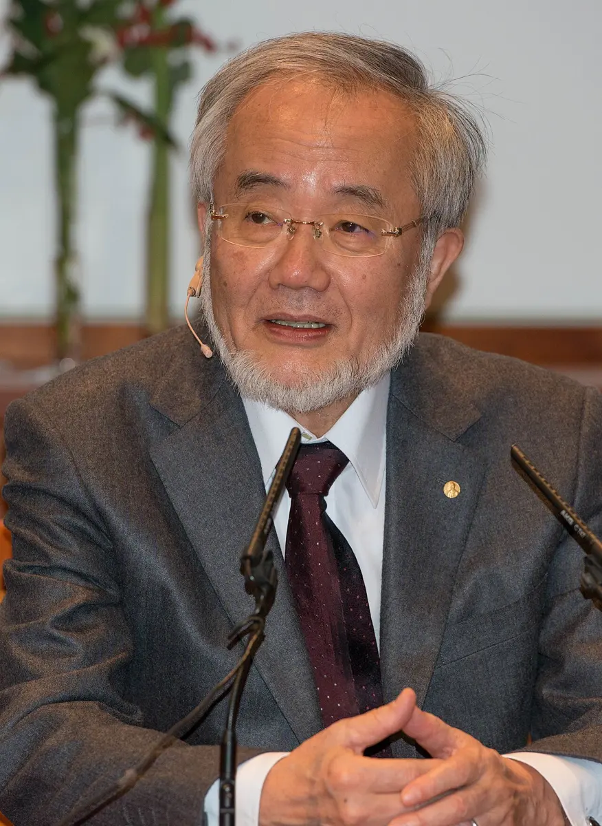
Yoshinori Ohsumi
born 1945 in Fukuoka, Japan
Started his own laboratory at 43, with himself as its only member and a basic light microscope, and chose a question almost nobody was studying.
In Kathmandu, a man with a cart calls out “khali sisi, purano kagaj!” (empty bottles, old paper!). He buys what people throw away, and it goes off to be broken down and made into new things. Nothing useful is wasted.
Every cell in your body has its own recycling system, and it works all the time. Old and broken parts are wrapped up, carried away, taken to pieces, and the pieces are used again. This is called autophagy, and the 2016 Nobel Prize in Physiology or Medicine went to Yoshinori Ohsumi, who found out how it works.
The problem: cells that wear out
A cell is a busy factory Grade 8: Parts of a cell. Its machines are proteins and small structures called organelles, such as the mitochondria that release energy from food. Like any machine, they wear out and break. Broken parts get in the way, and some do harm. A cell that never cleaned up would soon be clogged with rubbish.
Cells also face hard times. When food is short, a cell needs fuel and building materials from somewhere. One answer is to take apart some of its own parts and use the pieces.
Scientists knew part of the answer by the 1950s. In 1955 the Belgian scientist Christian de Duve discovered the lysosome, a tiny bag inside the cell full of enzymes that digest proteins, fats and sugars. He won a Nobel Prize for it in 1974 Grade 8: Lysosomes.
Then, in the 1960s, scientists looking through electron microscopes saw something strange. Some lysosomes contained whole pieces of the cell, even whole mitochondria, half digested. The cell seemed to be eating itself. In 1963 de Duve named this autophagy, from the Greek for “self-eating” (auto, self; phagein, to eat).
What does a lysosome do?
Pick an answer to see why.
How autophagy works
Today we know the steps. They happen in your cells right now.
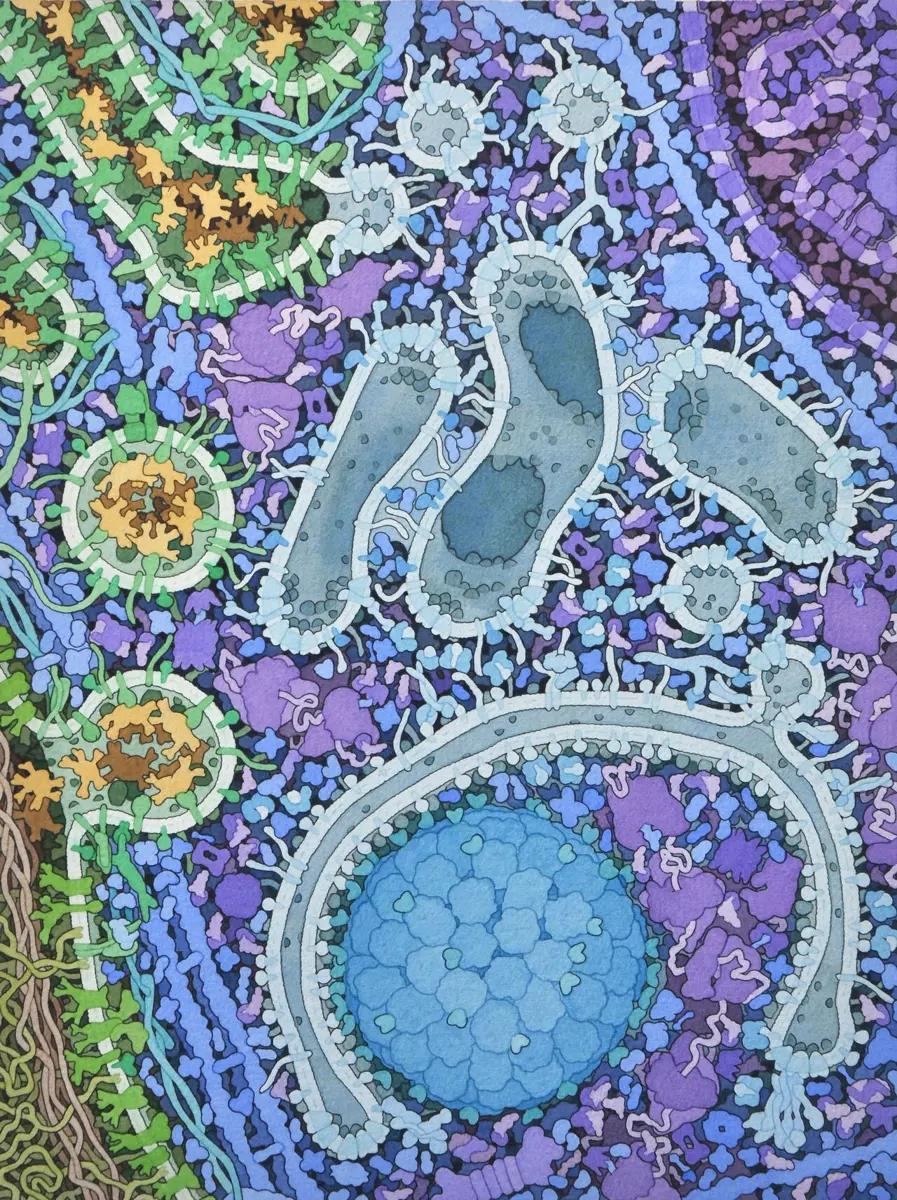
- A cup forms. A thin double layer of membrane (the same kind of thin skin that surrounds the whole cell) appears in the cytoplasm and grows, curving into a cup.
- It wraps the rubbish. The cup closes round whatever is nearby: old proteins, a worn-out mitochondrion. Now it is a sealed bag with two walls, called an autophagosome.
- It joins a lysosome. The bag travels to a lysosome, and the two join, like two soap bubbles becoming one.
- Digestion. The lysosome’s enzymes break everything down into small pieces: the building blocks of proteins, fats and sugars.
- Recycling. The pieces go back out into the cell, to be burnt for energy or built into new parts.
There is a second, separate system that breaks proteins down one at a time, the proteasome (its discovery won the 2004 Nobel Prize in Chemistry). But a proteasome can’t take apart a whole mitochondrion. For big jobs, the cell uses autophagy.
Why it was hard
For thirty years, autophagy stayed a mystery. By the late 1980s, fewer than twenty scientific papers a year were written about it.
- It was hard to see. An autophagosome is digested within minutes of reaching a lysosome, so at any moment a cell has only a few, and they look like many other small bags in the cell.
- The microscope gave only snapshots. An electron microscope shows tiny details, but only of cells that are dead and sliced. You can’t watch the process happen.
- Nobody knew the genes. Without knowing which genes control autophagy, there was no way to switch it off and see what it does, or to find out how it is switched on.
What he did, step by step
Ohsumi: watching yeast starve
In 1988 Yoshinori Ohsumi became an associate professor at the University of Tokyo and started his own laboratory. At first it had one member, himself, and very simple equipment, including a basic light microscope. He was 43. He decided to study something few others were working on: how cells break things down.
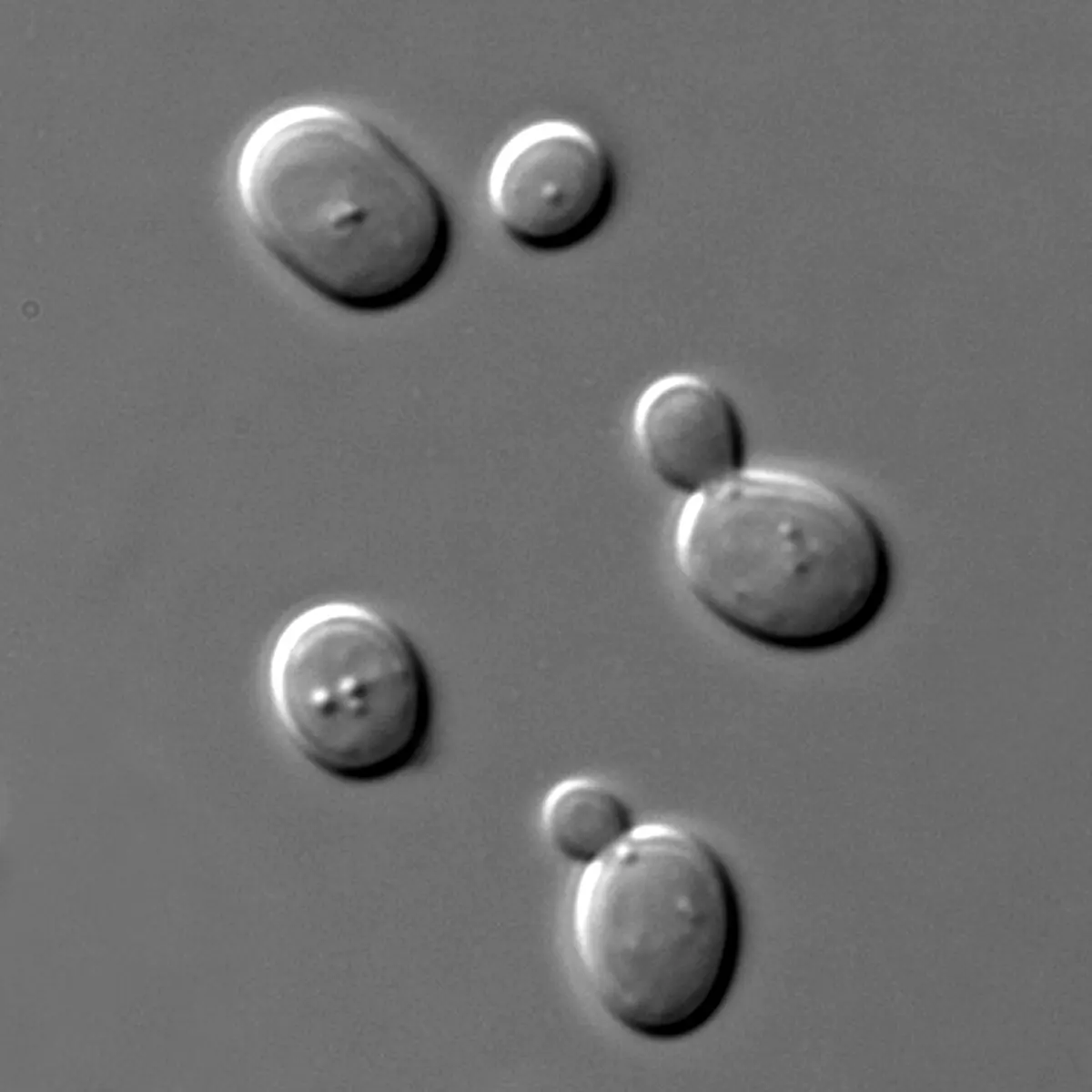
He chose baker’s yeast, the yeast that makes bread rise. Yeast is a fungus made of single cells, but its cells have a nucleus and organelles like ours Grade 9: Kingdom Fungi. In a yeast cell the job of the lysosome is done by a much bigger bag, the vacuole, which is large enough to see clearly under a light microscope.
- The idea. If autophagosomes are digested as soon as they reach the vacuole, nobody will ever see them. But what if the vacuole couldn’t digest? Then the bags would pile up inside it, and might become visible.
- The trick. Another scientist, Elizabeth Jones, had made yeast that lacked the vacuole’s digesting enzymes, and shared them with other laboratories. Ohsumi got some, and starved them of nitrogen, the element that every protein contains. A starving cell should switch on autophagy to find building materials.
- What he saw. Within 30 minutes, small round bodies appeared inside the vacuoles, jiggling about. Within a few hours they had filled the vacuoles. Ohsumi said he stared down the microscope for hours, unable to look away. Electron microscope pictures showed that each body was a bag of ordinary cell contents. Autophagy happened in yeast. He published it in 1992.
To find the genes, Ohsumi used a trick geneticists love. A gene is a stretch of DNA that carries the instructions for making one protein (Proteins do the work). A mutation is a change in a gene, and it can spoil the instructions so the protein is not made properly. So you break genes at random, then look for the cells that can no longer do the job. Whatever gene is broken in them must be one the job needs.
- Finding the genes. Now he had a test he could see. He used a chemical to make random changes (mutations) in the genes of many yeast cells, then starved them. His student Miki Tsukada checked thousands of yeast colonies, one by one, under the microscope. Any yeast whose vacuole stayed empty must have a broken gene needed for autophagy. Within a year they had found the first one. In 1993 they described more than a dozen genes that autophagy can’t work without.
- How the genes work. Through the 1990s his group worked out what each gene’s protein does: which ones sense that the cell is starving, which build the cup, which help it close.
- The same in us. Then came the big surprise: almost the same genes, doing the same jobs, are found in plants, animals and humans. What Ohsumi learned in yeast was true for our own cells.
In Ohsumi’s gene hunt, the yeast lacked digesting enzymes and was starved. Which result told him he had found a broken autophagy gene?
Pick an answer to see why.
How it works now: what autophagy does for you
Thanks to Ohsumi and those who followed him, we now know that autophagy is busy in almost everything your cells do.
- When food runs short. Starving cells turn up autophagy, taking apart some of their own spare parts for fuel and building blocks. This has been seen in yeast, in mice and in human cells grown in dishes. It helps living things survive hunger.
- Cleaning up. It removes damaged mitochondria and clumps of spoiled proteins, a daily quality check. Nerve cells, which must last your whole life, depend on it heavily.
- Fighting germs. It can wrap up bacteria and viruses that have got inside a cell and take them to the lysosome to be destroyed.
- Growing up. It helps an embryo develop and helps cells become specialized.
When it goes wrong, people get ill. Faults in autophagy have been linked to Parkinson’s disease and other illnesses of the nervous system, to type 2 diabetes, to some cancers, and to rare inherited diseases caused by changes in autophagy genes.
In your life today
- Medicines in testing. Many teams are testing medicines that speed autophagy up (to clear harmful protein clumps in the brain) or slow it down (because some cancer cells use it to survive). As of 2026, these are still mostly being tested.
- Fasting. In Nepal many people fast, for Teej, on Ekadashi, or in Ramadan. A body that goes without food does turn up autophagy; that has been measured in animals and in cells.
What came next
- Picking the rubbish. Autophagy was first thought to wrap up whatever is nearby. We now know cells can also choose: special proteins tag a broken mitochondrion or an invading bacterium so that it is wrapped up and nothing else.
- Aging. In worms, flies and mice, animals whose autophagy is boosted often live longer. Whether the same is true for people is an open question, and one of the most studied in biology today.
- The brain. Researchers are trying to find whether helping autophagy along can slow diseases such as Parkinson’s, in which clumps of protein build up in nerve cells.
In your science books
- Grade 8: parts of a cell, mitochondria, lysosomes and vacuoles.
- Grade 9: Kingdom Fungi, where yeast belongs.
- Going Further: how a gene makes a protein, and yeast without air.
Learn more
- The Nobel Prize’s own explanation: the press release (nobelprize.org), with pictures of the experiment.
- Ohsumi’s Nobel lecture, and his own life story.
The official reason
The prize was given “for his discoveries of mechanisms for autophagy.”