
The postal service inside your cells
Nobel Prize in Physiology or Medicine, 2013: James E. Rothman, Randy W. Schekman, and Thomas C. Südhof

James E. Rothman
born 1950 in the United States
As a boy he built electronics and launched model rockets, and taught himself trigonometry to work out how high they flew. He trained as a physicist before turning to biology.
Photo: US Embassy Sweden, CC BY 2.0
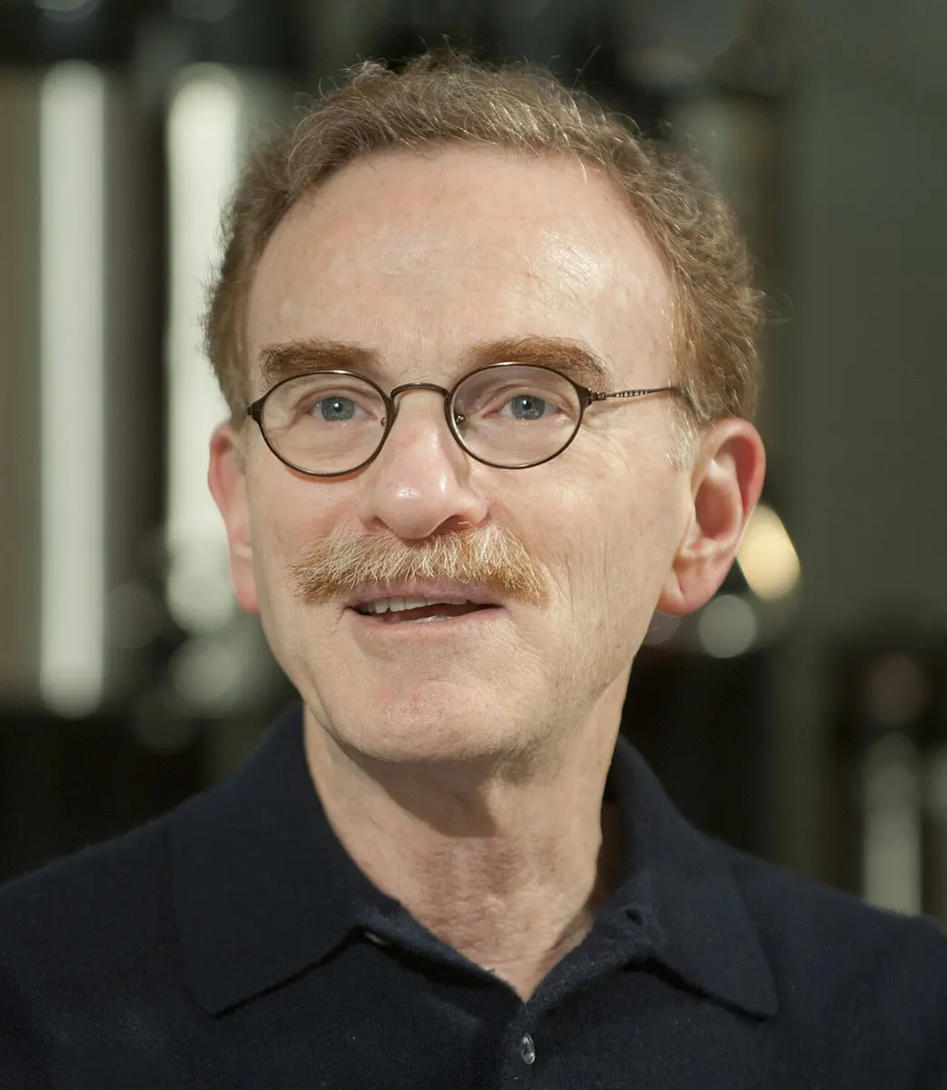
Randy W. Schekman
born 1948 in the United States
In seventh grade he saved money from mowing lawns for a real microscope. When his mother kept borrowing it, he rode his bicycle to the police station to complain, and got the microscope.
Photo: James Kegley, CC BY-SA 3.0

Thomas C. Südhof
born 1955 in Germany
He played the bassoon seriously as a teenager, and says his music teacher taught him more about life than most of his other teachers.
Photo: Juancamartos, CC BY-SA 4.0
Think of the post office or a courier company in your town. Parcels arrive from everywhere. Each one must be packed, labeled, put on the right van and handed over at the right door, and some, like a wedding invitation, must arrive on the right day. Mix up the labels and everything goes to the wrong place.
Every cell in your body runs a service like this, all day and all night. It packs molecules into tiny bubbles and delivers them, inside the cell or out of it, to exactly the right place at exactly the right moment. In 2013 the Nobel Prize in Physiology or Medicine went to three scientists who found out how the deliveries are addressed, docked and timed.
The problem: a cell full of rooms
A cell of your body is not just a bag of liquid. It is divided into many small compartments called organelles, each wrapped in its own thin skin, a membrane Grade 8: Parts of a cell. Think of them as the rooms of a factory:
- the nucleus holds the instructions, the DNA;
- the endoplasmic reticulum (ER), a maze of folded membranes, is where many proteins are made;
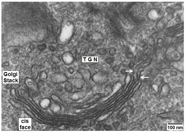
- the Golgi body, a stack of flat sacs, finishes the proteins, sorts them and packs them for delivery Grade 8: Golgi body;
- the cell membrane is the outer wall, with the outside world beyond it.
Many of the things a cell makes must leave the room where they were made. Insulin, the hormone that controls the sugar in your blood, is made inside cells of the pancreas and must be sent out into the blood Grade 9: Pancreas. Nerve cells send out chemicals called neurotransmitters to pass a message to the next cell. Most of these molecules are far too big to pass through a membrane by themselves.
How the delivery works
By the 1970s, scientists looking at cells through electron microscopes (which can see things far smaller than a light microscope can) had found the delivery vans. They are vesicles: tiny bubbles of membrane, each a few tens of nanometers across, with cargo inside.
A vesicle buds off from one membrane, like a small bubble pinching off a big soap bubble. It carries its cargo to another membrane and fuses with it: the two membranes join into one, and the cargo is let out on the other side. Proteins made in the ER travel in vesicles to the Golgi body; from the Golgi body, new vesicles carry them on to the cell membrane, and there the cargo is released outside the cell.
A cell has made a protein that must go out into the blood. How does it get out?
Pick an answer to see why.
Why it was hard
Seeing vesicles was one thing. Understanding how they work was another.
- How does a vesicle find the right door? A cell has many membranes. A vesicle from the ER must fuse with the Golgi body, not with the nucleus or the outer membrane. What is the address label, and who reads it?
- How do two membranes join? A membrane is a thin oily layer. Two of them don’t merge just by touching. Something has to pull them together and make them fuse.
- How is it timed? In a nerve cell, neurotransmitter must be released only when a message arrives, and then within a tiny fraction of a second. What holds the vesicles back, and what lets them go?
Photos from a microscope could not answer these questions. They show where things are, not which molecules make them happen.
What they did, step by step
The three laureates came at the problem in three different ways: genes (Schekman), proteins (Rothman) and nerve cells (Südhof).
Schekman: traffic jams in yeast
Randy Schekman, at the University of California, Berkeley, from 1976, chose an unusual creature: baker’s yeast, the single-celled fungus that makes bread rise. Yeast cells also send proteins out through vesicles. And yeast is easy to study through its genes: it grows fast, and you can look for cells with a faulty gene among millions.
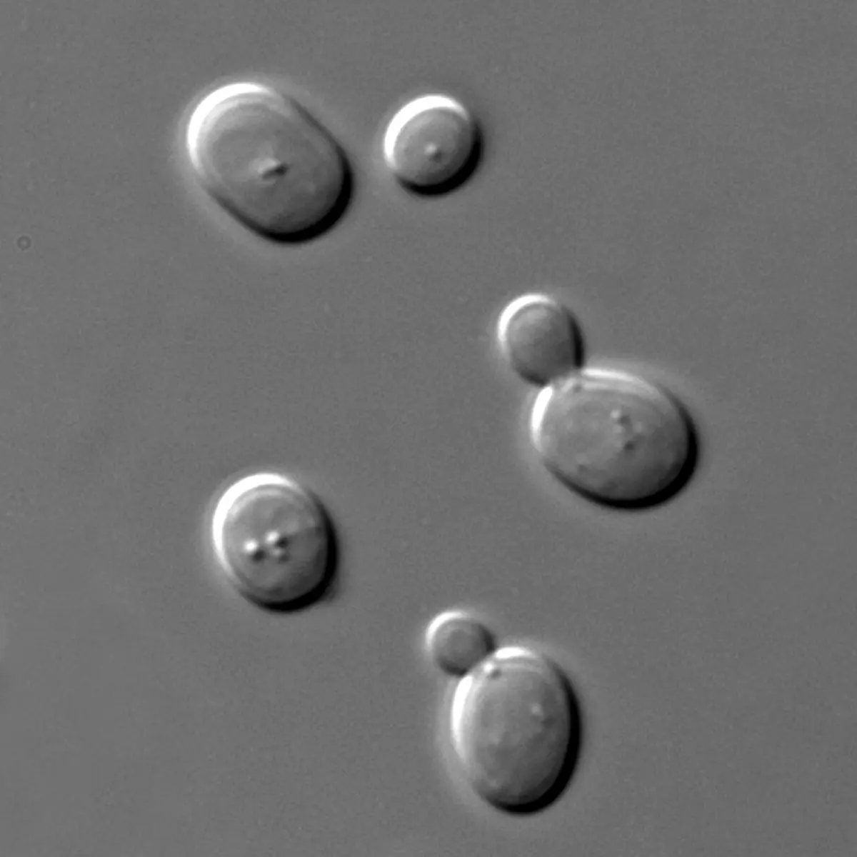
- A clever trick. A yeast cell whose delivery system is completely broken would die, and then there would be nothing to study. So Schekman looked for yeast with genes that fail only when the cells are warmed. At a normal temperature these yeast lived normally. Warmed up, their delivery system broke down.
- 1979, the first jam. With his student Peter Novick, he found yeast that, when warmed, stopped sending proteins out. Inside, the cells were full of vesicles that had piled up, like vans stuck in a traffic jam.
- 1980, 23 genes. They found 23 genes needed for the deliveries, which they named sec genes (for “secretion”, sending substances out). Where the jam happened showed what each gene did: some mutant cells were blocked at the ER, some at the Golgi body, and some at the cell membrane.
Rothman: the zip that joins two membranes
James Rothman, at Stanford University in California from 1978, tried the opposite approach. Instead of genes, he wanted the proteins themselves.
- Deliveries in a test tube. Rothman broke open animal cells and found a way to make vesicle deliveries happen outside the cell, in a test tube, between pieces of Golgi body. He used a protein from a virus that the cells had been infected with, which gets a special sugar label added when it reaches the next part of the Golgi body. If the label appeared, a delivery had happened. His key papers came out in 1984.
- Taking it apart. Now he could remove one ingredient at a time and see what stopped the deliveries. One by one he purified the proteins that were needed. The first, in 1988, was named NSF; the next, SNAP.
- The two discoveries meet. It turned out that one of Schekman’s yeast genes, sec18, carries the instructions for yeast’s own NSF. Another, sec17, matched SNAP. Yeast and mammals use the same machinery, so it must be very ancient, from before their ancestors split, a very long time ago.
- 1993, the SNAREs. Using NSF and SNAP as bait, Rothman fished out of brain tissue the proteins they hold on to, and called them SNAREs. One kind sits on the vesicle (a v-SNARE), and its partners sit on the target membrane (t-SNAREs).
- The idea. Rothman proposed that a v-SNARE and its matching t-SNARE grip each other and twist together like the two sides of a zip. As the zip closes, it pulls the vesicle and the target membrane so tightly together that they fuse. And because each v-SNARE fits only certain t-SNAREs, the SNAREs also work as the address: a vesicle can fuse only where its partner is waiting.
Later experiments by Rothman and others showed that SNAREs placed on artificial bubbles of membrane, in a test tube, are enough to make them fuse, and that only matching pairs do it.
How does a vesicle “know” which membrane to fuse with?
Pick an answer to see why.
Südhof: the trigger in nerve cells
Thomas Südhof trained as a doctor in Göttingen, Germany, and moved to Dallas, in Texas, in 1983. He wanted to understand how nerve cells talk to each other.
A nerve cell passes its message to the next cell across a tiny gap, called a synapse. When a nerve impulse, an electrical signal, reaches the end of the nerve cell, vesicles there fuse with the membrane and release neurotransmitter into the gap. The neurotransmitter reaches the next cell, which may then send an impulse of its own (Messages along nerves).
This has to be fast and exact. The vesicles must stay closed until the impulse arrives, and then open in less than a thousandth of a second. Scientists knew that calcium was the signal: when the impulse arrives, channels in the membrane open and calcium ions rush in. But what in the vesicle could sense calcium?
- 1990, a calcium sensor. Südhof’s team studied the proteins of the vesicles at nerve endings. One, which they named synaptotagmin, has parts that bind calcium.
- The test. In mice whose nerve cells lacked synaptotagmin-1, the fast release of neurotransmitter that follows an impulse was lost, though slower, untimed release went on. Calcium binding to synaptotagmin was what set off the fast release.
- The brake. Südhof also found complexin, a protein that holds the half-closed SNARE zip back until the calcium comes, and studied Munc18, the nerve cell’s version of one of Schekman’s yeast proteins, which turned out to be needed for every release.
In a nerve ending, what tells the waiting vesicles to release their neurotransmitter?
Pick an answer to see why.
How the system works now
Putting the three discoveries together:
- Packing. A vesicle buds from one membrane with its cargo inside, carrying v-SNAREs in its skin. (Schekman’s genes control the budding, the transport and the fusion.)
- Addressing. Its v-SNAREs can only pair with matching t-SNAREs, so it fuses only with the right membrane. (Rothman)
- Fusing. The SNAREs zip together and pull the membranes into one; NSF and SNAP then unzip the used SNAREs so they can work again. (Rothman, Schekman)
- Timing. Where release must happen on command, as in nerve cells and insulin-making cells, calcium sensors such as synaptotagmin hold the final step until the signal comes. (Südhof)
In your life today
- Every thought and movement. Each time a nerve cell passes a message, vesicles fuse at the synapse. Reading this sentence uses this machinery billions of times.
- Blood sugar. After you eat dal bhat, the sugar in your blood rises, and cells in the pancreas release insulin from vesicles. In type 2 diabetes, one of the problems is that this release doesn’t work well, and in muscle and fat cells, sugar transporters that insulin should bring to the surface in vesicles don’t arrive as they should Grade 10: Diabetes.
- Tetanus. The bacterium that causes tetanus lives in soil and can get into a deep wound, such as one from a rusty nail. Its poison cuts a SNARE protein inside certain nerve cells, whose job is to calm the muscles; without them, the muscles lock tight. In Nepal, babies are protected by the tetanus vaccine in their routine injections, and so are mothers during pregnancy.
- Botox. A related poison, from the bacterium that causes botulism, cuts the SNAREs at the nerve endings that tell muscles to move, so the muscles go limp. In tiny, carefully measured doses doctors use it as a medicine, for example to stop muscles that cramp or twitch, and to smooth wrinkles.
- Your defenses. Cells of the immune system release their weapons and signals from vesicles too.
What came next
- Seeing the zip. In 1998 the shape of the zipped SNARE bundle was worked out: four protein strands twisted together into one tight rope.
- Disease. Faults in vesicle proteins have been found in some kinds of epilepsy and other brain disorders, and in rare immune diseases, and scientists are studying how vesicle release goes wrong in diabetes.
- Open questions. Exactly how the zip pulls two membranes into one in less than a thousandth of a second, and how a synapse changes how readily it releases neurotransmitter as we learn, are still being worked out.
In your science books
- Grade 8: parts of a cell, including the endoplasmic reticulum and the Golgi body.
- Grade 9: the neuron and the pancreas, which releases insulin.
- Grade 10: diabetes.
- Going Further: messages along nerves.
Learn more
- The Nobel Prize’s press release: Machinery regulating vesicle traffic (nobelprize.org), and the committee’s longer scientific background.
- The three Nobel lectures: Rothman, Schekman and Südhof.
The official reason
The prize was given “for their discoveries of machinery regulating vesicle traffic, a major transport system in our cells.”