
The brain’s own GPS
Nobel Prize in Physiology or Medicine, 2014: John O’Keefe, May-Britt Moser, and Edvard I. Moser
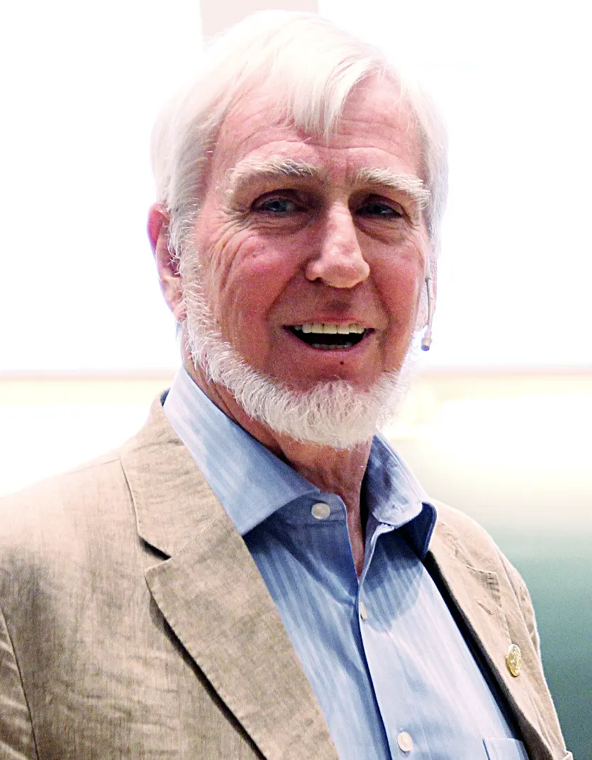
John O’Keefe
born 1939 in the United States
The son of Irish parents in New York. He has worked in London, at University College, since 1967.
Photo: Per Henning/NTNU, CC BY 2.0

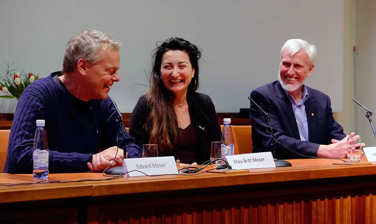
May-Britt Moser
born 1963 in Norway
She grew up on a small farm on an island on Norway’s west coast. She and Edvard studied psychology together in Oslo.
Photo: NTNU Trondheim, CC BY-SA 2.0

Edvard I. Moser
born 1962 in Norway
He and May-Britt married as students and built their lab together in Trondheim, starting in a basement.
Think of the walk from your home to school. You know where to turn, which lane is a short cut, and where the dog that barks lives. Wake up at night in your own room and you can find the door in the dark. Somewhere in your head there is a map, and something that marks where you are on it.
Your phone finds its place with signals from satellites. Your brain has no satellites, so how does it know? In 2014 the Nobel Prize in Physiology or Medicine went to three scientists who found the cells that do it: John O’Keefe, and the husband-and-wife team May-Britt and Edvard Moser.
The problem: where is the map?
People have wondered for a long time how we know where we are. In the 1700s the German philosopher Immanuel Kant argued that a sense of space is built into the mind from birth, before we learn anything.
In the 1940s an American psychologist, Edward Tolman, studied rats finding their way through mazes. A rat that had explored a maze could take a short cut it had never used before. Tolman said the rat must carry a cognitive map in its head: a picture of how the places fit together, not just a list of “turn left, then right”.
But nobody knew where in the brain such a map could be, or what it could be made of. The brain is made of nerve cells, or neurons Grade 9: Neuron or nerve cell. How could a cell “know” a place?
How a nerve cell talks
A neuron sends messages as tiny electrical pulses, called nerve impulses. Scientists say a neuron fires when it sends one. A busy neuron may fire many times a second; a quiet one hardly at all.
You can listen in. A very thin metal wire, thinner than a hair, is placed in the brain next to one neuron. Each time the neuron fires, the wire picks up a tiny jump in voltage. Joined to an amplifier and a loudspeaker, every impulse sounds like a click.
By the 1960s scientists could do this in animals that were awake. But usually the animal was held still, or trained to do one simple task, like pressing a lever. Nobody had simply let a rat wander about and listened to what its neurons were saying.
What does it mean when scientists say a nerve cell “fires”?
Pick an answer to see why.
Why it was hard
- A place is not a sense. There are cells for light in the eye and cells for sound in the ear. But there is no sense organ for “where am I?”. Your position has to be worked out by the brain from many clues: what you see, what you hear, how far you have walked and which way you turned.
- The brain is huge. A rat’s brain has about 200 million neurons, and yours has about 86 billion. A wire can listen to only one or a few of them at a time. Where should you look?
- The animal has to move freely. To see a cell respond to places, the rat must be allowed to go wherever it likes, for a long time, with the wires in its head and connected to the equipment.
What they did, step by step
John O’Keefe: cells that know a place
John O’Keefe grew up in New York and came to University College London in 1967. He had learned to record from single neurons in rats that could move about. He was so keen that he bought 50,000 feet (about 15 km) of fine wire for his electrodes.
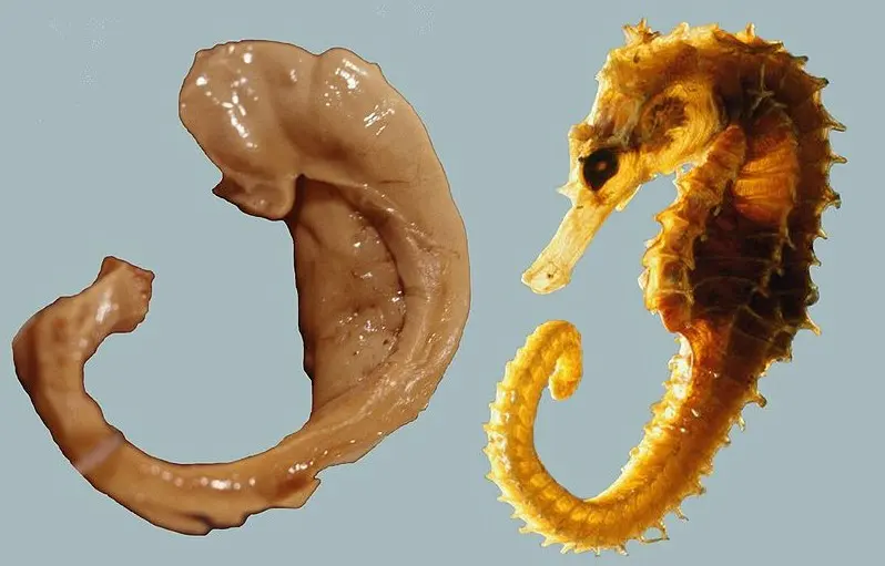
- 1970–71, listening to the hippocampus. With a student, Jonathan Dostrovsky, O’Keefe placed fine wires in the hippocampus of rats. The hippocampus is a curved part deep inside the brain, one on each side. It was already known to matter for memory. The rats walked about on a platform while the scientists listened.
- A strange pattern. Most of the cells they recorded were silent most of the time. Then, as the rat walked, one cell would suddenly fire, again and again, but only when the rat was in one particular spot on the platform. Wherever else the rat went, that cell stayed quiet. Another cell fired at another spot.
- Testing it. They changed things around the rat one at a time, and the cells mostly kept firing in the same spots. But when they took away the curtains around the platform, the cells’ firing changed completely. The cells were not responding to one smell or one sight. They were responding to where the rat was, worked out from everything around it. O’Keefe called them place cells, and published the first report in 1971.
- Many maps. O’Keefe found that each place cell has its own spot, so all of them together cover the whole room, like pins on a map. In a different room, the same cells fire in a new pattern: a new map. A memory of a room could be stored as a pattern of place cells. In 1978 he and Lynn Nadel wrote a book with a bold title: The Hippocampus as a Cognitive Map. Tolman’s map had been found.
Many scientists doubted it at first. Some thought the hippocampus was only for memory, not for space. It took years of experiments, by O’Keefe and others, before most were convinced.
A place cell in a rat’s hippocampus fires a lot. What does that tell you?
Pick an answer to see why.
May-Britt and Edvard Moser: a grid on the floor
May-Britt Moser and Edvard Moser met as students of psychology in Oslo. They became a couple and decided to study the brain together. For their first research they built a water maze: a round tank about 2 meters across, filled with water made cloudy with milk, where a rat learns to find a hidden platform to stand on. Every day they emptied it and bought new milk.
- 1990s, learning the method. They learned to record from single cells in O’Keefe’s lab in London. In 1996 they started their own lab at the university in Trondheim, in Norway, in an empty bomb shelter under the building.
- 2002, a question. Where do place cells get their information from? Many people thought the hippocampus worked out places by itself. The Mosers cut some of the connections inside the hippocampus, and the place cells still worked. So the signal could be coming from outside it.
- The next door. The main input to the hippocampus comes from a nearby part of the brain called the entorhinal cortex. In 2004 the Mosers found that cells there also fired at certain places. But each cell fired at several places, not one.
- 2005, the grid. They let the rats run in a much bigger space, a circle 2 meters across. Now they saw it: the spots where each cell fired formed a regular pattern of equilateral triangles, spreading over the whole floor. Joined up, any spot and its six nearest neighbors make a hexagon, like the cells of a honeycomb. The Mosers called these cells grid cells.
- Made by the brain. There were no triangles or hexagons on the floor; the pattern came from inside the brain. The Mosers turned out the lights, and the grid cells went on firing in their pattern. In the dark the rat could not see where it was, so its brain must have been working out its position from its own movement: how fast it was going, which way, and for how long. When they turned a large card on the wall of the arena, the grids turned with it: in the light, what the rat sees keeps the pattern in line with the room.
- Many sizes. Grid cells close together in the entorhinal cortex have patterns of the same size, shifted against each other, so that together they cover every point. Further along, the patterns get bigger, with the spots further apart. Some grids are fine and some are coarse, like the small and large squares on graph paper.
Grid cells kept firing in their pattern even in total darkness. What does that show?
Pick an answer to see why.
More pieces of the system
The entorhinal cortex has other helpers too. Head-direction cells, found by the American scientist James Ranck in the 1980s, fire when the head points one way: north, say. They work like a compass. In 2008 the Mosers’ team described border cells, which fire when the animal is next to a wall or an edge. O’Keefe and his colleagues had predicted cells like these.
How the inner GPS works now
Put the pieces together and you get a positioning system, which the Nobel committee called an “inner GPS”.
- As you walk, signals from your muscles, your balance organs in the ears and your eyes tell your brain how fast you are moving and which way you turn.
- Head-direction cells keep track of which way you face. Grid cells keep track of how far you have gone and in what direction, like the lines of latitude and longitude on a map.
- Border cells and the things you see, like walls and landmarks, keep correcting the grid so that small errors don’t build up.
- All this reaches the place cells in the hippocampus, which say “you are here, in this place”. The pattern of place cells is stored, so the next time you come back, you recognize the place.
Your phone’s GPS is different. It measures its distance from several satellites by timing their radio signals Grade 9: Global Positioning System. Your brain instead adds up its own movements and checks them against landmarks.
In your life today
- You have them too. Some people with severe epilepsy need brain surgery; before it, doctors place fine wires in their brains to find where the fits start. With the patients’ permission, scientists recorded from those wires while the patients found their way around a computer game town. They found place-like cells in the human hippocampus (2003) and grid-like cells in the entorhinal cortex (2013). Brain scans support this too.
- Learning a city. London’s taxi drivers must learn the city’s many thousands of streets by heart. Studies by Eleanor Maguire found that, after learning them, the back part of their hippocampus was bigger than in other people.
- Getting lost. In Alzheimer’s disease, which damages memory in many old people, the hippocampus and entorhinal cortex are among the first parts affected. One early sign is that a person gets lost in places they know well. Understanding the brain’s map may help us understand this, though there is no cure yet.
- Memory. The hippocampus is needed to store new memories of events (Store it for later). In sleeping rats, place cells fire again in the order of the route the rat ran earlier that day, as if replaying it. This may be part of how memories are stored.
What came next
- More cell types. In 2015 the Mosers’ team reported speed cells, which fire faster when the animal runs faster: just what grid cells need to measure distance. Other teams have found cells that respond to objects, and even cells in bats that work in three dimensions, for flying.
- How is the grid made? Nobody draws hexagons on the brain. Scientists think the grid pattern comes from how the grid cells are wired to each other, but exactly how is still being worked out.
- Maps of other things? Some experiments suggest the same cells may help us arrange ideas, not only places: a “map” of sounds, of people we know, or of memories. Whether the brain uses one system for both is an open question.
In your science books
- Grade 9: the neuron, the human nervous system and the brain.
- Grade 9: the Global Positioning System, the phone’s way of finding where it is.
- Going Further: messages along nerves and memory and the hippocampus.
Learn more
- The Nobel Prize’s press release: The brain’s navigational place and grid cell system (nobelprize.org), and the committee’s longer scientific background.
- The three Nobel lectures: O’Keefe, May-Britt Moser and Edvard Moser.
The official reason
The prize was given “for their discoveries of cells that constitute a positioning system in the brain.”