
MRI: pictures from magnets and radio waves
Nobel Prize in Physiology or Medicine, 2003: Paul C. Lauterbur and Peter Mansfield
-
Paul C. Lauterbur
1929–2007, born in Sidney, Ohio, USA
A chemist. As a boy he built his own laboratory in his parents’ basement. The idea for MRI came to him over dinner one evening in 1971.
-
Peter Mansfield
1933–2017, born in London, England
A physicist who left school at 15 to work for a printer, then went back to study at night school. He found how to make MRI pictures fast.
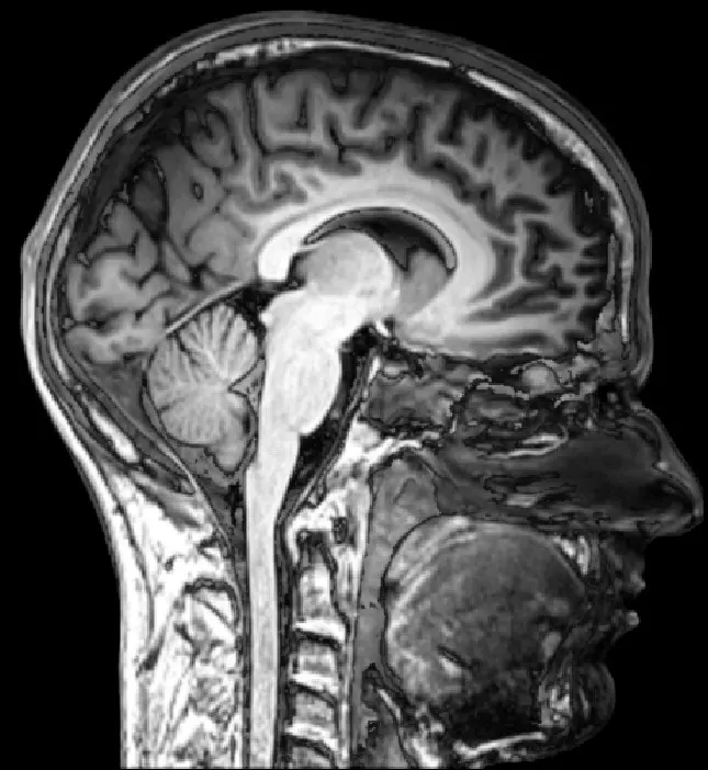
A footballer in Pokhara twists her knee, and it swells up. An X-ray shows the bones are fine. But the doctor wants to see the soft parts inside: the ligaments that hold the knee together and the cartilage that cushions it. X-rays pass through these almost without a trace. So the doctor sends her for an MRI scan. She lies still inside a large, noisy tunnel for about twenty minutes, and nothing touches her. Afterwards the doctor can see, slice by slice, a torn ligament inside her knee.
MRI stands for magnetic resonance imaging. It makes pictures of the inside of the body using a strong magnet and radio waves, with no X-rays at all. In 2003 the Nobel Prize in Physiology or Medicine went to Paul Lauterbur and Peter Mansfield, who discovered how to turn magnetic resonance into pictures.
The problem: seeing soft tissue
Doctors have long had ways to see inside the body Grade 9: Use of waves in diagnosing the body:
- X-rays (from 1895) show bones clearly, because bones stop X-rays and soft tissue lets most of them through. But the brain, muscles and ligaments look like gray shadows. X-rays are also ionizing radiation: in large doses they can damage cells Grade 9: X-rays.
- CT scans (from the 1970s) take X-rays from many directions and let a computer build a slice picture. They are much clearer, but they still use X-rays.
- Ultrasound uses sound echoes. It is safe, and good for a baby in the womb, but it cannot see through bone or air, so it can’t look inside the skull.
What doctors wanted was a safe way to see soft tissue clearly, anywhere in the body, including inside the skull and the spine.
How magnetic resonance works
Every hydrogen atom is a tiny magnet
About two-thirds of your body’s weight is water, and every water molecule (H₂O) has two hydrogen atoms. The nucleus of a hydrogen atom is a single proton Grade 9: Structure of an atom. Each proton spins, and that makes it a tiny magnet, like a very small compass needle.
Normally these billions of tiny magnets point every which way. Put the body in a strong magnetic field, and slightly more of them line up with the field.
Radio waves at just the right frequency
Each tiny magnet also wobbles around the direction of the field, like a spinning top that is tilted, at a fixed rate called its frequency. Send in a pulse of radio waves at exactly that frequency, and the protons soak up its energy and tip over. This matching of frequencies is called resonance. It is like pushing a child on a swing: small pushes work only if you push in time with the swing.
When the pulse stops, the protons swing back into line, and as they do they send out a faint radio signal of their own. An aerial around the body picks it up. This is nuclear magnetic resonance (NMR), discovered in 1946 by Felix Bloch and Edward Purcell (Nobel Prize in Physics, 1952).
The frequency depends on how strong the magnet is: the stronger the field, the higher the frequency. In a typical hospital scanner, with a field of 1.5 tesla, it is about 64 million waves per second (64 MHz), close to the FM radio stations you tune in to, which are between 88 and 108 MHz Grade 9: Radio waves.
Different tissues give back their signals in slightly different ways. Fat, muscle, brain and fluid differ in how much water they hold and in how quickly their protons swing back. Many diseases change the water in a tissue, so they show up.
In an MRI scan, which atoms send out the signal that makes the picture?
Pick an answer to see why.
Why it was hard
For 25 years, NMR was used by chemists, to find out which atoms are in a molecule and how they are joined. They put a small tube of liquid in an NMR machine, and got back a graph of frequencies.
But this told them what was in the tube, not where it was. The whole sample gave one signal. A picture needs to know where each signal comes from. Every textbook said that an NMR magnet must be as even as possible, the same strength everywhere, or the signal would be blurred.
In 1971 a doctor, Raymond Damadian, reported that some tumors taken from rats gave back their signals more slowly than healthy tissue. That raised a hope: perhaps NMR could find cancer. But to use it inside a living person, you would need to know where in the body each signal came from.
What they did, step by step
Lauterbur: a magnet that is stronger on one side
Paul Lauterbur was a chemistry professor at Stony Brook University, near New York, and an expert in NMR.
- September 1971, the idea. He watched NMR measurements on rat tumors. That evening, over dinner, he thought: what if the magnet were deliberately made uneven, a little stronger on one side than the other? Then protons on the strong side would answer at a higher frequency than protons on the weak side. The frequency of the signal would tell you where it came from. A field that changes steadily across the sample like this is called a gradient.
- Many directions. One gradient tells you only how much water lies along each line across the sample, a projection. So he would turn the gradient to many directions, and work the picture out from all the projections together, like guessing the shape of an object from its shadows. He tested the mathematics with pencil and paper first.
- The first picture. He used the chemistry department’s NMR machine, mostly at night. His test object was two thin glass tubes of ordinary water standing in a larger tube of heavy water (water made with a heavier kind of hydrogen, which answers at a different frequency and so stays invisible). He turned the sample by hand to four angles, measured each signal with a ruler, punched the numbers onto cards for a computer, and drew the picture by hand. It showed the two tubes of water. No other method could tell ordinary water from heavy water.
- March 1973, published. The journal Nature first rejected his paper. He asked them to look again, and it appeared in March 1973. Soon he made pictures of a small clam that his daughter had collected on the beach.
Why did Lauterbur make the magnetic field stronger on one side of the sample?
Pick an answer to see why.
Mansfield: from minutes to a fraction of a second
Peter Mansfield was a physicist at the University of Nottingham, in England, also an expert in NMR.
- 1972–1973, sharper pictures. He also used gradients, and worked out the mathematics to turn the signals quickly and exactly into a picture.
- Slices. He found how to pick out the signal from just one thin slice of the body, so that each picture is a clean cross-section.
- Too slow. Early pictures took 10 to 20 minutes each, and the patient had to keep perfectly still. A beating heart could not be pictured at all.
- 1977, a snapshot. Mansfield invented echo-planar imaging: by switching the gradients back and forth very fast, a whole slice could be recorded after a single radio pulse, in about a twentieth of a second. It took about ten more years for machines strong and fast enough to use it in hospitals.
How an MRI scan works now
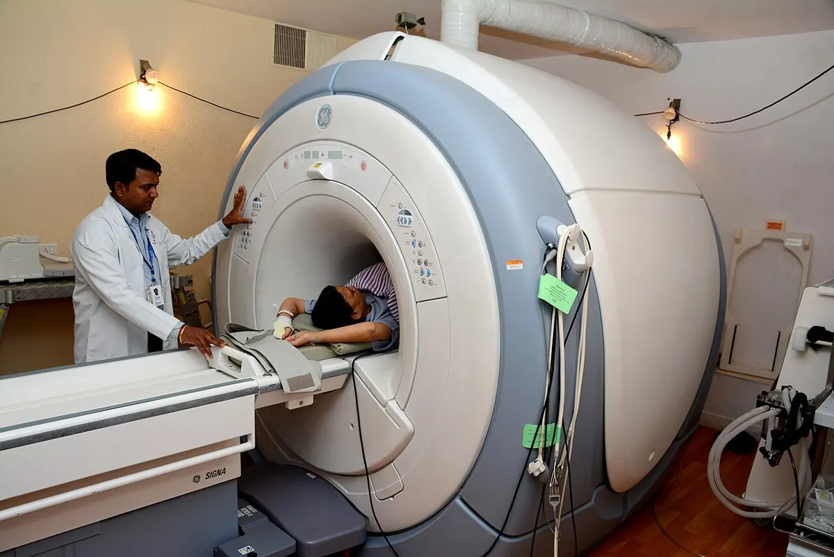
A modern scanner has three main parts:
- A very strong magnet, usually 1.5 or 3 tesla, tens of thousands of times stronger than the Earth’s field. It is a coil of superconducting wire cooled by liquid helium (the Nobel Prize in Physics of the same year explained how such wires survive strong fields).
- Gradient coils, which make the field a little stronger on one side, in any direction, and switch it on and off thousands of times a second. As they switch, they shake, and that makes the loud knocking and buzzing you hear in a scanner.
- Radio coils, which send the pulses and listen for the faint echoes, and a computer that turns them into pictures, one slice after another.
In your life today
- In Nepal. Large hospitals in Kathmandu and other cities have MRI scanners. Doctors use them for injuries to the knee and back, strokes and tumors in the brain, problems with the spine, and much more.
- Safe, with care. MRI uses no ionizing radiation, and as far as anyone knows it is harmless. But the magnet is always on. Steel objects such as keys, pins or a wheelchair can fly towards it, and people with a heart pacemaker or certain metal implants may not be scanned.
- Watching the brain think. A fast form of Mansfield’s echo-planar imaging can see which parts of the brain are working, because active brain areas get more blood with oxygen. This is functional MRI, used to study how we see, speak and remember.
- How common? Already in 2002, about 22,000 MRI scanners were in use around the world, and more than 60 million scans were done that year.
What came next
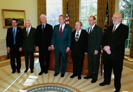
- Who else deserved it? Raymond Damadian, who in 1971 had found that tumors give different signals and who built an early whole-body scanner, was left out. He took out full-page newspaper advertisements protesting. The Nobel committee gave the prize for the discoveries that turn signals into pictures, which were Lauterbur’s and Mansfield’s. Scientists still argue about it.
- Faster and stronger. Scanners with 7-tesla magnets now show the brain in fine detail, and new methods make scans quicker, so that children need not lie still for so long.
- Cheaper scanners. An MRI scanner costs a great deal, and many people in the world live far from one. Engineers are building small, low-field scanners that can be wheeled to a patient’s bed. How good can a cheap, simple scanner be made? That is an open question.
In your science books
- Grade 8: uses of magnets, including the MRI scanner.
- Grade 9: the structure of an atom, radio waves, X-rays and the use of waves in diagnosing the body.
- Grade 10: the electromagnet.
- Going Further: is phone radiation dangerous? (why radio waves can’t break molecules).
Learn more
- The Nobel Prize’s press release: Magnetic resonance imaging (nobelprize.org).
- The laureates’ Nobel lectures: Lauterbur and Mansfield.
The official reason
The prize was given “for their discoveries concerning magnetic resonance imaging.”