
The address labels on proteins
Nobel Prize in Physiology or Medicine, 1999: Günter Blobel
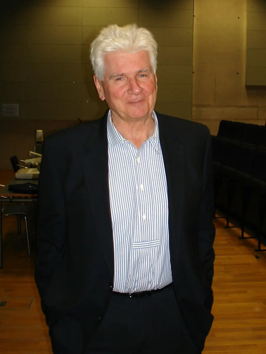
Günter Blobel
1936–2018, born in Germany, in a village that is now in Poland
At eight, fleeing the war with his family, he drove through the beautiful city of Dresden a few days before it was bombed. He gave all his prize money to help rebuild it.
Photo: Masur, CC BY-SA 4.0
Think of the letters and parcels that go through a post office in Kathmandu every day. Thousands of them, all mixed up in sacks, and yet nearly every one reaches the right house in the right town. It works because each one carries an address.
Each cell in your body has a far bigger delivery problem. It makes thousands of different proteins, about a billion protein molecules in all, and each must end up in the right place: some inside the nucleus, some in the mitochondria, some in the cell’s outer skin, and some sent out of the cell altogether. How does a protein know where to go? In 1999 the Nobel Prize in Physiology or Medicine went to Günter Blobel, who found that every protein carries its address written in its own chain.
A cell full of rooms
You have learned that a cell is not just a bag of jelly. Inside it are organelles, small parts with their own jobs, and most of them are wrapped in a thin skin, a membrane Grade 8: Cell components. They are like the rooms of a house, each with walls:
- the nucleus holds the DNA, the cell’s instructions Grade 8: Nucleus;
- the mitochondria release energy from food;
- the endoplasmic reticulum, or ER, is a maze of folded membranes. Part of it is covered with ribosomes, which makes it look rough Grade 8: Endoplasmic reticulum;
- the cell membrane is the outer wall of the whole cell Grade 8: The covering of the cell.
Proteins are made in one place
Proteins do most of the cell’s work. Some are enzymes, which speed up its chemical reactions; some are building blocks; some, like the hormone insulin, are sent out into the blood to carry messages. A protein is a long chain of small units called amino acids, joined in an exact order, from about 50 to several thousand of them, and then folded into a shape (Proteins do the work).
Every protein is built by a ribosome, a tiny grain that reads a copy of a gene, called messenger RNA, and joins the amino acids one by one in the order it says (Ribosomes, RNA). The ribosomes sit in the cytoplasm, outside all the rooms. So nearly every protein is made in the same “hall”, and then has to be sent to the room where it works.
Insulin is made in cells of the pancreas, but it does its job in the blood. What problem does the cell have to solve?
Pick an answer to see why.
Two puzzles
By the 1960s, scientists had two questions they could not answer.
- How does a new protein find the right room? The ribosome that makes insulin looks exactly like the ribosome that makes an enzyme for the mitochondria. Something must tell the cell which protein goes where.
- How does a protein get through a wall? A membrane is a very thin, oily layer, made mostly of fats. It is tightly sealed: that is its job. A protein is a huge, water-loving molecule. How could it pass through without tearing the membrane?
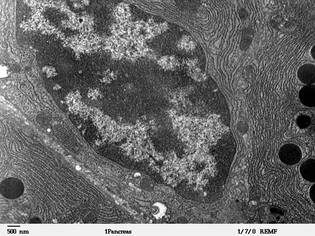
An important clue came from the laboratory of George Palade at the Rockefeller Institute in New York. Using the electron microscope, which shows things far smaller than a light microscope can, Palade and his team had followed the path of proteins made to be sent out of a cell. They are made on ribosomes stuck to the ER, travel through the ER and on through the Golgi body, and are finally released from the cell. Palade shared the 1974 Nobel Prize for this work. But nobody knew why some ribosomes stuck to the ER and others did not.
What Blobel did, step by step
Günter Blobel was a doctor from Germany. He had studied medicine, then decided he found the unsolved problems of medicine more interesting than treating patients. After a research degree at the University of Wisconsin in the USA, he joined Palade’s laboratory in New York in 1967.
1. An idea: an address at the front (1971)
In 1971, Blobel and his colleague David Sabatini put forward an idea. Perhaps a protein that must leave the cell carries a short extra piece at the front of its chain, the end that the ribosome makes first. As soon as this piece pokes out of the ribosome, it could make the ribosome stick to the ER.
2. A protein that was too long (1972)
By then, scientists had learned to make proteins in a test tube. They broke open cells, kept the ribosomes and the other parts needed for building proteins, and added the messenger RNA for one protein. The ribosomes then built that protein, outside any living cell.
In 1972, a team in Cambridge, England, led by César Milstein, did this with the messenger RNA for part of an antibody, a protein sent out of white blood cells. The protein made in the test tube was a little longer than the same protein found outside real cells. It had an extra piece at its front end. It looked just like the extra piece Blobel had imagined.
3. The signal hypothesis (1975)
In 1975, with Bernhard Dobberstein, Blobel set out the full idea, which he called the signal hypothesis (a hypothesis is an idea that experiments can test):
- A protein that must be sent out of the cell begins with a short extra piece of chain, the signal. It is made first, so it is the first part to stick out of the ribosome.
- The signal is recognized, and the ribosome, still building, is guided to the ER and attached to it.
- The growing chain is threaded through a channel, a narrow pore in the ER membrane, so it ends up inside the ER.
- Once it is through, the signal is cut off by an enzyme. That is why the finished protein is shorter than the one made in a test tube without any ER.
So the address is not added later, like a sticker. It is a part of the protein itself: a short stretch of amino acids in a particular order, written in the protein’s own gene. The signal peptide (a peptide is a short chain of amino acids) is usually about 15 to 30 amino acids long, and its middle is a stretch of oily amino acids, which can slip into an oily membrane.
4. The test: bits of ER in a test tube (1975)
An idea must be tested. Blobel and Dobberstein built a test-tube system from parts of three animals: the messenger RNA for the antibody protein from mouse cells, the ribosomes and protein-building parts from rabbit blood cells, and pieces of ER from the pancreas of dogs. When cells are broken up, the ER breaks into tiny closed bubbles, so the inside of the ER stays inside.
They did the experiment three ways.
- No ER. The protein came out long, with its signal still on. When they added an enzyme that digests proteins, it was all digested.
- ER present while the protein was being made. The protein came out the right, shorter length: the signal had been cut off. And it was inside the bubbles: the digesting enzyme, added outside, could not reach it.
- ER added only after the protein was finished. The protein stayed long, and outside. The ER could take a protein in only while it was being made, chain end first, as the hypothesis said.
They also tried the messenger RNA for globin, part of hemoglobin, a protein that stays in the cytoplasm of red blood cells. It has no signal, and it was never taken into the ER. The address was in the protein itself, not in the ribosome or the ER.
In the test tube, the protein made with pieces of ER present was not digested by a protein-digesting enzyme added from outside. What did that show?
Pick an answer to see why.
5. Finding the parts (1980s and after)
Over the next twenty years, Blobel and the people in his laboratory found the machinery, piece by piece. With his student Peter Walter, he found the carrier that recognizes the signal as it comes out of the ribosome and leads the ribosome to the ER. It is called the signal recognition particle. They also found the “docking place” on the ER that it fits. In 1991 his laboratory showed that the channel in the ER membrane really exists.
6. A label for every room (1980)
Blobel and other teams then found that this is not only true for proteins leaving the cell. Proteins for the mitochondria, for the chloroplasts of plants and for the nucleus carry their own kinds of signal too, each a different stretch of amino acids, and each read by its own machinery at its own room. Some signals tell a protein to stop half-way through a membrane and stay in it, as a part of the membrane. In 1980 Blobel described the general rule: each protein carries in its own chain the information that sends it to its proper place in the cell.
The rule turned out to be the same in yeast, plants and animals, and bacteria use the same kind of signal to send proteins out through their membrane. The system is very old: it must have been there in the ancestors of all of them.
When the label is wrong
If a mistake in a gene changes a protein’s signal, the protein can go to the wrong room, or be stuck on the way, even if the rest of it is perfect. Blobel’s work explained several inherited diseases in this way.
- Primary hyperoxaluria is a rare disease in which a liver enzyme, which should work in tiny organelles called peroxisomes, is sent to the mitochondria in some patients. It cannot do its job there, a substance called oxalate builds up, and the person gets kidney stones, even as a child.
- In some forms of familial hypercholesterolemia, an inherited disease with a very high level of cholesterol in the blood, the protein that should take cholesterol out of the blood does not reach the surface of the cells.
- In cystic fibrosis, a protein that should sit in the cell membrane is made, but most of it never gets there.
In your life today
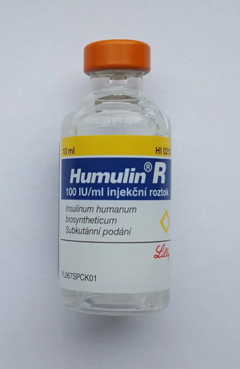
- Insulin. People with diabetes need insulin (Pancreas, Diabetes). Most insulin today is made by bacteria or yeast that have been given the human insulin gene. Insulin itself is made first with a signal at its front, which is cut off. Knowing how the signals work, engineers give such genes the right signal, so that the cells make the protein and send it out, ready to be collected.
- Medicines made by cells. The same is done for growth hormone, the factors that make blood clot (for people with hemophilia), and many newer medicines. Cells in large steel tanks work as protein factories, and the signal tells them to send the product out.
- Vaccines. The mRNA vaccines against COVID-19, given to many millions of people, carry instructions for the virus’s spike protein, which sits in the outer skin of the virus. The spike’s own signal sends it to the ER, so that our cells put it in their membrane, where the immune system can see it.

What came next
- The channel, seen. The channel that Blobel predicted in 1975 has since been seen in detail. Its parts were found in the 1990s, and electron microscopes can now show its shape atom by atom, with a chain passing through it.
- The next delivery. Once a protein is inside the ER, it is packed into tiny bubbles called vesicles and delivered on, to the Golgi body and to the cell membrane. How those deliveries find the right door won the 2013 Nobel Prize (The postal service inside your cells).
- Reading addresses from genes. Today the DNA of a living thing can be read, and computer programs can read a protein’s signal from its gene and predict where in the cell the protein will go. They are right most of the time, but not always. Some proteins carry two addresses, or change address when the cell needs them somewhere else, and nobody fully knows how a cell chooses.
A gene for a human medicine is put into yeast cells, but the protein stays inside the cells instead of coming out into the liquid. What should the engineers add to the gene?
Pick an answer to see why.
In your science books
- Grade 8: the cell and its components: ribosomes, the endoplasmic reticulum, the Golgi body and the nucleus.
- Grade 9: the pancreas and insulin.
- Grade 10: DNA and RNA, and diabetes.
- Going further: what proteins do and how a ribosome reads the message.
Learn more
- The Nobel Prize’s press release for 1999, with its two diagrams of the signals.
- Günter Blobel’s Nobel lecture, Protein targeting, and his own life story (nobelprize.org).
The official reason
The prize was given “for the discovery that proteins have intrinsic signals that govern their transport and localization in the cell.”