
How killer T cells find infected cells
Nobel Prize in Physiology or Medicine, 1996: Peter Doherty and Rolf Zinkernagel

Peter Doherty
born 1940 in Australia
A veterinarian who started out examining sick cattle and pigs in the Australian countryside, and became the first vet to win a Nobel Prize.
Photo: Vogler, CC BY-SA 4.0

Rolf Zinkernagel
born 1944 in Switzerland
A Swiss doctor who went to Australia for research, and was given a desk in Doherty’s laboratory because it was the only space left.
Photo: Aasemsj, CC BY-SA 3.0
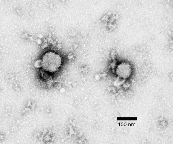
When you catch a cold or the flu, a virus gets inside the cells lining your nose and throat. Once inside, it is hidden. It uses the cell’s own machinery to make thousands of copies of itself, which burst out and infect more cells. A virus inside a cell is out of reach of most of the body’s defenses.
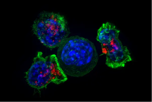
Yet in a week or so, you get better. Part of the reason is a group of white blood cells called killer T cells, which find the infected cells and destroy them, virus and all, before the virus can spread further. But how does a killer T cell tell an infected cell from a healthy one? They look the same from outside. In 1996 the Nobel Prize in Physiology or Medicine went to Peter Doherty and Rolf Zinkernagel, who found the answer.
The problem: how to find a hidden enemy
Your body is defended by its immune system: many kinds of white blood cells that find and destroy germs Grade 10: white blood cells. Two kinds of lymphocytes (say LIM-foh-sites), a type of white blood cell, were known by the early 1970s.
- B cells make antibodies: Y-shaped proteins that float in the blood and stick to germs, such as bacteria, marking them for destruction. Antibodies work well on germs outside cells.
- T cells (they grow up in the thymus, a small organ in the chest, hence the T) work differently. Some of them, the killer T cells, attack whole cells. But nobody knew how they recognized which cells to attack.
There was one strong clue, from transplants. If a kidney from one person is put into another, the patient’s T cells usually attack it and destroy it. This is called rejection. The T cells recognize certain molecules on the surface of the donor’s cells as foreign. These molecules were named after transplants: major histocompatibility antigens, or the MHC for short. Histo means tissue, and compatibility means fitting together. In people, they are called HLA.
Every person’s MHC molecules are slightly different from almost everyone else’s, except close family. That is why a kidney donor is usually a parent, a brother or a sister, and why their tissues are tested for a match first, a little like checking that a blood group matches before a blood transfusion Grade 10: why the blood group must match.
But this raised a puzzle. People don’t swap organs in nature. Why would the body have molecules whose main job seemed to be to cause trouble in transplants?
Why it was hard
- T cells can’t be watched at work. A killer T cell meets its target deep inside the body. To study it, scientists needed a way to test, in a dish, whether T cells had killed some cells.
- The right test needs the right cells. As you will see, the answer depended on which mice the T cells came from and which cells they were tested on. If the mice and the cells had been chosen differently, the discovery could easily have been missed.
- The answer turned an idea upside down. Most experts thought of the MHC as a barrier to transplants. Nobody expected it to be part of how T cells fight viruses every day.
What they did, step by step
Two people in one small lab (1971–1973)
Peter Doherty grew up in Brisbane, Australia, and trained as a veterinarian, an animal doctor. After working with farm animals and studying brain diseases in Scotland, he arrived at the John Curtin School of Medical Research in Canberra, Australia, at the end of 1971. Rolf Zinkernagel, a medical doctor from Basel in Switzerland, arrived in 1973. The only empty bench in the department was in Doherty’s small laboratory, so the two began working side by side.
A virus that kills by the immune response (1973)
- The virus. They studied a mouse virus called LCMV (lymphocytic choriomeningitis virus). When it was injected into a mouse’s brain, the mouse developed meningitis, a swelling of the thin layers that cover the brain, and died within about ten days.
- The surprise in the virus. The virus itself did little harm to the cells it infected. Mice that had no T cells survived the infection. It was the mouse’s own killer T cells, attacking the infected cells in the brain’s covering, that caused the deadly swelling.
- The killing test. Zinkernagel knew a neat test for killer T cells. Infected cells in a dish were first given a little radioactive chromium to soak up. Then T cells were added. If the T cells killed the infected cells, the dead cells leaked the chromium into the liquid, where it could be measured. More chromium meant more killing. Doherty collected T cells from the fluid around the brains of sick mice, and the test showed they killed infected cells.
The surprise (1973)
- More kinds of mice. Another team had just reported that mice of different strains (families bred to be all alike) fell ill from LCMV in different ways, and that this was linked to their MHC type. So Doherty and Zinkernagel infected mice of every strain they had in Canberra and, a week later, tested the T cells from their spleens (an organ full of lymphocytes) against infected cells in the dish.
- The strange result. All the mice fell ill and died. But only the T cells from some strains killed the infected cells in the dish. T cells from the other strains, which were fighting the same virus inside their own mice, did nothing to them.
- The clue in the cells. The infected cells in the dish came from a mouse cell line called L cells, kept in the laboratory for decades. By luck, these cells had the same MHC type as the strain of mice used most in the department, the CBA mice. And it was exactly the mice with that same MHC type whose T cells killed them. T cells from mice with a different MHC type, such as BALB/c mice, did not.
- Testing it the other way. To be sure, they infected cells taken from mice of the other strains. Now each strain’s T cells killed infected cells of its own MHC type, and ignored infected cells of a different type, even though the virus was the same.
- The conclusion (1974). A killer T cell must recognize two things at once: a piece of the virus, and the cell’s own MHC molecule. The virus alone is not enough, and the MHC alone is not enough. They published this in the journal Nature in 1974.
T cells from BALB/c mice, which were fighting LCMV, did not kill LCMV-infected L cells. Why not?
Pick an answer to see why.
Altered self (1974–1975)
Doherty and Zinkernagel suggested two ways this might work. Perhaps the T cell had two receivers, one for the virus and one for the MHC. Or perhaps the virus somehow changed the cell’s MHC, so that the T cell saw “altered self”: the body’s own molecule with something foreign about it. They also saw the big meaning of their result. The MHC is not there to make transplants fail. Its real job is to help T cells find infected cells. Transplants are rejected only because a donor’s different MHC molecules look, to a T cell, like the patient’s own MHC changed by an infection.
Their ideas were attacked at first, because they turned what experts believed upside down. Doherty traveled round the United States giving the same talk at about 20 institutions. Within a few years, the evidence had convinced the world.
How it works now
Later research showed that the “altered self” idea was close to the truth. Here is what happens inside every cell of your body, every minute.
- Chopping up proteins. Every cell keeps chopping up a sample of the proteins it makes into short pieces, called peptides (say PEP-tides), each about nine amino acids long (how a gene makes a protein).
- Showing them off. Each MHC molecule has a groove on top, like the palm of an open hand. It picks up one peptide, holds it in the groove, and carries it to the cell’s surface. A cell’s surface is covered with these MHC molecules, each showing one piece of what is going on inside. In 1987 a team at Harvard University worked out the shape of an MHC molecule and saw a peptide sitting in its groove.
- The check. Killer T cells move through the body, touching cell after cell. Each T cell has its own T-cell receptor, which fits only one particular combination: one particular peptide sitting in one of the body’s own MHC molecules.
- Healthy or infected? In a healthy cell, the peptides are all pieces of the body’s own proteins, and no T cell reacts. In an infected cell, some MHC molecules show pieces of virus protein. A T cell whose receptor fits that combination attaches, and kills the cell.
That is why the T cells in the experiment ignored infected cells with the wrong MHC type: the virus piece was there, but held in an MHC molecule the T cell was not built to recognize.
How does each T cell learn to work with its own body’s MHC? Young T cells are tested in the thymus. Only those whose receptors can grip the body’s own MHC molecules lightly survive, and those that grip a normal body peptide too strongly are destroyed, so that they won’t attack healthy cells.
A killer T cell touches a healthy liver cell. Why doesn’t it kill it?
Pick an answer to see why.
In your life today
- Transplants. Before a kidney transplant, doctors test the HLA types of the patient and the donor, and test whether the patient’s blood already holds antibodies against the donor’s cells. In Nepal, kidney transplants are carried out at the Shahid Dharmabhakta National Transplant Centre in Bhaktapur and at other hospitals, and patients take medicines for the rest of their lives that calm their T cells, so the new kidney is not rejected.
- Better vaccines. Knowing exactly which pieces of a virus the T cells see helps scientists design vaccines that train T cells as well as antibodies.
- Fighting cancer. Cancer cells also show unusual peptides in their MHC molecules. Newer treatments help a patient’s own T cells find and kill them.
- When T cells attack the body. In some diseases, such as type 1 diabetes, multiple sclerosis and rheumatoid arthritis, T cells attack the body’s own cells. People with certain HLA types are more likely to get them, and this discovery helps explain why.
What came next
- Why are we all so different? There are thousands of different versions of the HLA genes in people. Each version holds a different set of peptides well. So in a big epidemic, some people’s MHC molecules will show the new virus well to their T cells, and they will survive. Being different from each other protects the human species as a whole. This also explains why finding a matching donor is so hard.
- Helper T cells. A second family of MHC molecules shows peptides to helper T cells, which direct the rest of the immune system. The same idea of “peptide in MHC” turned out to explain them too.
- Open questions. Why do some HLA types make a person more likely to get a disease in which T cells attack the body? Which peptides set off type 1 diabetes or multiple sclerosis? And can vaccines against cancer or HIV be built from the peptides T cells see? These are still being studied.
In your science books
- Grade 10: white blood cells, the functions of blood, and why the blood group must match, another kind of matching before blood or tissue is given.
- Grade 9: lymph, the liquid that carries lymphocytes.
- Grade 8: viruses and the harm microorganisms do.
- Going further: How does a gene make a protein?, the chains of amino acids that cells chop into peptides.
Learn more
- The prize at nobelprize.org: The Nobel Prize in Physiology or Medicine 1996, with the press release and the presentation speech.
- The two Nobel lectures, which tell the story of the experiments: Doherty and Zinkernagel.
The official reason
The prize was given “for their discoveries concerning the specificity of the cell mediated immune defence.”