
The ribosome, the cell’s protein factory
Nobel Prize in Chemistry, 2009: Ada E. Yonath, Venkatraman Ramakrishnan, and Thomas A. Steitz
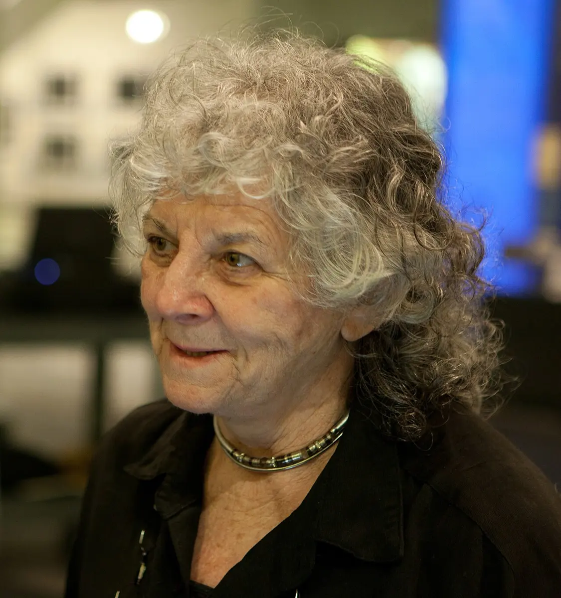
Ada E. Yonath
1939–2026, born in Jerusalem
At five she piled a table, a chair and a stool on top of each other to measure the height of the balcony, fell into the yard and broke her arm. She spent twenty years on the ribosome while other scientists called her a dreamer.

Venkatraman Ramakrishnan
born 1952 in Chidambaram, India
He didn’t get into the Indian Institutes of Technology, studied physics on a national science scholarship instead, and switched to biology after his PhD.
Photo: The Royal Society, CC BY-SA 4.0
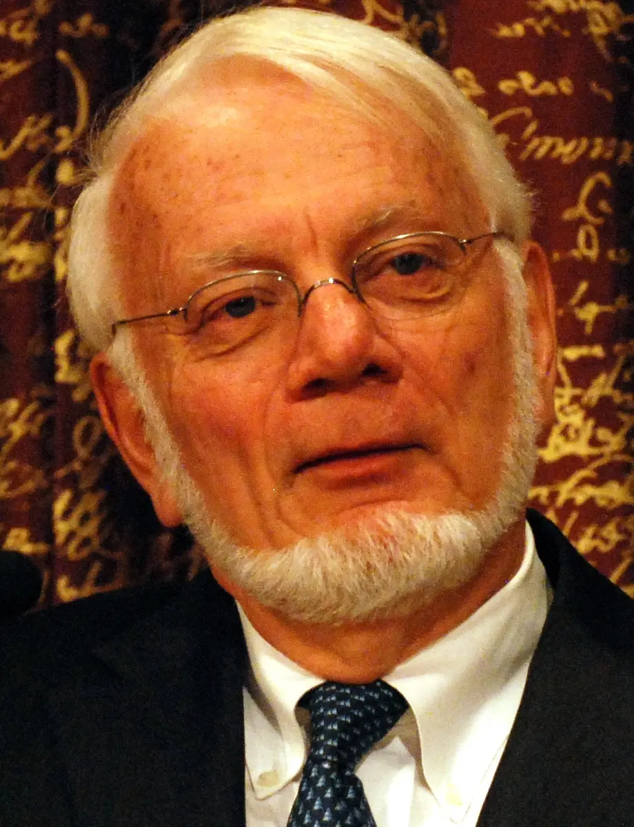
Thomas A. Steitz
1940–2018, born in the United States
A professor at Yale who solved the hardest part of the puzzle, turning blurry spots into the first sharp picture of the ribosome’s large part.
Photo: Prolineserver, CC BY-SA 3.0
When you eat dal, your body breaks its protein down into small pieces called amino acids. Your cells then join those amino acids, in a new order, into your own proteins: the keratin in your hair, the hemoglobin in your blood, the muscle in your arm. Right now, in every cell of your body, thousands of tiny machines are doing this joining. Each one is called a ribosome.
Scientists had known about ribosomes since the 1950s, but nobody could see how one worked. It is far too small, and it is one of the most complicated objects in a cell. The 2009 Nobel Prize in Chemistry went to three scientists who mapped it atom by atom: Ada Yonath, Thomas Steitz and Venkatraman (Venki) Ramakrishnan.
The problem: a machine nobody could see
By the 1960s scientists knew the outline of the story (How does a gene make a protein?).
- Your genes are written in DNA, in a four-letter alphabet: A, T, G and C Grade 10: DNA.
- DNA stays in the nucleus. To make a protein, a cell copies one gene into a messenger, mRNA, which carries the message out to the ribosomes in the cytoplasm Grade 10: RNA.
- A protein is a chain of amino acids, of 20 kinds. The ribosome reads the mRNA three letters at a time. Each group of three, a codon, stands for one amino acid.
So the ribosome is a translator. It turns the language of the genes into the language of proteins. But how? How does it pick the right amino acid, again and again, without mistakes? How does it join them? In 1964 James Watson, who had found the shape of DNA (Nobel Prize in Medicine 1962), wrote that we cannot describe how a molecule works “unless we know first its structure”. The ribosome’s structure would take until 2000.
How a ribosome works
A ribosome has two parts, called subunits, which clamp around the mRNA like the two halves of a bun around a filling Grade 8: Ribosomes.
- The small subunit holds the mRNA and reads it.
- The large subunit joins the amino acids together.
The amino acids are carried in by small helper molecules called transfer RNA, or tRNA. Each tRNA carries one kind of amino acid at one end, and has three letters at the other end, its anticodon. A tRNA can only stay in the ribosome if its anticodon pairs with the codon being read, the way A pairs with U and G pairs with C. So each codon gets the right amino acid.
A ribosome is fast and careful. In bacteria it joins up to about 20 amino acids every second; in your cells, about 5 to 10. It makes only about one mistake in every 10,000 to 100,000 amino acids (estimates differ). A ribosome is about 25 nanometers across, where a nanometer is a millionth of a millimeter: you could line up about 4,000 of them across the width of one hair.
What does the small subunit of a ribosome do?
Pick an answer to see why.
Why it was hard
- Too small to see. A light microscope can’t show anything much smaller than the wavelength of light, about a fifth of a thousandth of a millimeter. A ribosome is about ten times smaller. Electron microscopes of the time could show its outline as a blurry blob, but not its atoms.
- The only way: X-ray crystallography. X-rays have a wavelength about as small as an atom. If you shine them through a crystal, where the same molecule is repeated millions of times in neat rows, the X-rays scatter into a pattern of spots. From the pattern, the positions of the atoms can be worked out. This had worked for DNA and for small proteins.
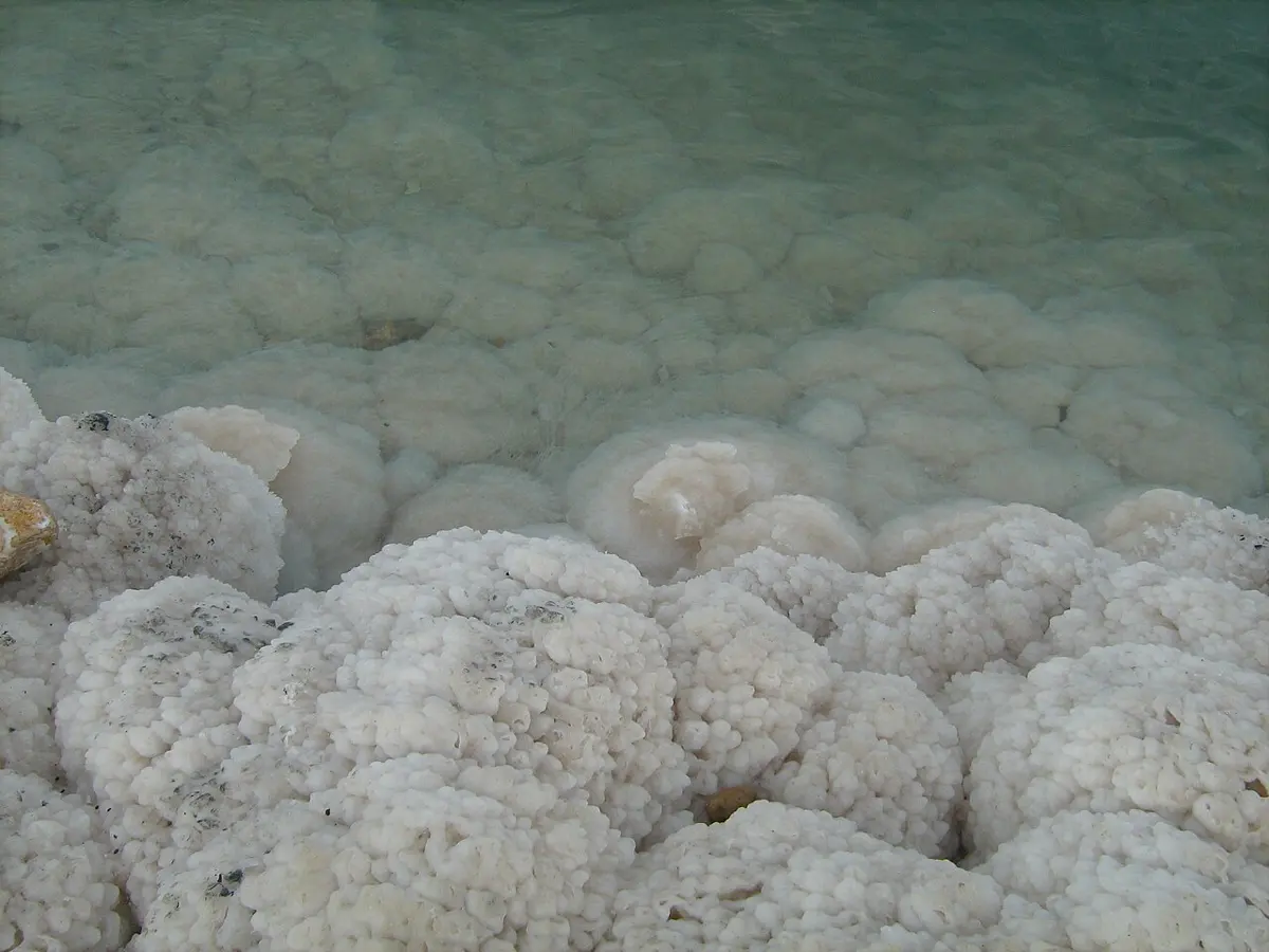
- But you need a crystal. Salt water left to dry slowly in the sun gives fine crystals; boiled dry in a pan, it leaves a dull crust. Big, floppy molecules are much harder to crystallize than salt, and the ribosome is enormous: a bacterium’s ribosome has more than 50 proteins and three RNA molecules, hundreds of thousands of atoms in all. Most scientists thought a good crystal of it was impossible.
- The spots aren’t enough. Even with a crystal, the spots tell you only how bright each scattered ray is, not how its waves line up with the others. Without that, called the phase, you can’t turn the spots back into a picture. This is the phase problem, and for something as big as a ribosome it was very hard to solve.
What they did, step by step
Yonath: twenty years of crystals
Ada Yonath grew up poor in Jerusalem. Her father died when she was eleven, and she worked as a cleaner, babysitter and tutor to help her mother. She studied chemistry, and set up Israel’s first laboratory for working out the shapes of proteins with X-rays.
- Late 1970s, the idea. At the Weizmann Institute in Israel, and with a team at the Max Planck Institute in Berlin, she decided to crystallize the ribosome. She later wrote that she was called “the village fool”.
- The bears. She read that bears pack the ribosomes in their cells into neat, orderly sheets before they hibernate, and that the ribosomes stay healthy for months. Perhaps ribosomes from living things that survive hard conditions would be tougher, and crystallize better. She chose bacteria from hot springs and from the Dead Sea, one of the saltiest lakes in the world.
- 1980, the first crystals. She made the first crystals of the ribosome’s large subunit. They were tiny and far from perfect, but they were crystals.
- Freezing. The strong X-rays quickly damaged the crystals. She froze them in liquid nitrogen, colder than −180 °C, so they lasted much longer under the beam. This trick is now used in laboratories all over the world.
- 1980s and 1990s. Step by step, with better crystals and better X-ray machines, the spots became sharper. By the mid-1990s other teams, using her methods, joined the race.
Steitz: solving the phase problem
Thomas Steitz worked at Yale University in the United States with Peter Moore, who had studied the ribosome for years.
- 1998, the first picture. Steitz used blurry pictures of the ribosome taken with an electron microscope by Joachim Frank (who later shared the Nobel Prize in Chemistry in 2017) to work out how the ribosomes sat inside the crystal. With that, and with heavy atoms soaked into the crystal as landmarks, he could solve the phase problem. His first map of the large subunit was blurry: no single atoms could be seen, but the long RNA molecules could be traced. It was the breakthrough.
- August 2000, atom by atom. Steitz and Moore’s team published the large subunit of the Dead Sea microbe’s ribosome in enough detail to place its atoms.
Ramakrishnan: the reading head
Venki Ramakrishnan was born in Chidambaram, a temple town in Tamil Nadu, and grew up in Baroda (now Vadodara), in Gujarat, where his parents were scientists. He studied physics in Baroda and did a PhD in physics in the United States. Then he decided that biology was more exciting, and went back to being a student.
- 1980s, a hard start. He began working on the ribosome in Peter Moore’s laboratory at Yale. Afterwards he applied to about 50 universities in the United States for a job, and none of them offered him one. He kept working on the ribosome in a government laboratory.
- 1999, Cambridge. He moved to the Laboratory of Molecular Biology in Cambridge, England.
- September 2000, the small subunit. His team, and separately Yonath’s, published the small subunit, from a hot-spring bacterium, atom by atom.
- How it checks. Ramakrishnan then crystallized the small subunit with mRNA and tRNA in place. He found a kind of ruler: a few letters of the small subunit’s RNA reach out and feel the shape of the pairs between codon and anticodon. If the pairs don’t fit exactly, the tRNA falls off. The ribosome checks twice, which is why it so rarely makes a mistake.
Why did Ada Yonath use ribosomes from bacteria that live in hot springs and the Dead Sea?
Pick an answer to see why.
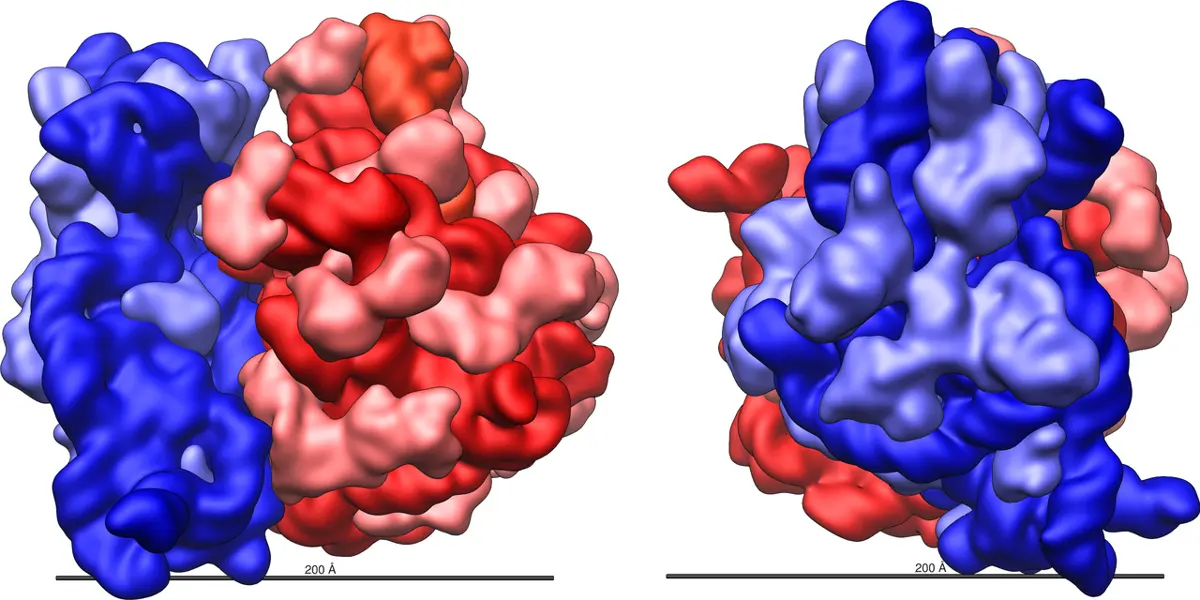
What the pictures showed
The maps showed things that nobody could have guessed from the outline.
- The joining is done by RNA. In Steitz’s large subunit, there was no protein at all near the spot where amino acids are joined. The work is done by the ribosome’s RNA. Almost every other chemical job in a cell is done by proteins called enzymes. Here, RNA does the most important one.
- A tunnel. The new protein chain leaves the ribosome through a tunnel in the large subunit, long enough to hold about 30 to 40 amino acids. Yonath had first seen signs of it in the 1980s.
- The check. Ramakrishnan’s ruler explained how a ribosome can be both fast and careful.
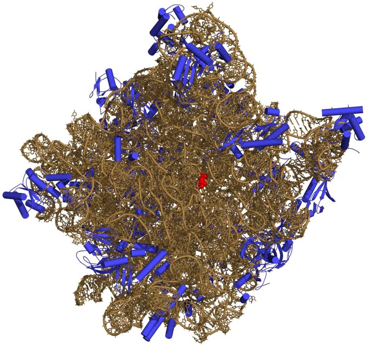
In your life today: antibiotics
Bacteria have ribosomes too, and they need them to live. A bacterium’s ribosome is a little different from yours. So a medicine that jams a bacterium’s ribosome, but not yours, can kill the germ and leave you unharmed. Many of the antibiotics doctors use work exactly this way (What an antibiotic does).
All three laureates crystallized ribosomes with antibiotics stuck in them, and saw where each one fits. Yonath’s team mapped more than twenty.
You may know some of these names. Azithromycin, one of the most commonly used antibiotics in Nepal and around the world, plugs the tunnel. Doxycycline, a kind of tetracycline, stops new tRNAs coming in.
The problem is that bacteria change. A bacterium with a small change in its ribosome, so that the drug no longer sticks, survives and multiplies. This is antibiotic resistance, and it is growing fast, especially where antibiotics are bought without a doctor’s advice or stopped too early. Knowing exactly where and how each drug fits helps chemists design new ones that still fit, or that fit somewhere new.
Why can an antibiotic like azithromycin kill bacteria without killing your own cells?
Pick an answer to see why.
What came next
- Whole ribosomes in action. Ramakrishnan, Steitz, Yonath and others went on to map the whole ribosome, with tRNAs and mRNA in place, frozen at different moments of its work, like frames of a film.
- Human ribosomes. With the new cryo-electron microscopes, scientists have since mapped human ribosomes too, and the ribosomes inside our mitochondria. These look rather like bacterial ones, which explains why a few antibiotics have side effects.
- New medicines. Companies use the maps to design antibiotics for bacteria that resist the old ones. Steitz helped found one.
- An open question. Because the heart of the ribosome is RNA, many scientists think that the first living things may have used RNA for everything, before proteins existed: an RNA world. How did the very first ribosome begin? Nobody knows yet.
In your science books
- Grade 8: ribosomes, the cell’s smallest organelles, and bacteria.
- Grade 10: DNA and RNA.
- Going Further (Grades 6–8): what happens to the dal bhat you eat, where your ribosomes build your own proteins.
- Going Further (Grades 9–12): How does a gene make a protein?, with a ribosome to play with, and Why do antibiotics stop working?
Learn more
- The Nobel Prize’s own explanation: popular information on the 2009 prize (nobelprize.org).
- The laureates’ Nobel lectures: Ramakrishnan, Steitz and Yonath.
- Venki Ramakrishnan’s book Gene Machine (2018) tells the story of the race from the inside.
The official reason
The prize was given “for studies of the structure and function of the ribosome.”