
GFP: the jellyfish’s glowing protein
Nobel Prize in Chemistry, 2008: Osamu Shimomura, Martin Chalfie, and Roger Y. Tsien
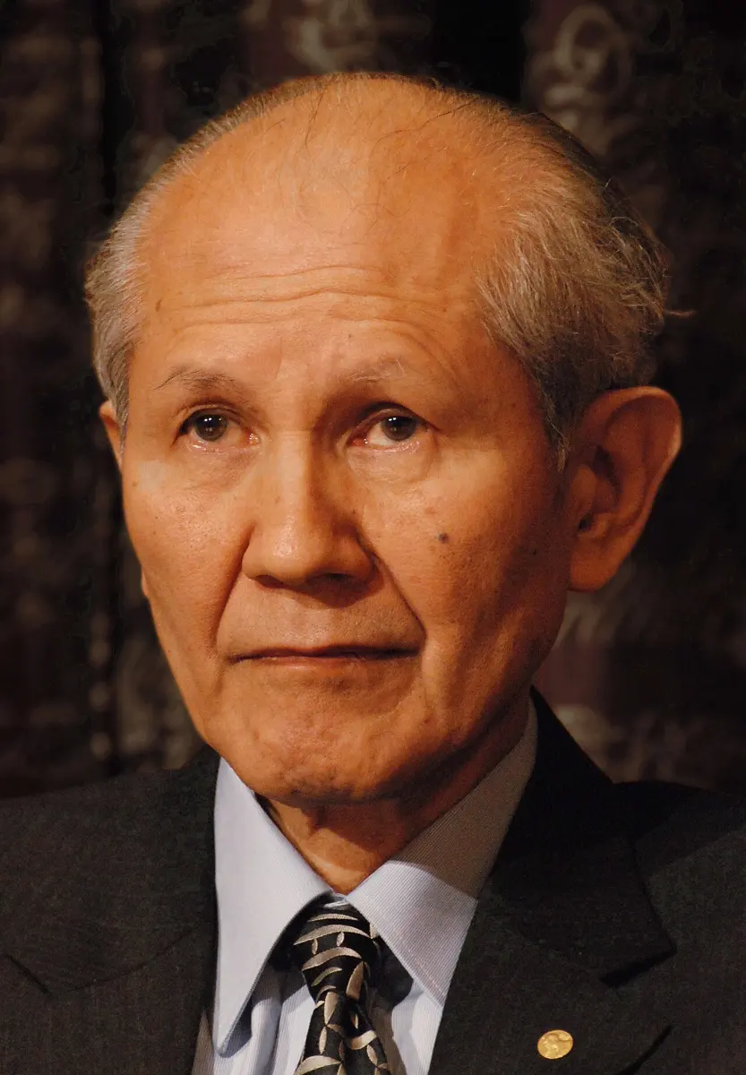
Osamu Shimomura
1928–2018, born in Japan
As a 16-year-old factory worker near Nagasaki he saw the flash of the atomic bomb. He spent the rest of his life studying living things that make light.
Photo: Prolineserver ( talk ), CC BY-SA 4.0

Martin Chalfie
born 1947 in the United States
After a summer of failed experiments in college he nearly gave up biology. He slept through the phone call telling him he had won.
Photo: Duncan.Hull, CC BY-SA 4.0
-
Roger Y. Tsien
1952–2016, born in the United States
A boy who did chemistry experiments at home, partly with chemicals his father brought from work. He grew up to give biologists a whole box of colored lamps.
On a summer night in Nepal you may see fireflies (जुनकिरी) blinking in the fields. Their light is not made by heat or electricity. It is made by a chemical reaction inside their bodies. Living things that make their own light are called bioluminescent.
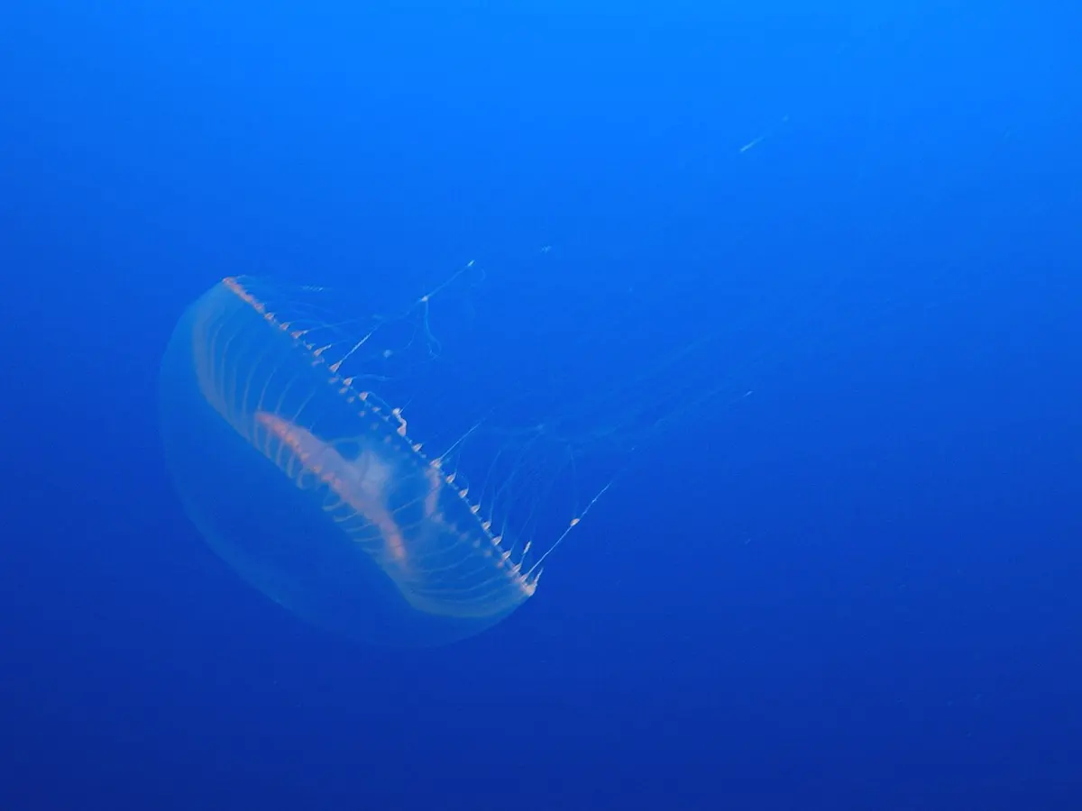
In the sea, many animals glow. One of them, a small jellyfish off the west coast of North America, glows green round the edge of its umbrella. A Japanese chemist spent years finding out why, simply because he wanted to know. The green protein he found became one of the most useful tools in biology. In 2008 the Nobel Prize in Chemistry went to Osamu Shimomura, Martin Chalfie and Roger Tsien for the green fluorescent protein, GFP.
The problem: cells you cannot watch
Your body is made of many millions of millions of cells Grade 8: Cell. A typical cell is about 0.02 mm across, far too small to see without a microscope. Inside each cell, almost all the work is done by proteins: they build things, carry things, send messages and speed up reactions. There are tens of thousands of different proteins in your body.
Biologists want to know: in which cells is a protein made? When? Where does it go inside the cell? What does it meet there?
Those are hard questions. Under an ordinary microscope, a cell’s proteins are invisible, and almost all of them are colorless. You can color a cell with a dye, but most dyes kill it, so you see only a dead cell, frozen at one moment. Biologists needed a way to put a lamp on one protein inside a living cell and watch it move.
How GFP makes its light
Fluorescence
Some materials soak up light of one color and give out light of another. This is called fluorescence. The ink in a highlighter pen is fluorescent: it soaks up invisible ultraviolet light and some blue light from daylight, and gives back more yellow-green light than plain paint could, which is why it looks so bright. Light of shorter wavelength, such as blue or ultraviolet, carries more energy than green light Grade 9: Electromagnetic spectrum. A fluorescent material takes in a bigger packet of energy and gives out a slightly smaller one, losing the difference as a tiny amount of heat. So the light that comes out is always of a longer wavelength than the light that went in: blue in, green out, never green in, blue out.
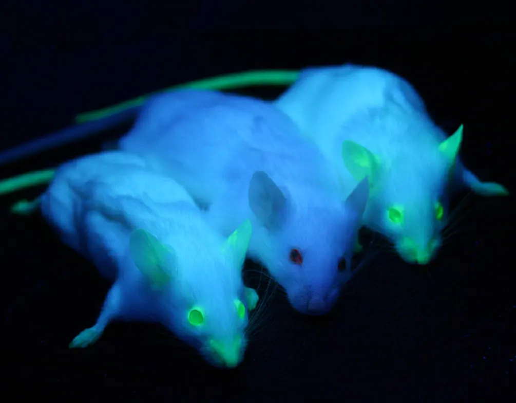
GFP is a fluorescent protein. Shine blue or ultraviolet light on it, and it glows green.
Proteins, and the gene for each one
A protein is a long chain of smaller pieces called amino acids, of 20 kinds, joined one after another and then folded into a shape (Proteins do the work). The order of the amino acids is written in a gene, a stretch of the cell’s DNA Grade 10: DNA. The cell reads the gene and builds the protein. Every cell has a set of gene switches, too: each gene is switched on only in certain cells, at certain times.
That is the key to the whole story. If you have the gene for GFP, you can give it to a living thing, and its own cells will build the glowing protein.
A fluorescent material is lit with blue light. Which color could it give out?
Pick an answer to see why.
What they did, step by step
Shimomura: 850,000 jellyfish
Osamu Shimomura grew up in wartime Japan. In 1945 he was working in a factory near Nagasaki, and on 9 August he saw the blinding flash of the atomic bomb dropped on the city. Both colleges in Nagasaki had been destroyed; he studied pharmacy at a temporary campus.
- 1956, a glowing sea creature. At Nagoya University his professor gave him a task others had failed at: find the substance that makes a tiny glowing sea animal, the sea-firefly, shine. He purified it and made it into crystals for the first time.
- 1960–61, the jellyfish. That work took him to Princeton University in the United States, to study the glowing jellyfish Aequorea victoria with Frank Johnson. In the summer of 1961 they collected jellyfish at Friday Harbor, on the coast of Washington State, cut off the glowing edges and pressed them through a filter.
- The sink. Searching for the light-making substance, Shimomura poured some of the extract into a sink, and it flashed brightly. Seawater had got into the sink. He worked out that calcium in seawater set off the light. The light-making protein, which he named aequorin, gave blue light, not green.
- 1962, the green protein. In their report on aequorin, Shimomura and Johnson also described a second protein from the jellyfish. It looked a little greenish in sunlight, and glowed bright green under ultraviolet light. It was later named the green fluorescent protein.
- 1970s, the light-making part. Shimomura showed that GFP holds a small light-making group inside it, which soaks up energy and gives out green light. In the jellyfish, aequorin makes blue light, and GFP turns that energy into green. That is why the jellyfish glows green.
That last point is what made GFP special. Aequorin, like the chemical in a firefly, needs a fuel to make light, and a cell would have to be fed it. GFP needs nothing but light. Shine blue light on it, and it glows.
Between 1961 and 1988, Shimomura and his family drove or flew across America to Friday Harbor 19 times, catching about 850,000 jellyfish, often thousands a day, with his wife and children helping. He wanted aequorin, not GFP. He did not set out to make a tool for biology.
Chalfie: making a worm glow
Martin Chalfie at Columbia University in New York studied a tiny worm, Caenorhabditis elegans, about 1 mm long. It is see-through, so its cells can be watched under a microscope, and its body has only 959 cells.
- 1988, a seminar. At a talk about glowing animals, Chalfie heard about GFP and was excited. If he could put the GFP gene into the worm, its cells might glow, and he could see which cells were using a gene.
- The gene. Another scientist, Douglas Prasher, at Woods Hole in Massachusetts, had just found (cloned) the GFP gene. In 1992 he sent a copy to Chalfie.
- About a month later, glowing bacteria. Chalfie’s graduate student Ghia Euskirchen put the gene into ordinary bacteria. Under ultraviolet light they glowed green. Many experts had expected this to fail: they thought the jellyfish needed special helper proteins to build GFP’s light-making part. It needed none. GFP builds itself.
- February 1994, the glowing worm. Chalfie put the GFP gene behind a gene switch that is on only in the worm’s six touch cells, the nerve cells it feels touch with. The worms that hatched had six glowing green cells. The picture went on the cover of the journal Science.
Tsien: a box of colors
Roger Tsien, at the University of California, San Diego, was a chemist who loved designing molecules that let you watch what happens inside living cells.
- 1994, how the lamp forms. Tsien worked out how GFP makes its light-making part. In the chain of 238 amino acids, three neighbors, numbers 65, 66 and 67, react with each other to form it. The reaction needs oxygen but no other protein.
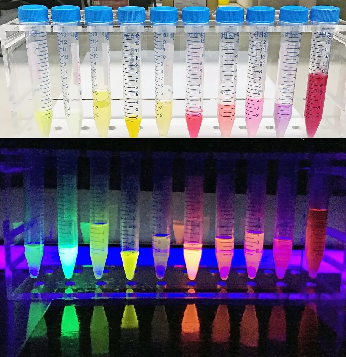
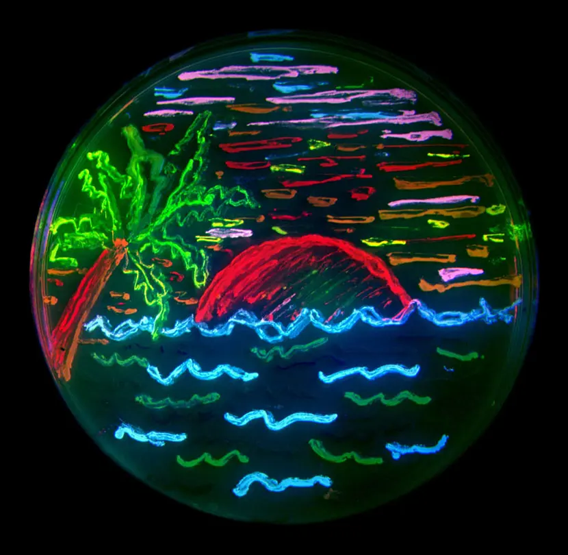
- 1994–98, brighter and new colors. By changing single amino acids, his group made GFP glow brighter and last longer under the microscope (one change, in 1995, made it much brighter under blue light), and made new colors: blue, cyan (blue-green) and yellow. The shape of GFP, worked out in 1996, showed why it is so tough: the chain folds into a can of 11 strands, with the light-making part sealed safely in the middle.
- Red. Red was the color he could not get from GFP, and red is the most useful, because red light passes through living tissue more easily. Two Russian scientists, Mikhail Matz and Sergei Lukyanov, found a red fluorescent protein in a coral. It came as a clump of four chains, too bulky to use as a tag, so Tsien’s group reshaped it into a single chain. From it they made orange and red proteins they named after fruit: mCherry, mStrawberry, mOrange, mPlum.
With several colors, scientists can tag different proteins, or different cells, in different colors at once, and watch how they meet.
Why was red the most wanted color of all?
Pick an answer to see why.
In your life today
- Medicine research. GFP and its cousins are used in thousands of laboratories. Scientists have watched cancer cells spread and build blood vessels, nerve cells grow, and viruses get into cells. Many new medicines are tested with glowing cells.
- The brain. In “Brainbow” mice, nerve cells glow in about a hundred different colors, mixed from three or four proteins like inks in a printer, so each cell can be traced through the tangle.
- Calcium lamps. Shimomura’s other protein, aequorin, glows when calcium is present. Scientists use it, and newer calcium sensors built from GFP, to watch nerve and muscle cells switch on.
- Clean water tests. Scientists have made bacteria that glow green when there is arsenic in water. Arsenic in tube-well water is a real problem in parts of South Asia, including parts of Nepal’s Terai, though Nepal’s wells are usually tested with simple chemical kits.
What came next
- Many more colors. Since 2008, scientists have made fluorescent proteins that glow in the far red and near infrared, which see deeper into the body; proteins that switch on and off with light; and proteins that change color when a cell does something, such as a nerve cell firing.
- Sharper pictures. Switchable fluorescent proteins made it possible to see details smaller than an ordinary light microscope can show. That earned another Nobel Prize in Chemistry, in 2014 (super-sharp microscopes).
- An open question. Nobody knows why the jellyfish glows. Many sea animals use light to frighten enemies, find food or attract a mate, but what Aequorea victoria gains from its green ring is still a mystery.
In your science books
- Grade 8: the cell and its parts, which GFP lets scientists watch.
- Grade 9: the electromagnetic spectrum, visible light and ultraviolet rays.
- Grade 10: DNA, RNA and genetic technology.
- Going Further (Grades 9–12): how a gene makes a protein, and how a protein chain folds.
Learn more
- The Nobel Prize’s own explanation: The green fluorescent protein (nobelprize.org).
- The three laureates’ Nobel lectures: Shimomura, Chalfie and Tsien.
The official reason
The prize was given “for the discovery and development of the green fluorescent protein, GFP.”