
Seeing the shapes of viruses and chromosomes
Nobel Prize in Chemistry, 1982: Aaron Klug
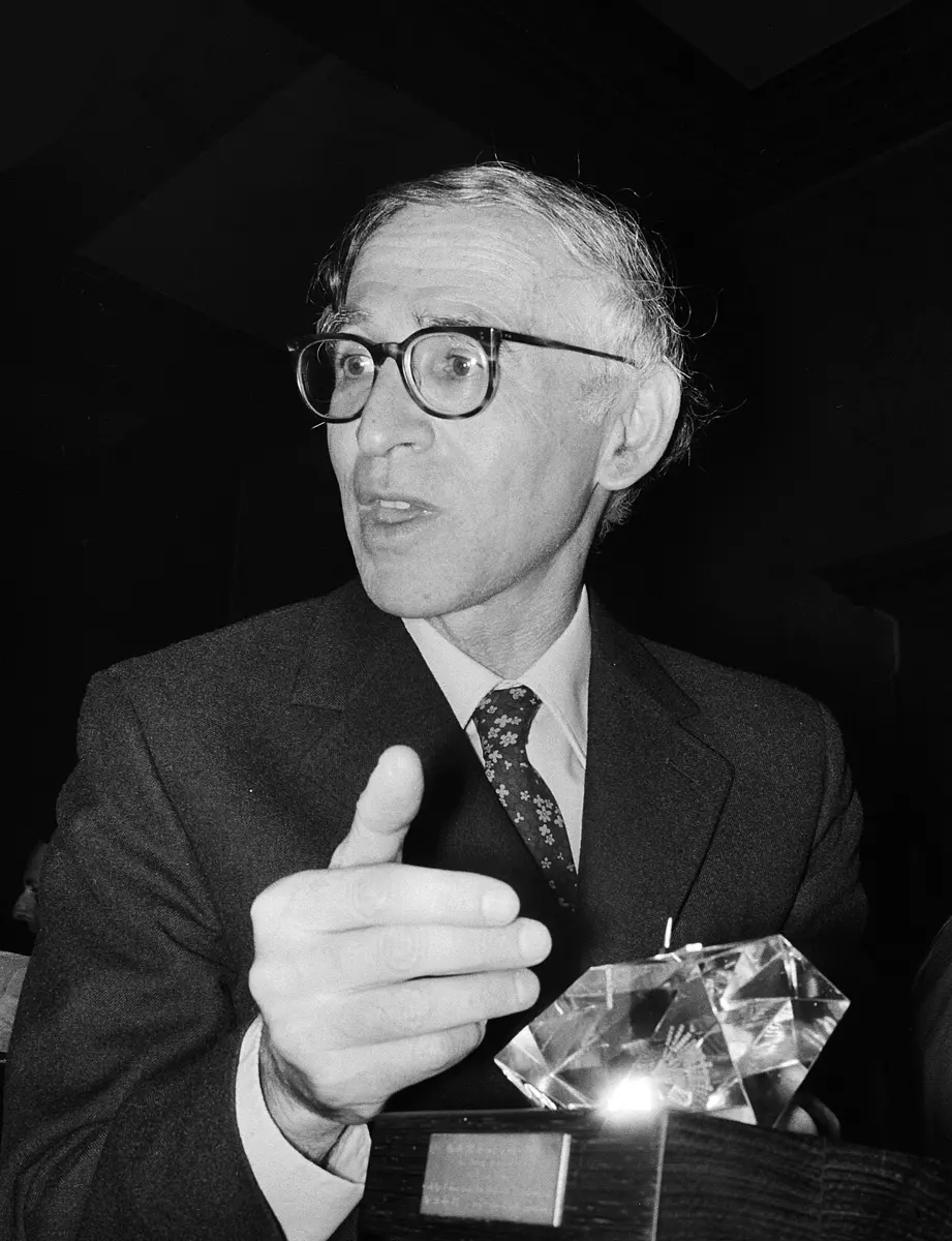
Aaron Klug
1926–2018, born in Lithuania
He grew up in Durban, South Africa, and chose science after reading a book about the first germ hunters. He spent most of his life at a laboratory in Cambridge, England.
Take the DNA out of just one cell of your body and stretch it out, and it would be about 2 meters long, taller than any adult. Yet it fits inside the cell's nucleus, which is about 6 thousandths of a millimeter across. That is like packing 20 kilometers of thread into a tennis ball, and then being able to find and read any part of it.
How is it packed? And how does a virus, which is far too small to see with any ordinary microscope, build its neat coat around its genes? To answer questions like these you need to see the shapes of very small things made of protein and DNA. The 1982 Nobel Prize in Chemistry went to Aaron Klug, who found a way.
The problem: too small to see clearly
An ordinary light microscope, like the one in your school laboratory, can't show anything much smaller than about 0.0002 mm (200 nanometers). The waves of light are simply too long to show finer detail. Most viruses are smaller than that.
The electron microscope, invented in the 1930s, uses a beam of electrons instead of light, and can show much smaller things. But for the molecules of life it had three big problems:
- Too little contrast. Living things are made mostly of light atoms (carbon, hydrogen, oxygen and nitrogen), which hardly stop electrons. A virus looks like a pale ghost. So scientists coated their samples with a stain, a salt of a heavy metal such as uranium, which can squash or change the shape a little.
- The beam does damage. A strong electron beam breaks the molecules while you look at them. A weak beam gives a dim, grainy, “noisy” picture.
- Everything is piled up. An electron microscope picture is like an X-ray photo of your hand: the front and the back of the object are squashed together into one flat picture, a projection. You can't tell which part was in front.
There was another way to find shapes. In X-ray crystallography, X-rays are shone through a crystal of the molecule, and the pattern they make shows where every atom is (the 1962 Nobel Prizes for the shapes of proteins and DNA came from it). But it needs a crystal, a perfectly regular stack of millions of identical copies. Big, floppy groups of molecules, like the DNA and proteins of a chromosome, seldom form crystals.
Why does an electron microscope picture of a virus not show its 3D shape directly?
Pick an answer to see why.
Viruses and chromosomes: protein and DNA together
Klug studied things built of two kinds of giant molecule: nucleic acids (DNA and its cousin RNA, which carry the genes) and proteins (Genes are recipes for proteins).
- A virus Grade 8: Virus is little more than a set of genes inside a coat of protein. It has no cell of its own; it gets into a living cell and makes the cell copy it.
- A chromosome Grade 10: Chromosome is a long thread of DNA wound around proteins called histones. This mixture of DNA and protein is called chromatin.
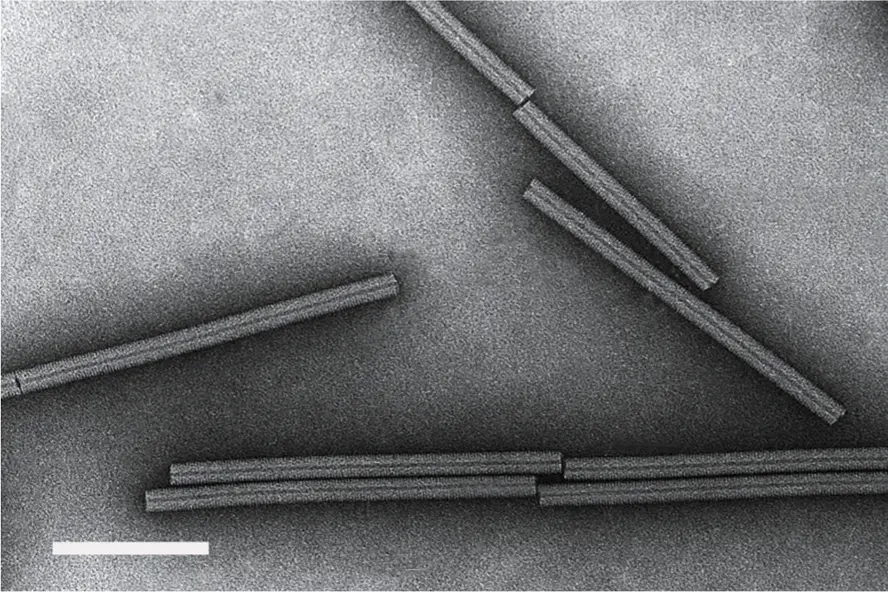
His first virus was tobacco mosaic virus, or TMV. It infects tobacco, tomato and pepper plants and gives their leaves a patchy, mottled look, like a mosaic. A TMV particle is a stiff rod about 300 nanometers long and 18 nanometers thick. Inside it is a single thread of RNA, and around it are 2,130 copies of the same protein, fitted together in a spiral like the steps of a spiral staircase.
Why it was hard
Klug needed to know how thousands of protein molecules fit together, and where the RNA or DNA lies among them. But the electron microscope gave him only flat, grainy pictures with the layers piled up, and these things would not form good crystals for X-rays. Nobody had a way to turn many flat pictures back into one solid shape.
What he did, step by step
Klug: from flat pictures to solid shapes
- The long way round to science. Klug was born in 1926 in Lithuania and taken to South Africa at the age of two. At his school in Durban, the cleverest boys were expected to study Latin, not science. A book called Microbe Hunters, about the scientists who found germs, made him start medicine; then he moved to chemistry, then to physics. At the University of Cape Town he learned X-ray crystallography, and in 1949 he went to Cambridge in England.
- 1954: Rosalind Franklin's virus. In London he met Rosalind Franklin, whose X-ray photos had helped show that DNA is a double helix. She had just turned to TMV, and her beautiful photos fascinated him. With two students, Kenneth Holmes and John Finch, they worked out the outline of the virus: the protein spiral, with the RNA tucked between its turns. Franklin died of cancer in 1958, aged 37. In his Nobel lecture Klug said that, had she lived, she “might well have stood in this place on an earlier occasion”.
- Round viruses like footballs. Many viruses, like the one that causes polio, are round. They are built from many identical proteins arranged on a shape with 20 triangle faces, an icosahedron. In 1962 Klug and the American scientist Donald Caspar explained how a large round shell can be built from many copies of one protein. Their idea was helped by the geodesic domes of the American architect Buckminster Fuller, huge round buildings made of many triangles. Like a football, with its pattern of pentagons and hexagons, such a shell needs exactly 12 five-sided points.
- 1962: a new laboratory. Klug moved to the new MRC Laboratory of Molecular Biology in Cambridge, where he stayed for the rest of his career.
- Finding the signal in the noise. A virus coat and many other structures repeat the same unit over and over. Klug found that a fuzzy picture of such a thing holds much more information than the eye can see. By measuring the picture and using mathematics (and at first, by shining laser light through the photographic film), he could separate the regular, repeated pattern from the random grain. The hidden detail appeared. This meant weaker electron beams, less damage and less stain.
- 1968: from flat pictures to 3D. With David DeRosier, Klug showed how to rebuild a solid shape from flat pictures taken from different directions. Each picture is a projection, a view from one side. A computer combines enough of them into a 3D map (Figure 2). Their first result was the tail of a virus that attacks bacteria. The same mathematics works for CT scanners in hospitals, which build a 3D picture of your body from X-ray views taken all round it.
- How a virus builds itself. In 1955 other scientists had found that if you separate TMV's protein from its RNA and mix them again, they put themselves back together into working viruses: the virus self-assembles. But it took many hours, too slow for a real infection. Klug's team found why the real virus is faster. The proteins first gather into a disc of two rings, 17 proteins each. A loop of the RNA threads into the hole in the middle of a disc, and the disc snaps into the first turns of the spiral. With discs, a complete virus formed in 10 to 15 minutes. And because only TMV's own RNA has the right loop, the protein coats its own genes and no others.
- The 1970s: how DNA is packed. Klug's team turned to chromatin. Taking DNA and histones out of cells, they found that the DNA is wound around little spools, each made of 8 histone proteins. In 1974 Roger Kornberg, then working in Klug's laboratory, proposed this bead model, the nucleosome. Klug's team grew crystals of the bead and found its shape: a flat disc about 11 nanometers across, with 146 pairs of DNA bases wound about 1¾ times around it. With Finch, Klug also showed how the string of beads coils up into a thicker fiber, about 30 nanometers thick.
Why did Klug need pictures of the same kind of object taken from several directions?
Pick an answer to see why.
How DNA is packed now
We now know that the packing has several levels, each like winding a thread on a reel, and then winding the reels together (Figure 3):
- The DNA double helix is about 2 nanometers thick Grade 10: DNA.
- It wraps about 1¾ turns around each spool of 8 histones, making a nucleosome. A short length of “linker” DNA joins one nucleosome to the next, like beads on a string.
- The beads are packed together into thicker fibers and loops.
- Before a cell divides Grade 10: Cell division, the loops fold up tightly into the short, thick chromosomes you can see in a light microscope.
The packing is not just tidiness. A gene wound tightly on its spools is hard for the cell to read, and an unwound one is easy. So the way the DNA is packed helps decide which genes a cell uses: why a skin cell and a nerve cell, with exactly the same DNA, are so different.
In your life today
- Pictures of viruses. The 3D models of viruses you see in the news, round shells with spikes, come from electron microscopes and the mathematics Klug began. In 2017 the Nobel Prize in Chemistry went to three scientists who took it further with cryo-electron microscopy, which freezes the molecules instead of staining them (Freezing molecules to take their picture). One of them, Richard Henderson, worked in Klug's laboratory in Cambridge.
- Vaccines and medicines. Knowing the exact shape of a virus's coat helps scientists design vaccines and medicines that block it. During the COVID-19 pandemic, the shape of the coronavirus's spike was worked out in this way within weeks.
- Zinc fingers. In 1985 Klug's team discovered zinc fingers: small loops of protein, each held in shape by one zinc atom, which grip DNA at a particular sequence. Many of the proteins that switch our genes on and off use them. Scientists later joined zinc fingers to “scissors” proteins to cut DNA at a chosen place, an early way to edit genes.
What came next
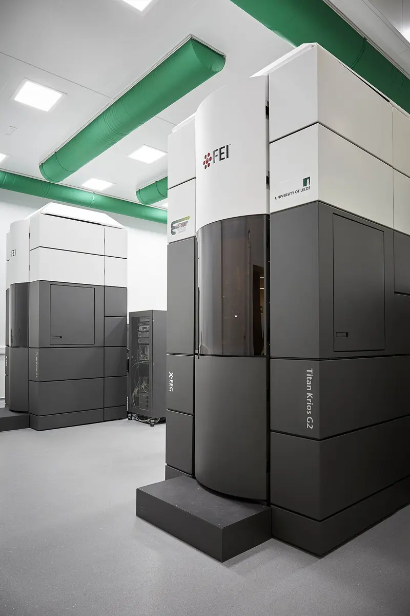
- Sharper and sharper. From about 2013, new electron cameras and computers let cryo-electron microscopes show big molecules in almost atom-by-atom detail, with no crystals and no stain. Klug's dream of seeing the machinery of life without damaging it came true.
- Packing in a living cell. Klug's 30-nanometer fiber is easy to make in a test tube. But when scientists look inside living cells, they often don't find neat fibers; the beads seem to be packed more loosely and irregularly. How DNA is really folded inside a living nucleus, and how a cell finds the right gene in all that thread, is still being worked out.
- Brain diseases. In later life Klug studied the protein tangles found in the brains of people with Alzheimer's disease. Electron microscopes now show the exact shapes of such tangles, which may help in finding treatments.
What is a nucleosome?
Pick an answer to see why.
In your science books
- Grade 8: viruses and the nucleus of a cell.
- Grade 9: X-ray imaging, which, like a CT scanner, makes a picture from rays passing through the body.
- Grade 10: DNA, chromosomes and cell division.
- Going Further: genes are recipes for proteins.
Learn more
- The Nobel Prize's announcement: press release (nobelprize.org).
- Klug's Nobel lecture: From macromolecules to biological assemblies.
The official reason
The prize was given “for his development of crystallographic electron microscopy and his structural elucidation of biologically important nucleic acid-protein complexes.”