
Microscopes that see viruses and atoms
Nobel Prize in Physics, 1986: Ernst Ruska, Gerd Binnig, and Heinrich Rohrer
-
Ernst Ruska
1906–1988, born in Heidelberg, Germany
He built his first electron microscope as a student, and waited 55 years for the prize. He was 79 when he received it.
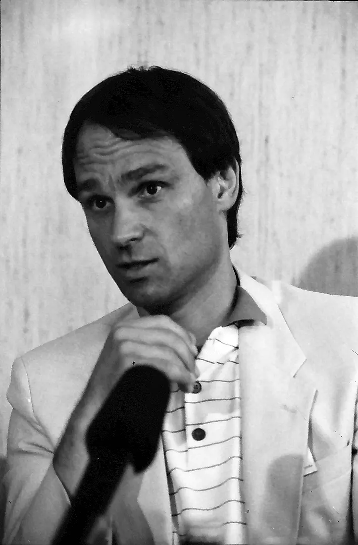
Gerd Binnig
born 1947 in Frankfurt, Germany
As a student he began to doubt physics and spent more time playing in a beat band. He was 39 when he won the prize.
Photo: Willy Pragher, CC BY 4.0
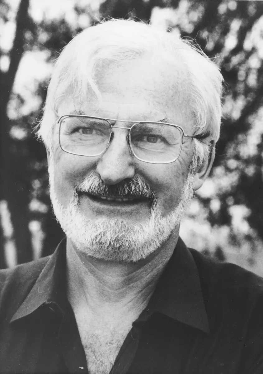
Heinrich Rohrer
1933–2013, born in Buchs, Switzerland
Born half an hour after his twin sister. He decided on physics only when he had to register at university.
In your school’s science room there may be a microscope. With it you can see onion cells, or the red blood cells in a drop of blood. But you can’t see a virus, the thing that gives you flu. And no light microscope, however good, will ever show you a single atom.
In 1986 the Nobel Prize in Physics was given for two new kinds of microscope that broke through this limit. Ernst Ruska built the first electron microscope, which looks with electrons instead of light. Gerd Binnig and Heinrich Rohrer built the scanning tunneling microscope, which doesn’t look at all: it feels a surface, atom by atom.
The problem: light is too coarse
A light microscope uses glass lenses to bend light and make a magnified image Grade 10: Lens. By the end of the 1800s, lens makers had become so good that their microscopes were almost perfect. Yet they still couldn’t show anything much smaller than about a quarter of a thousandth of a millimeter.
The reason is that light is a wave. In the 1870s the German physicist Ernst Abbe showed that a microscope can’t show details much smaller than about half the wavelength of the light it uses. Visible light has a wavelength of about half a thousandth of a millimeter Grade 9: Visible light. Think of waves in a pond: a big rock changes the waves, but a thin stick standing in the water hardly disturbs them. In the same way, light waves pass round things much smaller than their wavelength without “seeing” them.
Bacteria, a few thousandths of a millimeter long, could just be seen. But viruses are ten to a hundred times smaller, and atoms are about a thousand times smaller than the wavelength of light. To see them, scientists needed something with a much shorter wavelength than light.
Why can’t a better light microscope, with better lenses, show a single atom?
Pick an answer to see why.
Electrons: a light with a shorter wave
Electrons are the tiny negative particles in every atom Grade 9: Structure of an atom. Heat a metal wire in a vacuum, and electrons boil off it. Pull them with a high voltage, and they shoot off in a beam. Old television sets worked like this: a beam of electrons hit the screen and made it glow.
In 1924 the French physicist Louis de Broglie suggested something strange: moving electrons also behave as waves. Their wavelength is tiny. For the fast electrons in a microscope, it is about 100,000 times shorter than the wavelength of light. A microscope that used electrons instead of light could, in principle, show things far smaller.
But you can’t focus electrons with glass. They don’t pass through it. So how do you make a lens for electrons?
Why it was hard
- No glass lenses. Electrons are stopped by glass, so a completely new kind of lens was needed.
- No air. Electrons are scattered by the molecules in air after a short distance. The whole microscope had to be a sealed tube with the air pumped out.
- The beam burns things. A strong electron beam heats whatever it hits. Living cells can’t survive in a vacuum anyway, so specimens had to be dried and cut very thin.
- Nobody believed in it. Many biologists thought the beam would destroy anything worth looking at, and that the whole idea was useless.
What they did, step by step
Ruska: lenses made of magnetism
Ernst Ruska was an electrical engineering student at the Technical College in Berlin, working with Max Knoll, who was building better tubes for measuring fast electrical signals.
- 1929: a coil as a lens. A current in a coil of wire makes a magnetic field Grade 10: The magnetic field around a solenoid. A magnetic field pushes sideways on moving electrons. Another scientist, Hans Busch, had worked out that a short coil should bend a beam of electrons to a focus, just as a convex lens focuses light. Ruska tested this and showed that it worked. He also found that wrapping the coil in iron, with only a narrow gap, squeezed the field into a small space and made a much stronger lens.
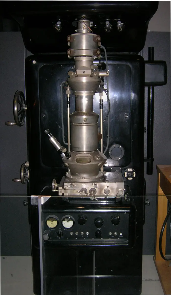
- April 1931: two lenses. Ruska put two coil lenses one after the other, like the two lenses of a light microscope. They made a picture of a tiny metal grid magnified 14.4 times. That is less than a cheap magnifying glass, but it was the first electron microscope. (The beam was so strong that it burnt the grid to cinders.)
- 1931: the good news. That summer Ruska heard about de Broglie’s idea. At first he was disappointed: if electrons are waves, they too must have a limit. Then he worked out their wavelength, and was delighted: the limit was about a hundred thousand times smaller than for light.
- 1933: better than light. With stronger iron-clad lenses, Ruska built a new microscope that magnified 12,000 times. For the first time an electron microscope showed finer detail than any light microscope. He built it while living on a small grant, and joked that it was the cheapest electron microscope a German science fund ever paid for.
- 1939: microscopes for everyone. Ruska moved to the company Siemens, where he and Bodo von Borries designed electron microscopes that could be built and sold. The first went on sale in 1939. Ruska’s brother Helmut, a doctor, used them to make some of the first pictures of viruses and of the viruses that attack bacteria.
Binnig and Rohrer: a finger that feels atoms
By the 1970s electron microscopes could almost show single atoms, but only in very thin samples. They could not show the arrangement of atoms on the surface of a solid. Yet surfaces matter: computer chips are built on the surface of silicon, and many reactions happen on surfaces.
Heinrich Rohrer worked at IBM’s research laboratory near Zurich, in Switzerland. In 1978 he hired a young German physicist, Gerd Binnig. Their first idea was to study tiny spots on thin layers. It grew into a new kind of microscope.
Their idea used a strange effect of quantum physics called tunneling. Because electrons behave as waves, an electron near the surface of a metal is not completely trapped inside it. Its wave leaks out a tiny distance. So if two pieces of metal are brought very close, closer than about a millionth of a millimeter, but not touching, a few electrons can jump across the gap. In ordinary physics this is impossible, like a ball rolling through a hill instead of over it. With a small voltage, these electrons make a tiny current, the tunneling current.
The tunneling current changes enormously with the gap. Make the gap just 0.1 nm wider (about half the width of an atom), and the current becomes about ten times smaller. That makes it a wonderfully sensitive ruler.
- The plan (1978–1979). Bring a very sharp metal tip close to a surface, close enough for a tunneling current. Move the tip across the surface, line by line. Wherever the surface rises, even by a fraction of an atom, the current jumps. A control circuit then lifts the tip to keep the current steady. Record how the tip goes up and down, and you have a map of the surface: a scanning tunneling microscope (STM). In January 1979 they described the idea for a patent.
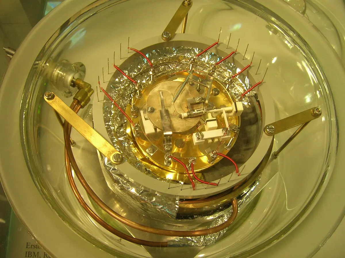
- Stillness. The gap had to be held steady to about a hundredth of the width of an atom. A footstep in the building or a voice in the room would shake the tip far more than that. Their first working set-up floated on magnets over a bowl of superconducting lead, cooled with liquid helium, held together, as they wrote, with “lots of Scotch tape”.
- Moving by tiny amounts. To move the tip by less than the width of an atom, they used piezoelectric crystals, which get very slightly longer or shorter when a voltage is applied to them.
- 16 March 1981. Working at night, “hardly daring to breathe”, so as not to shake anything, they measured a tunneling current that changed with the gap exactly as the theory said. The microscope worked.
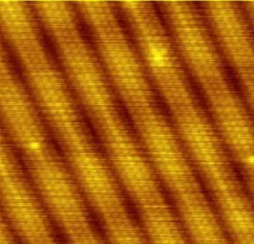
- 1982–1983: atoms. They turned it on a famous puzzle: how the atoms are arranged on the surface of a silicon crystal, which nobody had been able to work out. The STM showed the pattern directly, atom by atom.
In an STM, the gap between the tip and the surface becomes 0.1 nm wider. What happens to the tunneling current?
Pick an answer to see why.
How they are used now
Electron microscopes and STMs work in different ways, and each does what the other can’t.
- A transmission electron microscope, Ruska’s kind, sends electrons through a very thin slice. It shows the inside of cells, viruses, and the atoms inside materials. The best ones today can show details smaller than a tenth of a nanometer.
- A scanning electron microscope moves a fine beam of electrons over the outside of a specimen, and gives pictures with a strong 3D look, such as close-ups of insects’ eyes.
- An STM maps the surface of materials that conduct electricity, atom by atom. In 1986 Binnig and others also invented its cousin, the atomic force microscope, which feels a surface with a tiny spring and works on any material, even living cells in water.
In your life today
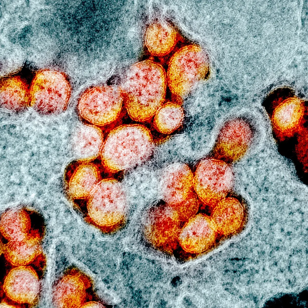
- Medicine. When a new disease appears, an electron microscope is often one of the first tools used to see the germ that causes it. The pictures of the COVID-19 virus that you saw in 2020 were made this way.
- Phones and computers. The chips in a phone have parts only a few tens of atoms across. Chip makers check them with electron microscopes and atomic force microscopes.
- New materials. Chemists use these microscopes to study catalysts, the surfaces where reactions happen, and new materials such as graphene, a sheet of carbon one atom thick.
What came next
- Molecules frozen in ice. Biologists wanted to see proteins in their natural shape, not dried out. Freezing them very quickly in thin ice and imaging them with electrons led to the Nobel Prize in Chemistry in 2017 (Freezing molecules to take their picture).
- Building with atoms. Since the IBM letters, scientists have built tiny structures atom by atom, and studied how electrons behave in them.
- An open question. We can now see and move single atoms on a surface. But building useful machines and devices one atom at a time is extremely slow. Can we find ways to make atoms and molecules arrange themselves into the structures we want?
In your science books
- Grade 8: the structure of an atom.
- Grade 9: the structure of an atom, waves and wavelength and visible light.
- Grade 10: lenses and the images they form; the magnetic field around a solenoid and the electromagnet, the ideas behind Ruska’s coil lenses.
- Going Further: what a molecule looks like.
Learn more
- The Nobel Prize’s own explanation: the press release and the speed read (nobelprize.org).
- The Nobel lectures: Ruska, on the early history of the electron microscope, and Binnig and Rohrer, “Scanning tunneling microscopy: from birth to adolescence”.
The official reason
The prize was given “for his fundamental work in electron optics, and for the design of the first electron microscope.”
The prize was given “for their design of the scanning tunneling microscope.”