
Growth factors: signals that make cells grow
Nobel Prize in Physiology or Medicine, 1986: Rita Levi-Montalcini and Stanley Cohen
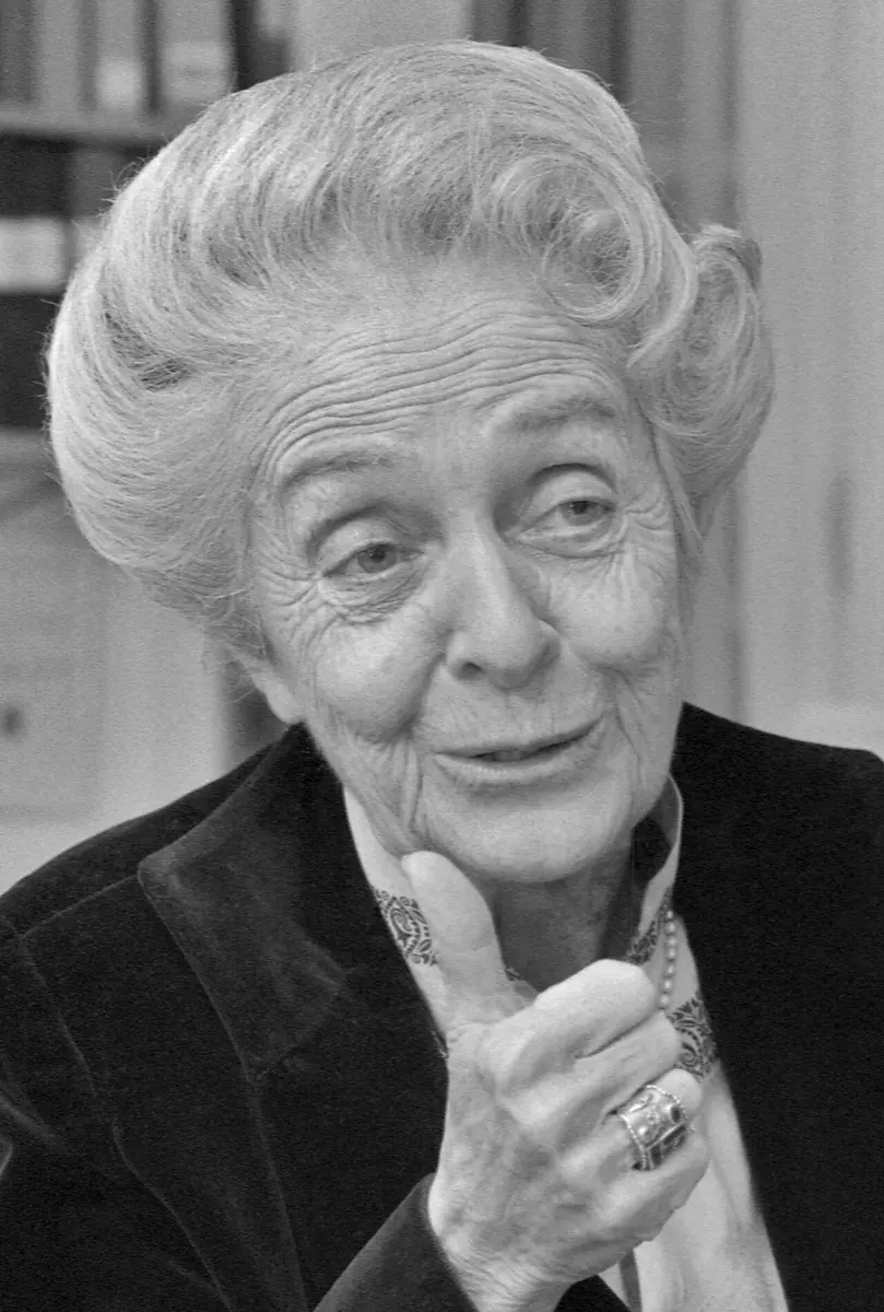
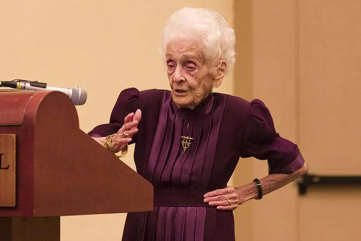
Rita Levi-Montalcini
1909–2012, born in Italy
Her father thought university was not for girls; at 20 she talked him round. Barred from her university by Mussolini’s race laws, she did her experiments in her bedroom. She lived to 103, still working.
Photo: Kurt Hagblom, Lund University Library, Public domain
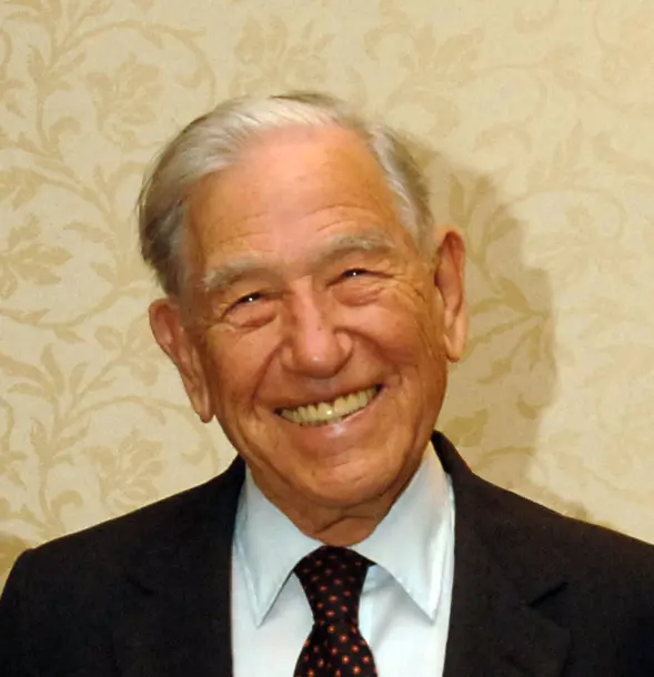
Stanley Cohen
1922–2020, born in the United States
The son of a tailor, he went to a free city college. For his doctorate he collected more than 5,000 earthworms from the university lawn at night.
Photo: NICHD, National Institutes of Health, Public domain
Cut your finger, and within a week the cut has closed, with new skin over it. Somehow the cells around the cut know they must divide and fill the gap, and they also know when to stop.
You began as a single cell. Today your body has tens of millions of millions of cells, of more than 200 kinds, each in its place. Nerve fibers reach the tip of every finger and the skin of every toe. Something must tell each cell when to grow, when to divide, which way to send its fibers, and even when to die.
The 1986 Nobel Prize in Physiology or Medicine went to the two people who found the first of these messages: growth factors, which cells send to their neighbors to make them grow.
The problem: what tells a cell to grow?
By the 1940s, scientists knew about hormones: chemical messages made by glands such as the pituitary gland at the base of the brain, carried in the blood to the whole body. Growth hormone, from the pituitary, makes a child grow taller. But growth hormone doesn’t act on cells directly, and a baby grows inside its mother without it. Something else was controlling how the body’s parts form.
The nervous system was a puzzle of its own. A growing embryo makes more nerve cells than it needs. In the chick embryos that biologists studied, many young nerve cells sent out fibers and then died, while others lived. Nobody knew what decided which.
Scientists also knew that cells grown in a dish (in tissue culture) grew better if a little blood or a mashed-up organ was added. Something in the blood helped. But what? Nobody could say.
How cells send messages
To follow the story, you need one idea: cells talk to each other with signal molecules.
A cell sends out a signal molecule, often a small protein. Another cell receives it with a receptor, a protein that sits in its outer covering, the cell membrane, and fits that one signal like a lock fits its key. When the signal fits into the receptor, the receiving cell changes what it does: it may grow, divide, move, or make something.
A hormone travels all over the body in the blood. A growth factor is usually made by cells close by and acts on their neighbors. It works in very tiny amounts.
How does a cell “hear” a signal molecule sent by another cell?
Pick an answer to see why.
Why it was hard
Growth factors are made in very tiny amounts, mixed with thousands of other substances. One billionth of a gram of NGF in a milliliter of liquid is enough to make nerve fibers grow. To find such a substance, you need:
- a source that has a lot of it;
- a test: a quick way to see whether a sample has it, so you can tell which parts of a mixture to keep;
- the skill to purify it, step by step, until only the one substance is left.
At the start, Levi-Montalcini and Cohen had none of these.
What they did, step by step
Levi-Montalcini: from a bedroom to Rio de Janeiro
Rita Levi-Montalcini trained as a doctor in Turin, in Italy, and studied how the nervous system forms.
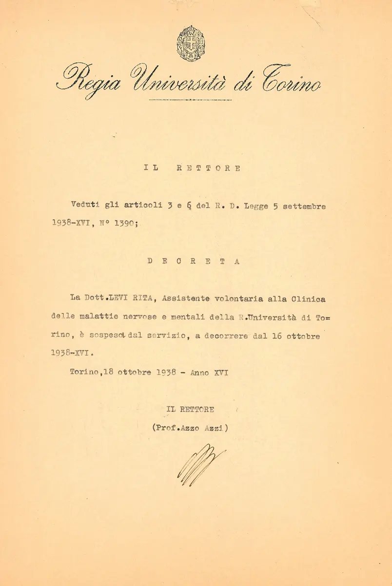
- 1938–1945, the bedroom laboratory. In 1938 Mussolini’s government passed laws that barred Jews from universities and professions, and she lost her post. She set up a small laboratory in her bedroom and worked on chick embryos, with her old teacher Giuseppe Levi as her only assistant. When bombing drove the family out of Turin, she rebuilt it in a cottage in the countryside. She found that when a young embryo’s wing bud was removed, the nerve cells that should have supplied it were made but then died. Nerve cells seemed to need something from the tissue they grew towards.
- 1947, America. The embryologist Viktor Hamburger invited her to Washington University in St. Louis to repeat this work. She meant to stay for a year. She stayed for 30.
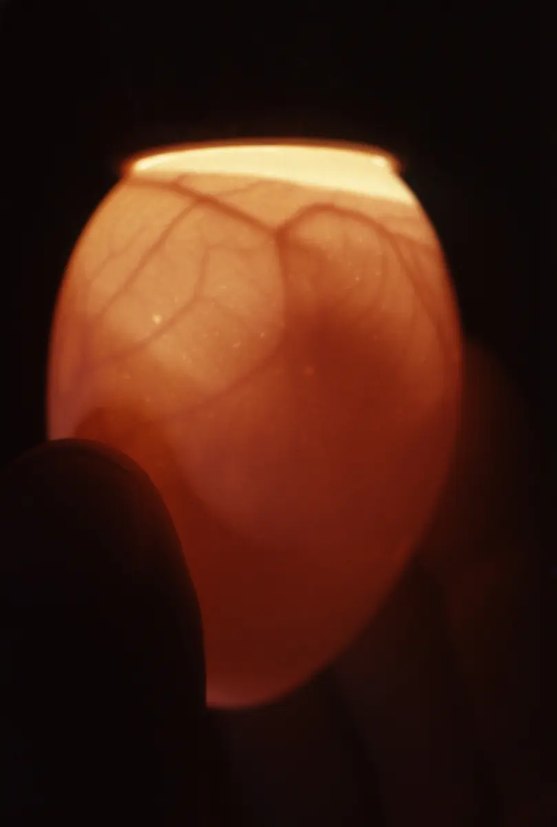
- About 1950, the tumor. A former student of Hamburger, Elmer Bueker, had put a piece of a mouse tumor (a mass of cancer cells) into chick embryos and found that nerve fibers grew into it. Levi-Montalcini repeated this and saw much more: the nerve fibers grew into the tumor in thick masses, the nerve clusters supplying it swelled, and fibers even grew into the embryo’s blood vessels. Then she put the tumor outside the embryo, on the membrane around it, where only blood connected the two. The nerves still grew wildly. So the tumor had to be releasing a substance that traveled in the blood.
- 1952, the halo. St. Louis had no tissue culture laboratory, so she flew to Rio de Janeiro in Brazil, to a friend, Hertha Meyer, who had one, carrying two mice with tumors in her handbag. There she put a small cluster of nerve cells from a chick embryo, a ganglion, near a piece of tumor in a dish. Within a day, nerve fibers grew out of the ganglion like the rays of the Sun: a halo. This was the quick test she needed. The unknown substance became known as nerve growth factor, NGF.
Cohen: snake venom and a salivary gland
In 1953 a young biochemist, Stanley Cohen, joined the group to find out what NGF was made of.
- The tumor extract. He purified an active part of the tumor. It contained protein and nucleic acid (the family of DNA and RNA). Which one was NGF?
- The snake venom surprise. To find out, he added a little snake venom, because venom contains an enzyme that destroys nucleic acid. If the halo vanished, NGF was nucleic acid. Instead, the halo grew denser. The venom alone, with no tumor at all, made huge halos: 6 millionths of a gram of venom worked as well as 15,000 millionths of a gram of tumor. Venom was a far richer source of NGF.
- 1958, a better source. A snake’s venom glands are its salivary glands, changed. So Cohen tested the salivary glands of mice, and those of male mice were richer still. From them he purified NGF and found it was a protein.
- The proof. Cohen made antibodies against NGF, proteins of the immune system that stick to it and block it. When the antibodies were injected into newborn mice, most of their sympathetic nerve cells (the nerves that control the heart, blood vessels and gut without our thinking) disappeared. These nerve cells cannot live without NGF.
Cohen added snake venom to destroy the nucleic acid in his tumor extract. What happened, and what did it show?
Pick an answer to see why.
Cohen again: eyelids that opened early
- A side effect. When Cohen injected the salivary gland extract into newborn mice, something unexpected happened: their eyelids opened at 6–7 days old instead of 12–14, and their teeth seemed to come through early. This had nothing to do with nerves.
- 1959 onwards, a second factor. At Vanderbilt University he tracked down the substance responsible and purified it in the early 1960s. It made the epidermis, the outer layer of skin, grow and thicken, so he called it epidermal growth factor, EGF. It also speeded up the healing of wounds in the clear front of the eye, the cornea.
- Early 1970s, its structure. He found that mouse EGF is a small protein, a chain of 53 amino acids.
- 1978, the receptor. Cohen found the EGF receptor on cells. When EGF fits into it, the inside part of the receptor, an enzyme, starts putting small chemical tags, phosphate groups, on proteins, including itself. By 1980 his group found that the tags go onto one particular amino acid, tyrosine. This was the first time anyone saw how a growth signal outside a cell is passed inside it.
How it works now
We now know that NGF does three things for the nerve cells that need it:
- It keeps them alive. Young nerve cells compete for it; those that get enough survive.
- It makes their fibers grow.
- It guides them: fibers grow towards the place where NGF comes from.
In a growing embryo, the tissues that need nerves, such as the skin and the heart, give out small amounts of NGF. Nerve fibers grow towards it. A fiber that reaches the target takes NGF in at its tip, and the NGF is carried back along the fiber to the cell body. That cell lives. A nerve cell whose fiber goes the wrong way gets no NGF, and dies. In this way, each tissue ends up with about as many nerves as it needs.
NGF and EGF were only the first. Since then, dozens of growth factors have been found: ones that make blood vessels grow, ones from blood platelets that help wounds heal, ones that make blood cells. Many of their receptors work just like EGF’s, by putting phosphate tags on tyrosine.
Growth factors also explained something about cancer. Some cancer-causing genes turned out to be faulty copies of a growth factor or its receptor. A receptor that is stuck “on” tells the cell to divide again and again, even with no growth factor present.
Why does a faulty EGF receptor that is stuck “on” lead to cancer?
Pick an answer to see why.
In your life today
- Cancer medicines. Since about 2000, medicines that block the EGF receptor, or a close relative of it called HER2, have been used to treat some cancers of the lung, the bowel, the head and neck, and the breast. They work only for cancers whose cells depend on that receptor, so the cancer is tested first.
- Eye drops made of NGF. In 2017, eye drops of human NGF were approved in Europe, and later in the United States, for a rare disease in which the nerves of the cornea are damaged and the eye’s surface cannot heal.
- Wound healing. EGF and other growth factors are what make your skin close over a cut. Scientists are testing them to help wounds that heal badly, such as foot ulcers in people with diabetes.
What came next
- A family of nerve factors. In 1982, a second factor that keeps nerve cells alive, BDNF (brain-derived neurotrophic factor), was found in the brain. It and NGF belong to a family now called the neurotrophins.
- The brain. NGF also acts on some nerve cells in the brain, among them cells that are lost in Alzheimer’s disease. The hope that NGF could treat dementia has not yet come true, partly because a large protein like NGF cannot easily get from the blood into the brain.
- Repair. Growth factors guide nerve fibers in an embryo. Could they make fibers grow again after the spinal cord is injured, so that a person who cannot walk could walk again? This is one of the big open questions of medicine today.
In your science books
- Grade 8: the cell and its covering, where receptors sit.
- Grade 9: the nerve cell, the autonomic nervous system (the sympathetic nerves), epithelial tissue (the epidermis), and hormones, the body’s long-distance messages.
- Grade 10: cell division.
- Going further: from one cell to trillions, proteins do the work and messages along nerves.
Learn more
- The Nobel Prize’s press release for 1986, with the halo test, and its short speed read.
- The two Nobel lectures: Levi-Montalcini’s The nerve growth factor: thirty-five years later and Cohen’s Epidermal growth factor (nobelprize.org).
The official reason
The prize was given “for their discoveries of growth factors.”