
Every one of us began the same way: as a single cell, about 0.12 mm across, smaller than the full stop at the end of this sentence. That cell had no skin, no bones, no brain and no heartbeat. Today you are made of something like 30 to 37 trillion cells, and they are wildly different from one another. A red blood cell is a tiny dented disc with no nucleus. A nerve cell running from your spine to your big toe can be a meter long. A fat cell is one big drop of oil. Your skin’s outer cells are dead, flat flakes.
Yet almost all of them carry exactly the same DNA, copied from that first cell. So two questions. How did one cell become trillions? And how did cells with the same instructions become skin, bone and brain? This topic answers both, and shows how the same two processes keep you going every day: your body is making about 4 million new cells every second as you read this.
From one cell to trillions
You met cell division in Grade 10 Grade 10: Mitosis: a cell copies its chromosomes, then divides into two daughter cells with the same chromosomes as itself. The zygote, the cell made when a sperm joins an egg, divides by mitosis into 2 cells, then 4, then 8. Each round of division doubles the number of cells.
Doubling grows astonishingly fast. Fold a sheet of paper in half and it is 2 layers thick; fold it again and it is 4. After 10 folds it would be 1,024 layers, and you couldn’t fold it any more. That 2¹⁰ = 1,024 ≈ 1,000 is a handy shortcut: every 10 doublings multiply the number by about a thousand.
The first week
Something surprising happens in the first few days: the cells divide, but the embryo does not grow. It stays inside the egg’s tough coat, the zona pellucida, about 0.15 mm across, and every division just shares the same material between more and smaller cells. This first stage of division without growth is called cleavage.
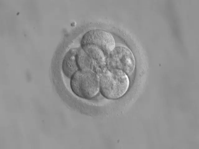
By day 5 the embryo is a hollow ball, the blastocyst, and its cells are no longer all alike. About 40 cells form a thin outer layer, which will grow into part of the placenta, the organ that will feed the baby from the mother’s blood. About 20 cells form a small clump inside, the inner cell mass. Everything in your body came from those 20 or so cells (some of them also make the yolk sac and other wrappings around the embryo).
Around day 6 or 7, the blastocyst squeezes out of its coat and begins to sink into the soft lining of the uterus. Only now does the embryo start to grow, fed by the mother.
During the first four days, a human embryo goes from 1 cell to about 16. What happens to its size?
Pick an answer to see why.
Not just doubling
After the first week, a body is not built by everyone doubling together. Different groups of cells divide at different speeds, some stop dividing for good, and some die on purpose. This planned cell death is called apoptosis, and it is as much a part of building a body as division is.
Same DNA, different cells
Here is the puzzle. Look at a few of the cells from one body, drawn to the same scale.
They are different in size, shape and job. In Grade 9 you saw how similar cells group into the four kinds of animal tissue: epithelial, muscular, connective and nervous Grade 9: Animal tissue. A cell becoming one particular kind, with the shape and job of that kind, is called differentiation, and the finished cell is a specialized (or differentiated) cell.
Why are they different, when they all came from one zygote by mitosis, which copies the chromosomes exactly? For a long time it was a real possibility that cells lose the genes they don’t need as they specialize: that a skin cell keeps only the skin instructions. The way to test it is to see whether a specialized cell still has all the instructions to make a whole body.
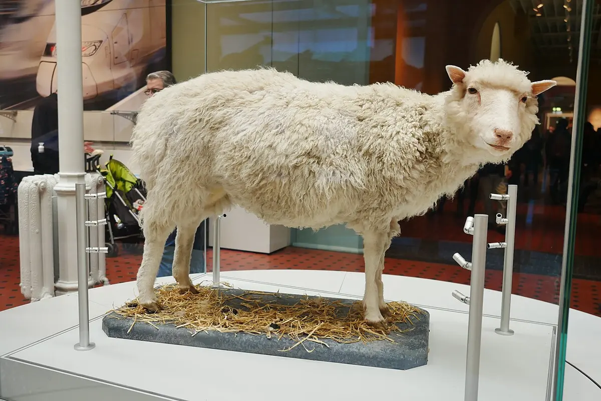
Other experiments said the same. In 1958 the American botanist F. C. Steward grew whole carrot plants from cells taken from a carrot root, an early success of the tissue culture you met in Grade 8 Grade 8: Tissue culture. And on 5 July 1996, at the Roslin Institute in Scotland, a lamb was born from an egg given the nucleus of a cell from an adult sheep’s udder. She was Dolly, the first mammal cloned from an adult cell.
Genes switched on and off
So every cell carries the same genes, about 20,000 of them, each the instructions for one or more proteins. What makes cells different is which genes they use. A gene that a cell is using is said to be switched on (biologists say it is expressed); a gene it isn’t using is switched off. A cell’s kind is decided by its own set of switched-on genes.
Think of a recipe book with 20,000 recipes, and a copy in every kitchen in Bhaktapur. The sweet shop uses the pages for sel roti and laddu, the momo shop the pages for momo and achar, the juju dhau maker one page only. Same book, different pages, different shops. And some pages, like “light the stove” and “wash the pots”, every kitchen uses: in a cell these are the housekeeping genes, for releasing energy from food, building proteins and copying DNA.
How a gene is read to build its protein is a story of its own: How Does a Gene Make a Protein? Here what matters is the result. The hemoglobin genes are switched on strongly only in the cells that become red blood cells; they make so much hemoglobin that it ends up as about 97% of the cell’s dry mass. Only the beta cells of the pancreas make insulin for the blood. A rod cell in your eye makes rhodopsin, which catches light. Each of these cells also has the other genes, silent.
What decides which genes a cell switches on? Mostly signals from its neighbors, and where it is in the embryo. Cells send chemical messages to the cells around them, and a cell reads the messages it receives, together with its own past, to decide what to become. Each decision narrows the choices for its daughter cells: a cell on its way to becoming skin no longer has the choice of becoming blood.
A nerve cell and a muscle cell from the same person look and work completely differently. Why?
Pick an answer to see why.
Stem cells
A specialized cell such as a nerve cell or a red blood cell usually can’t divide any more. So where do new cells come from? From cells that have stayed unspecialized: stem cells.
A stem cell is a cell that can do two things:
- divide to make more stem cells like itself, and
- make cells that go on to specialize.
You met the plant version in Grade 9: the meristem, the patches of dividing cells at the tips of stems and roots that let a plant grow all its life Grade 9: Meristematic tissue. Animals have stem cells too, but they differ in how many kinds of cell they can make.
| Cell | Can become | Example |
|---|---|---|
| zygote | every cell of the body and the placenta | the fertilized egg |
| embryonic stem cell | every kind of cell in the body (but not the placenta) | the inner cell mass of a blastocyst |
| adult stem cell (tissue stem cell) | only the few kinds of cell of its own tissue | blood stem cells in the bone marrow; stem cells at the base of the skin and in the lining of the gut |
As an embryo develops, its cells’ choices narrow step by step. In the third week, the cells of the inner cell mass move and fold into three layers, the germ layers you met in Grade 10 Grade 10: Germ layers. Every tissue of the body comes from one of them.
By the end of the eighth week every major organ has begun to form, though the embryo is only about 3 cm long. From then until birth it is called a fetus, and most of what happens is growth: more division, and the finishing of cells that are already on their way. At birth, about 38 weeks after fertilization, a baby weighs about 3 kg.
Which cells could, on their own, become every kind of cell in the body?
Pick an answer to see why.
Keeping a body going
Growth stops in your late teens, but mitosis doesn’t. Most of your cells wear out, and stem cells replace them. How fast depends on the tissue:
| Cells | How fast they are replaced |
|---|---|
| lining of the small intestine | every 3 to 5 days |
| outer layer of the skin | every one to two months |
| red blood cells | every 120 days (4 months) |
| fat cells | about 10% of them each year |
| heart muscle cells | about 1% a year at age 25, 0.5% at 75 |
| nerve cells in the outer brain (cortex) | almost never: as old as you are |
The fastest are the cells that face the harshest life: the gut lining, scoured by food and digestive juices, and the skin, rubbed and dried by the world. In the scientists’ best estimate, your body makes about 330 billion new cells a day, most of them blood cells, with a mass of about 80 g, more than a hen’s egg, every day.
The balance
A stem cell that divides makes two cells. To keep a tissue the same size, on average one of the two must stay a stem cell and the other must go on to specialize. If too many stay stem cells, the tissue grows; if too few, the stem cells run out. How exact must the balance be?
In this model, even 51% instead of 50% more than triples the stem cells in four months. A tissue can only stay the same size if its cells keep the balance almost perfectly, and the body does it with the kind of feedback you saw in How Does Your Body Stay at 37 °C?: signals from neighboring cells tell a stem cell when to divide and what its daughters should become, and a cell whose DNA is badly damaged destroys itself.
When those controls break, because of mutations in the genes that control division, a cell can divide when it shouldn’t, again and again. Its daughters do the same. That is cancer: cells dividing out of control, crowding out and invading the tissues around them. Cancer is mitosis gone wrong.
Why does a tissue with stem cells stay the same size year after year?
Pick an answer to see why.
Stem cells in medicine
The first stem cell treatment, and still the most common, is the bone marrow transplant. A patient with leukemia, a cancer of the blood, is given strong treatment that destroys their diseased bone marrow; then blood stem cells from a matching donor (or from the patient, collected beforehand) are dripped into a vein. They find their way into the bones and start making all the kinds of blood cell again. The American doctor E. Donnall Thomas, who developed it, shared the Nobel Prize for it in 1990. Nepal’s first bone marrow transplant was done at the Civil Service Hospital in Kathmandu in August 2016.
In 2006 the Japanese scientist Shinya Yamanaka showed that you can run differentiation backwards. By adding just four genes to connective-tissue cells (fibroblasts) from mice, he turned them into cells like embryonic stem cells, able to become any kind of cell. In 2007 his team and another in the USA did it with human cells. These are induced pluripotent stem cells (iPS cells; pluripotent means able to become many kinds of cell). Yamanaka shared the 2012 Nobel Prize with John Gurdon: the man who showed a specialized cell keeps all its genes, and the man who switched them back on. Scientists now grow iPS cells into heart muscle, nerve cells and retina cells, to study diseases in a dish and test treatments. In 2026 Japan gave the first conditional approvals to treatments made from them: nerve cells for Parkinson’s disease and sheets of heart muscle for heart failure, approved on small trials and still being checked as more patients are treated.
Investigate: grow a root from a stem
A plant shows you differentiation happening in a glass of water.
Questions
A man’s skin cell and his blood stem cell are compared. Which statement is true?
Pick an answer to see why.
Which is the correct order in the development of a human embryo?
Pick an answer to see why.
Explain the difference between cell division and cell differentiation, and why a body needs both.
Answer
Cell division (mitosis) makes more cells: one cell becomes two with the same chromosomes. Differentiation makes cells different: a cell switches on a particular set of genes and takes on the shape and job of one kind of cell, such as a muscle fiber or a nerve cell. Division alone would give a ball of trillions of identical cells, unable to see, move or digest. Differentiation alone, without division, would give just one cell. A body needs division to make enough cells, and differentiation to turn them into tissues and organs that do different jobs.
In Gurdon’s experiment, the nucleus of a tadpole’s intestine cell was put into a frog’s egg whose own nucleus had been destroyed, and the egg grew into a frog. What did this show? Why was it important to destroy the egg’s own nucleus first?
Answer
It showed that a fully specialized cell (a gut cell) still has all the genes needed to make a whole frog: specializing had switched genes off, not removed them, and the egg could switch them back on. The egg’s own nucleus had to be destroyed so that every gene in the new frog must have come from the gut cell’s nucleus. If the egg’s nucleus had been left in, the frog could have grown using the egg’s genes, and the experiment would prove nothing.
A human zygote is about 120 µm across. Assuming the embryo doesn’t grow, find the average width of its cells at the 16-cell stage (∛16 = 2.52). Why do the cells get smaller in the first days, when later in life cells grow before they divide?
Answer
d = 120 µm ÷ ∛16 = 120 ÷ 2.52 = 48 µm. In the first days the embryo is inside its coat and takes in very little from outside: it can’t settle into the uterus and draw food from the mother until it has hatched, around day 6–7. So the divisions share the egg’s material among more and more cells, and each is smaller. Once the embryo is attached and fed, cells grow between divisions, and the embryo grows.
Use the cell family tree in Figure 19.4 as a model to explain why an embryonic stem cell is more useful for research than a blood stem cell, and why a blood stem cell is still useful to a leukemia patient.
Answer
In the model, each step down the tree narrows a cell’s choices. An embryonic stem cell, from the inner cell mass, sits near the top: it can become any kind of body cell (muscle, nerve, liver…), so it can be used to study or make any tissue. A blood stem cell sits far down the mesoderm branch: it can make only blood cells. But that is exactly what a leukemia patient needs: after the diseased marrow is destroyed, transplanted blood stem cells settle in the bones and make every kind of blood cell for the rest of the patient’s life.
The lining of the small intestine is replaced every 3 to 5 days, but the cortex of the brain keeps the same nerve cells for life. Suggest a reason for the difference, and one problem each pattern causes.
Answer
The gut lining faces a harsh life: rubbed by food, attacked by digestive juices and acid, and exposed to whatever we swallow. Cells there wear out fast, so constant replacement keeps the lining whole. Nerve cells in the cortex store what we have learned in their connections; replacing them would wipe those connections, so they last a lifetime. Problems: the gut’s fast-dividing cells are hurt first by cancer drugs and radiation, which attack dividing cells, causing sickness and diarrhea during treatment. The brain’s cortex can make almost no new nerve cells, so damage from a stroke or an injury there is largely permanent.
In the model of Figure 19.4 and the Keep the balance widget, what must happen at each stem cell division for a tissue to stay the same size? Explain, with this idea, how a cancer can start.
Answer
On average, one of the two daughter cells must stay a stem cell and the other must specialize (or, over many divisions, half the new cells stay stem cells). Then the number of stem cells stays constant while a steady stream of new specialized cells replaces those that are lost. The balance must be almost exact: even a 51 : 49 split, kept up for months, multiplies the cells several times over. Normally, signals from neighboring cells keep it. If mutations damage the genes that control division, a cell can ignore these signals and keep dividing, its daughters inherit the same fault, and their number grows out of control: a tumor, which is how a cancer starts.
Where to go next
- How Does a Gene Make a Protein? What “switching on a gene” actually means: how a cell reads a gene to build hemoglobin, keratin or insulin.
- How Does Your Body Stay at 37 °C? The feedback loops that keep a body, made of all these different cells, steady inside.
- How Does Food Become Energy, and Become You? Where the energy and the material for 330 billion new cells a day come from.