
The markers that say “this is me”
Nobel Prize in Physiology or Medicine, 1980: Baruj Benacerraf, Jean Dausset, and George D. Snell
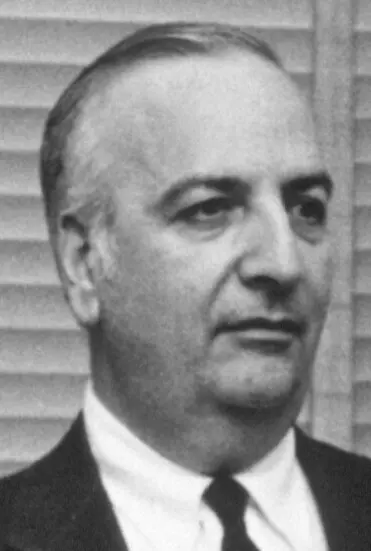
Baruj Benacerraf
1920–2011, born in Caracas, Venezuela
He had asthma as a child, which made him curious about how the body reacts to things. For a while he ran a bank in New York at the same time as his laboratory.
Photo: Unknown author, Public domain
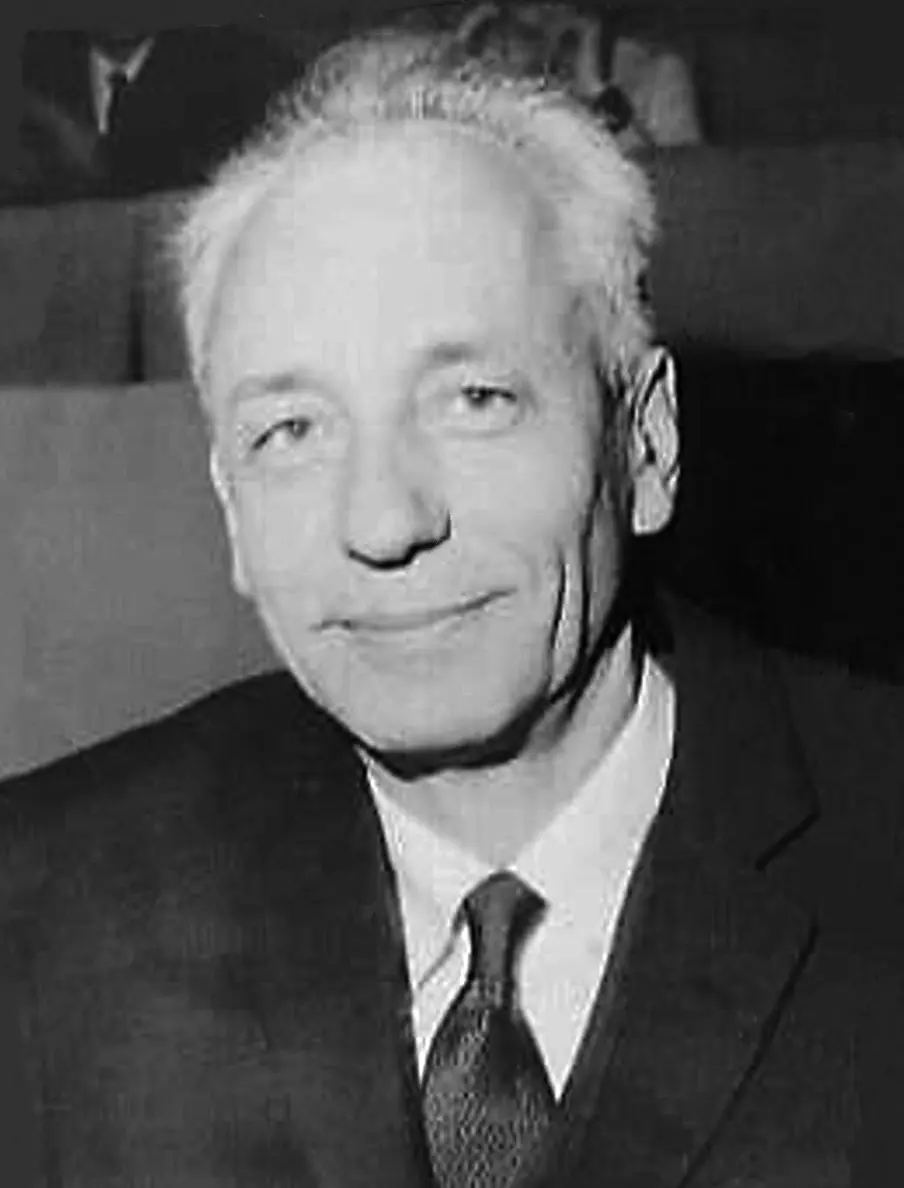
Jean Dausset
1916–2009, born in Toulouse, France
As a young doctor in the Second World War he gave blood transfusions to wounded soldiers in North Africa, which led him to study blood.
Photo: Agip, CC BY-SA 3.0 nl
-
George D. Snell
1903–1996, born in Bradford, Massachusetts, USA
A mouse geneticist who, from 1935, spent the rest of his long career at one laboratory on an island in Maine.
If someone needs blood, a doctor first checks their blood group. Give the wrong group, and the patient’s body attacks the new blood as if it were a germ Grade 10: why the blood group must match.
A kidney, a piece of skin or a bone marrow transplant is the same, only much harder to match. Our cells carry another set of markers on their surface, and they come in so many versions that two unrelated people almost never have the same set. In 1980 the Nobel Prize in Physiology or Medicine went to three scientists who found these markers and the genes that make them: George Snell in mice, Jean Dausset in people, and Baruj Benacerraf, who showed that the same genes control how strongly the body fights an infection.
The problem: the body throws out other people’s tissue
By the early 1900s surgeons could stitch a kidney or a patch of skin from one person into another. The operation often went well. But a week or two later the new tissue swelled, turned dark and died. The body had rejected it.
Doctors noticed something important: skin taken from your own body, and moved to another place, healed perfectly. Skin from someone else did not, unless that someone was your identical twin. So rejection had to do with what you inherit: something in other people’s cells was “foreign”, and something about it was decided by genes.
How the body tells “me” from “not me”
Your white blood cells guard your body Grade 10: white blood cells. One kind, the lymphocytes, recognize things that don’t belong: a bacterium, a virus, or a cell from another person. They do it by feeling the shapes of molecules on the outside of things. A molecule that a lymphocyte can recognize is called an antigen.
You already know one set of antigens: the A and B markers on red blood cells, which give your blood group. The 1980 prize was for finding a much bigger set, on almost every cell of the body. These markers are now called the MHC, short for major histocompatibility complex (histo means tissue; “compatibility” means getting along). In people, they are also called HLA.
Skin moved from one part of your arm to another heals well. Skin from a classmate is rejected. Why?
Pick an answer to see why.
Why it was hard
Nobody could see these markers. They are molecules on the cell surface, far too small for a microscope. The only clue was whether a transplant lived or died, and that depends on many genes at once. In people it was even harder: you cannot do transplant experiments on people just to find out how they work, and people are not bred in families of identical individuals the way laboratory animals can be.
What they did, step by step
Snell: mice that accept or reject (1930s–1950s)
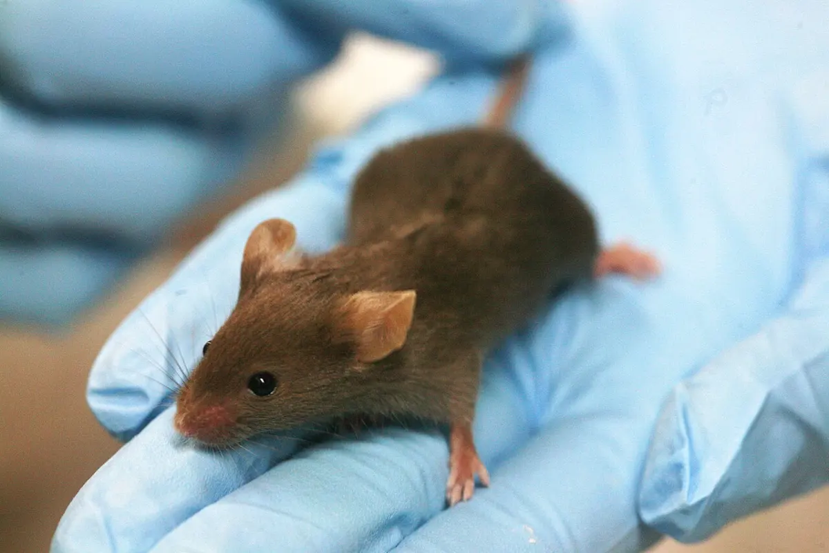
- Identical mice. In 1935 George Snell joined the Jackson Laboratory in Bar Harbor, Maine, in the United States. Its scientists had mated brothers with sisters for many generations, until every mouse in a family, or strain, had the same genes, like identical twins.
- Tumors as a test. Within one strain, a tumor from one mouse grew in any other. Moved to a mouse of a different strain, it was rejected. Normal tissue, like skin, followed the same rules. Snell called the genes that decided this histocompatibility genes, or H genes.
- A clue in the tail (1940s). Snell crossed the strains with mice that carried a gene for a kinked tail. He found that whether a mouse accepted a tumor usually went together with the shape of its tail. That meant the H gene sat on the same chromosome, close to the tail gene, so the two were passed on together.
- The strongest gene. A British scientist, Peter Gorer, came to work with him. In 1936 Gorer had found a marker on mouse blood cells that he called “antigen II”. Testing the same mice, they found that Gorer’s marker and Snell’s gene were one and the same. In 1948 they named it H-2: the strongest H gene of all.
- Patient breeding. To study each H gene alone, Snell bred new strains that differed from each other in just one piece of chromosome. Each took at least 14 generations of careful crossing. A great fire in 1947 destroyed the first set of these crosses, and he had to start again.
Dausset: the markers in people (1950s–1960s)
- A puzzle in the blood. Jean Dausset was a doctor in Paris who studied blood. He noticed that some patients who had received many blood transfusions made antibodies that destroyed white blood cells. At first he thought they were attacking their own cells. But their own white cells were unharmed; only the white cells of their blood donors were destroyed.
- A marker that some people have (1958). He realized that the patients were reacting to markers on the donors’ white cells, which they did not have themselves. In 1958 he described the first one. He called it Mac; today it is called HLA-A2, and it is one of the most common of these markers in many parts of the world.
- Mothers’ antibodies. A baby inherits half of its markers from its father, so during pregnancy a mother’s body can meet markers it does not have. Women who had had several children often carried antibodies against them. Dausset used blood from such mothers, and from transfused patients, to sort people by their markers.
- One family of genes. By testing parents and children, he showed that all these markers are made by a group of genes on one chromosome (in people, chromosome 6), passed on together. This was the human version of Snell’s H-2. It was named HLA, for human leucocyte antigens (leucocyte means white blood cell).
- Proof with skin (1960s). Volunteers agreed to receive small skin grafts. The better their HLA markers matched the donor’s, the longer the grafts survived.
- Working together. Dausset helped organize meetings where laboratories from many countries compared their results, swapped samples and agreed on names, so that every hospital could type its patients in the same way.
Why did Dausset find useful antibodies in women who had given birth to several children?
Pick an answer to see why.
Benacerraf: genes that decide how strongly we respond (1960s)
Baruj Benacerraf worked at New York University and later at Harvard. He gave guinea pigs very simple man-made substances to see if their bodies would make antibodies against them. Some animals responded strongly. Others, given the same substance, made no response at all.
Breeding them showed that whether an animal responded was inherited, decided by single genes that he called immune response genes (Ir genes). And when their position was found, the Ir genes turned out to sit right inside the MHC, among the transplant genes.
So the MHC was not just about transplants. The same group of genes helps decide how well your body fights each germ.

How it works now
Why would our bodies have markers whose job is to make transplants fail? They don’t. Transplants were invented by doctors only recently. The real job of the MHC was found in the 1970s and 1980s.
Each MHC molecule is like a small display stand on the cell’s surface. It holds up a short piece of whatever proteins the cell is making inside. If the cell is healthy, the pieces are all from its own proteins, and the lymphocytes pass by. If a virus is inside the cell, some of the pieces are from the virus, and a lymphocyte that fits them kills the cell (see our page on the 1996 prize). Your MHC version decides which pieces can be shown well, which is why it affects how strongly you fight each germ, as Benacerraf found.
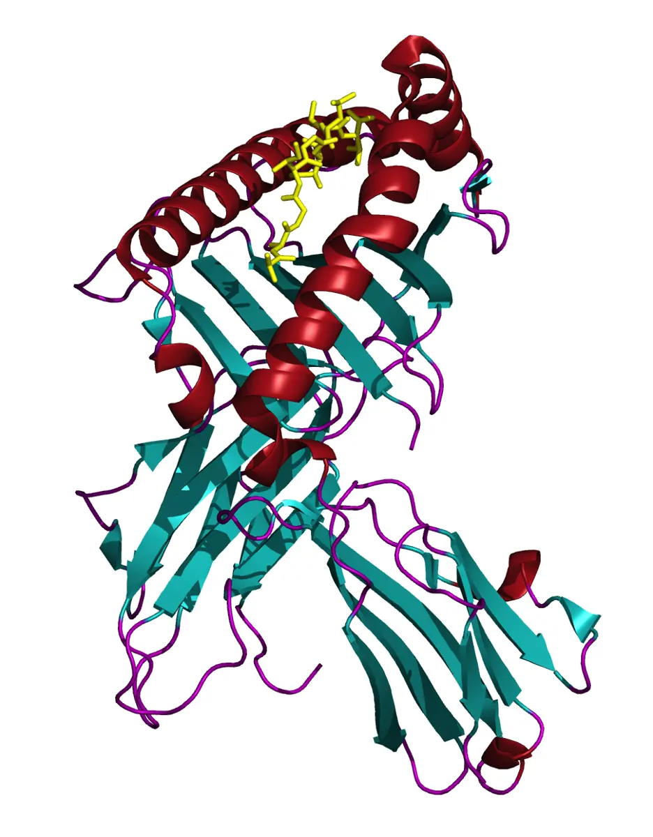
There are many genes in your HLA set, and each comes in a very large number of versions; scientists have now found tens of thousands. You inherit one HLA set from your mother and one from your father, on your two copies of chromosome 6. Because the set is passed on as one piece, brothers and sisters have a simple chance of matching.
In your life today
- Transplants. Before a kidney or bone marrow transplant, doctors test the HLA of the patient and the donor. The closer the match, the less the body fights the new organ, and the smaller the dose of medicines needed to calm the immune system. In Nepal the law allows a living person to give a kidney only to a family member, and the national transplant center is in Bhaktapur (our page on transplants).
- Bone marrow registries. For bone marrow, a close match is essential. Millions of volunteers around the world have had their HLA typed so that a patient with no matching brother or sister can find an unrelated donor.
- Diseases. Some HLA versions are linked to particular illnesses, such as type 1 diabetes and a disease that stiffens the spine. HLA tests help doctors recognize them.
What came next
- The shape of the stand. In 1987 scientists at Harvard worked out the shape of an HLA molecule and saw the groove that holds the protein piece.
- Self and germ together. Peter Doherty and Rolf Zinkernagel showed that lymphocytes check the MHC and the germ at the same time (the 1996 prize).
- Better transplants. Matching, along with medicines that quieten the immune system, has made kidney transplants ordinary operations around the world.
- Open questions. Why are some HLA versions linked to diseases in which the body attacks itself? And could we one day “teach” a patient’s body to accept a donor’s organ for good, without lifelong medicines?
In your science books
- Grade 10: white blood cells, blood groups and why they must match, chromosomes and Mendel’s laws.
- Going further: how a gene makes a protein.
Learn more
- The Nobel Prize’s press release and presentation speech for 1980.
- The laureates’ Nobel lectures: Snell, Dausset and Benacerraf.
The official reason
The prize was given “for their discoveries concerning genetically determined structures on the cell surface that regulate immunological reactions.”