
Restriction enzymes: scissors for DNA
Nobel Prize in Physiology or Medicine, 1978: Werner Arber, Daniel Nathans, and Hamilton Smith
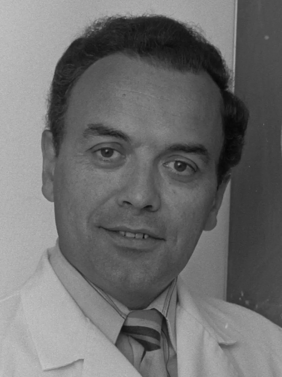
Werner Arber
born 1929 in Gränichen, Switzerland
He studied chemistry and physics, and came into biology through a job looking after an electron microscope. When he won, his ten-year-old daughter explained his work as a fairy tale.
Photo: This image is from the collection of the ETH-Bibliothek and has been published on Wikimedia Commons as part of a coopera, CC BY-SA 4.0
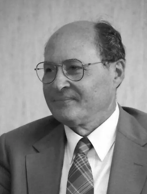
Daniel Nathans
1928–1999, born in Wilmington, Delaware, USA
The youngest of nine children of Jewish immigrants from Russia, he worked after school from about the age of ten. He was the first to use the new scissors to map a virus’s genes.
Photo: Willy Pragher, CC BY 3.0 de
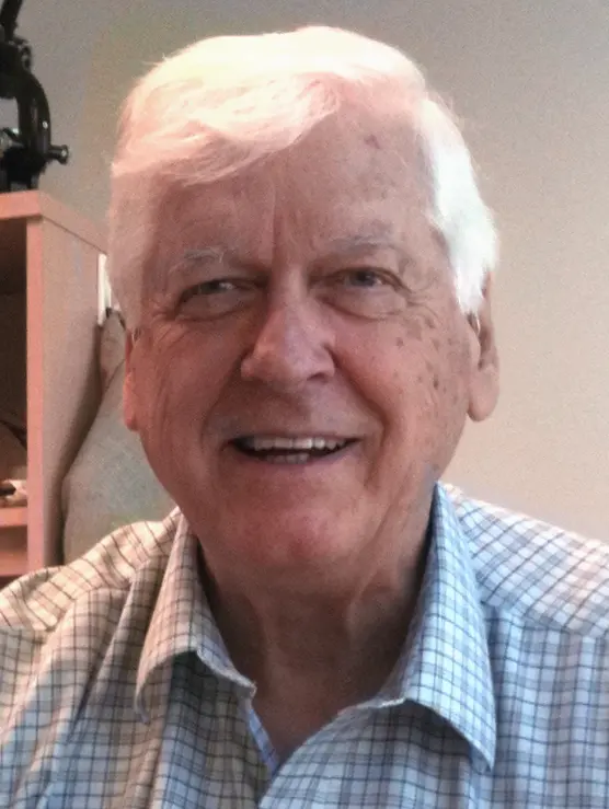
Hamilton Smith
1931–2025, born in New York, USA
A doctor who found his way into research late. His discovery began with an experiment that went wrong, and a drop in how thick a liquid was.
Photo: Jane Gitschier, CC BY 2.5
Every cell in your body carries a set of instructions, written in DNA. If you could print the instructions in one of your cells as a book, it would have about a million pages and fill 100 meters of shelf. One wrong letter on one page can cause a disease.
Fifty years ago, scientists could not read this book at all. The DNA in a cell is one enormously long thread, and there was no way to cut it into pieces small enough to study. The 1978 Nobel Prize in Physiology or Medicine went to three scientists who gave biology its first pair of scissors for DNA: restriction enzymes. They were found in bacteria, which had been using them for millions of years to fight off viruses.
The problem: a thread too long to read
You know that DNA is a long molecule made of two strands twisted into a double helix, and that its “letters” are four bases: A, T, G and C. A always pairs with T, and G with C, so each step of the ladder is a base pair Grade 10: DNA. A gene is a stretch of DNA, usually thousands of base pairs long, that carries the instructions for one protein.
The trouble is the size. Even the DNA of a small virus is thousands of base pairs long; that of a bacterium, millions; yours, about three billion in each set. In the 1960s, scientists could break DNA, by stirring it hard or by enzymes that chew it at random, but every molecule broke in different places. That gave a useless mixture, like tearing a book into random scraps and trying to find page 300.
What they needed was a tool that cuts DNA at exactly the same places every time: wherever a particular short “word” of letters appears. The presentation speech in 1978 put it this way: the pages of the book were glued together, and nobody could separate them without destroying the text.
Why was it useless to break DNA at random places?
Pick an answer to see why.
A war between bacteria and viruses
The scissors came from a strange war. There are viruses that attack only bacteria. They are called bacteriophages, or phages for short (“bacteria eaters”). A phage lands on a bacterium and injects its own DNA, like a syringe. The bacterium’s machinery is tricked into copying the phage’s DNA, and soon the bacterium bursts, releasing a hundred new phages.
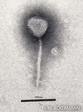
In the early 1950s, scientists noticed something odd. A phage grown in one strain of the bacterium E. coli grew very badly when it was moved into a different strain. It was restricted. But the few phages that did manage to grow in the new strain were suddenly fine there, and now grew badly in their old one. It was as if each strain of bacteria could tell “our” phages from “foreign” ones.
What they did, step by step
Werner Arber: the bacteria’s guard
Arber, at the University of Geneva in Switzerland, wanted to know how this worked.
- 1960–1962, the foreign DNA is cut. With his student Daisy Dussoix, Arber showed that when a restricted phage injects its DNA, the bacterium quickly breaks the DNA up. The phage dies because its instructions are destroyed.
- Two enzymes, one word. Arber proposed that a bacterium has two enzymes that recognize the same short sequence of letters in DNA. One, a restriction enzyme, cuts any DNA that contains the word. The other, a modification enzyme, protects the bacterium’s own DNA by putting a small chemical tag on the same word.
- 1963 onwards, the tag is found. Arber and others showed that the tag is a methyl group (one carbon and three hydrogen atoms) added to one of the bases. DNA with the tag is left alone; DNA without it is cut.
So the bacterium tags every copy of the word in its own DNA, and cuts up any DNA that comes in without the tags. A phage that happened to get tagged before the cutting enzyme struck was “passed” as one of the family, which explained the old puzzle.
In 1968 the first two such enzymes were purified, from E. coli. But they were disappointing as tools. They did recognize a particular word, but then cut the DNA at a random place far away from it. The scissors existed; they were just not yet precise.
Why doesn’t a bacterium’s restriction enzyme cut up the bacterium’s own DNA?
Pick an answer to see why.
Hamilton Smith: a cut in exactly the right place
Smith, at Johns Hopkins University in Baltimore, USA, found precise scissors by accident.
- Spring 1968, a missing virus. With a young student, Kent Wilcox, Smith was studying how the bacterium Haemophilus influenzae takes up DNA. In one experiment they fed it DNA from a phage, and the DNA disappeared. Smith had just read about the new enzymes from E. coli, and he guessed that the bacterium was restricting it.
- The next day, two viscometers. A long DNA molecule makes water thick and slow to flow, like syrup; cut DNA flows easily. A viscometer measures how thick a liquid is. Smith and Wilcox set up two: one with the bacterium’s own DNA, one with phage DNA, and added a juice made of broken bacteria to each. The bacterium’s own DNA stayed thick. The phage DNA got thinner and thinner. Something was cutting foreign DNA only.
- 1968–1970, the enzyme and its word. They purified the enzyme. Then Smith and Thomas Kelly worked out the letters at the ends of the pieces. Every piece began and ended the same way. The enzyme cut wherever it found one six-letter word, for example GTTAAC, and it cut right in the middle of it. They published in 1970.
This was the first restriction enzyme known to cut at a fixed place. Look at the word on both strands of DNA: read from left to right on the top strand it is GTTAAC; on the bottom strand, read the other way, it is also GTTAAC. It reads the same from both directions, a bit like the word “level”. Most restriction enzymes recognize words like this.
Daniel Nathans: the first map made with scissors
Nathans, in the same department as Smith, was in Israel in spring 1969 when a letter from Smith arrived about the new enzyme. He saw at once what it could do.
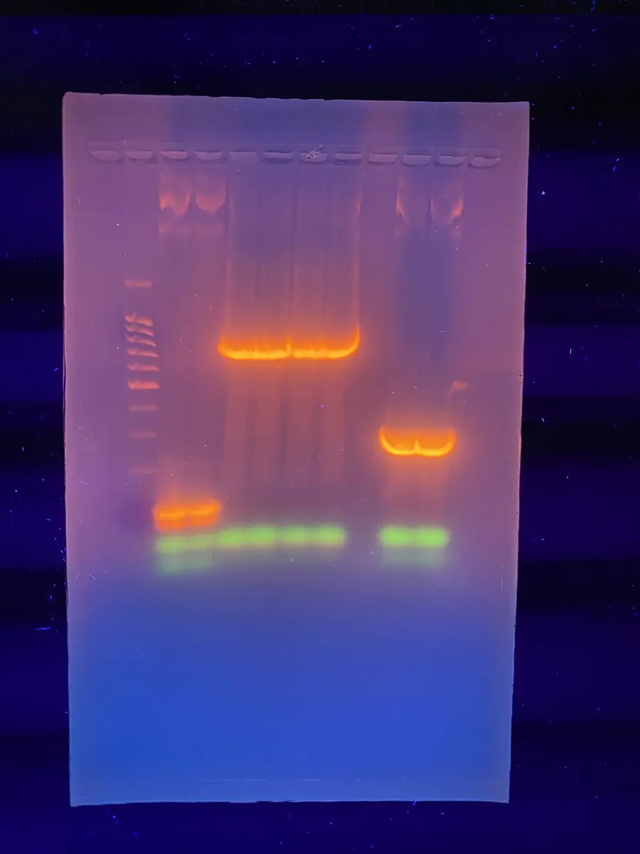
- 1969–1971, cutting a virus into pieces. Nathans chose a small virus of monkeys, SV40, whose DNA is a closed ring of about 5,200 base pairs. With his student Kathleen Danna he cut it with Smith’s enzyme and sorted the pieces by size in a gel, a slab of jelly through which an electric current pulls the DNA. Small pieces slide through the jelly faster than big ones. The ring always fell into the same 11 pieces.
- 1971, a prediction. In the same paper Nathans described what else the scissors could be used for, and he predicted much of what came later.
- 1973, the map. With two more restriction enzymes, each cutting at different words, the team found how the pieces overlapped and put them in order around the ring. It was the first map of a virus’s DNA made by chemical methods. They could then show which piece held which gene.
Why it was hard
Arber’s work looked like an obscure detail of bacteria. Why a phage grows in one strain and not another seemed to matter to nobody but phage specialists. He began it as a side question in a project on radiation damage, and later wrote that if he had known it would take him so many years, he might not have felt he could justify it.
The chemistry was hard too. In the 1960s there was no machine to read DNA letters. To find the word their enzyme cut, Smith and Kelly labeled the ends of the pieces with radioactive phosphorus and broke them down letter by letter, identifying one, then two, then three letters at a time, over many months. Others had tried similar experiments before Smith and failed.
How it works now
Today scientists know thousands of restriction enzymes, each from a different bacterium, and many can be ordered from a catalog like any chemical. Each is named after the bacterium it comes from: EcoRI from E. coli, HindIII from Haemophilus influenzae. (Smith and Nathans suggested these names in 1973.) Each recognizes its own word, usually 4 to 8 letters long.
With them, DNA can be:
- cut into the same pieces every time, so that a gene can be found, studied and copied;
- compared: DNA from two people, cut with the same enzyme, gives pieces of slightly different sizes where their letters differ. This was the first method of DNA fingerprinting, used from the 1980s to identify people in crime cases and to test who a child’s parents are Grade 10: DNA testing in investigations;
- joined: an enzyme like EcoRI leaves sticky ends. Cut a human gene and a small ring of bacterial DNA with the same enzyme, mix them, and their sticky ends pair up. Another enzyme, a ligase, seals the joins. In 1973 Stanley Cohen and Herbert Boyer did this and put the new DNA into bacteria, which copied it every time they divided. This was the start of genetic engineering.
Why must the plasmid and the human DNA be cut with the same restriction enzyme?
Pick an answer to see why.
In your life today
- Insulin. People with type 1 diabetes need insulin every day. It used to be extracted from the pancreases of cows and pigs. In 1978 scientists put the gene for human insulin into bacteria, and in 1982 insulin made this way became the first medicine from genetic engineering to be sold (insulin from bacteria). Most insulin used today, in Nepal too, is made by genetically engineered microbes.
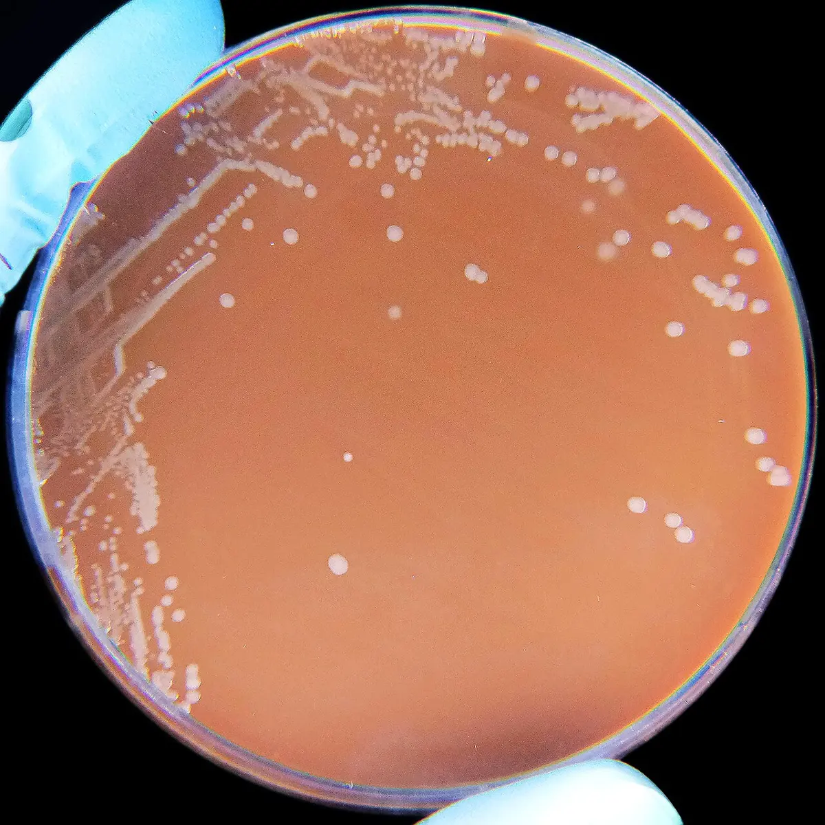
- Vaccines for babies. Every baby in Nepal is offered the pentavalent vaccine, which protects against five diseases. Its hepatitis B part is a protein made by genetically engineered yeast. Its Hib part protects against Haemophilus influenzae type b, another type of the very bacterium in which Smith found his scissors.
- Crops and medicines. Bt cotton, many cancer medicines and tests for diseases all rest on the ability to cut and join DNA.
What came next
- Reading every letter. Restriction enzymes made it possible to cut DNA into pieces small enough to read letter by letter. In 1995 Hamilton Smith was part of the team that read every letter of a bacterium’s DNA for the first time: that of Haemophilus influenzae, the bacterium of his enzyme.
- New scissors. In 2012 scientists learned to program another bacterial weapon against viruses, CRISPR, to cut any DNA sequence they choose (the 2020 prize). It is now used for gene editing.
- An endless war. Phages and bacteria are still at war: bacteria evolve new guards, phages evolve ways past them. Doctors are now testing whether phages could be used to kill bacteria that antibiotics no longer can. Whether this “phage therapy” will work widely is an open question.
In your science books
- Grade 8: viruses and bacteria.
- Grade 10: DNA, genetic technology and DNA testing in investigations.
- Going further: changing genes directly, in “How did farmers make our crops?”, and one language for all life, in “How does DNA show that all life is related?”
Learn more
- The Nobel Prize’s press release and presentation speech.
- The laureates’ Nobel lectures: Arber, Nathans and Smith.
- Werner Arber’s own story, which ends with Silvia’s tale.
The official reason
The prize was given “for the discovery of restriction enzymes and their application to problems of molecular genetics.”