
The CT scanner: X-rays that see in slices
Nobel Prize in Physiology or Medicine, 1979: Allan Cormack and Godfrey Hounsfield
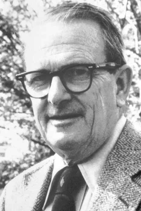
Allan Cormack
1924–1998, born in Johannesburg, South Africa
A nuclear physicist who worked out the mathematics of CT in his spare time. When he published it, almost nobody wrote back.
Photo: Nobel Foundation, Public domain
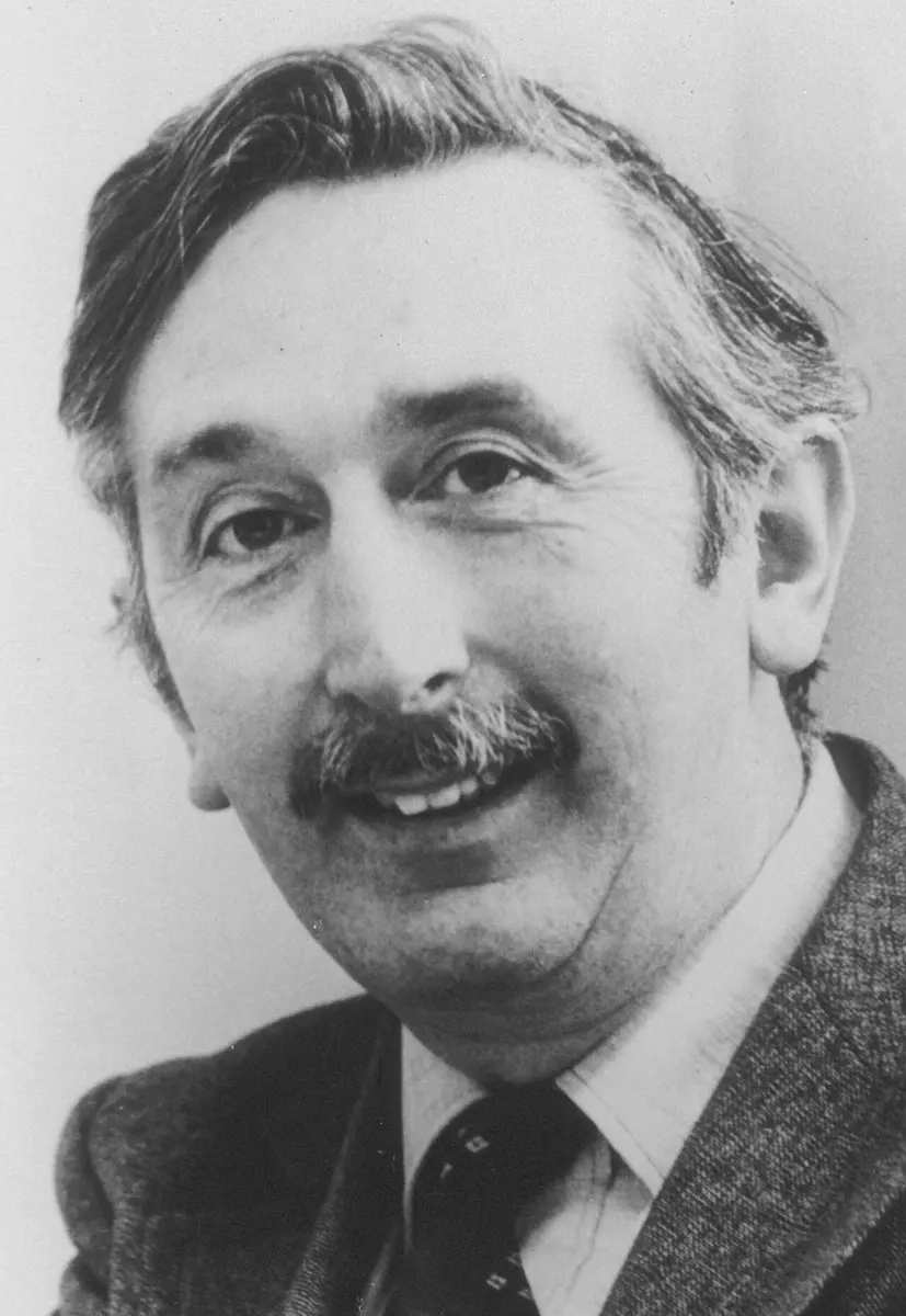
Godfrey Hounsfield
1919–2004, born in Nottinghamshire, England
A farm boy who taught himself by building gadgets, and an engineer without a university degree. He built the first CT scanner.
Photo: Unknown author, Public domain
After a bad fall or a road accident, the doctor may send a patient for a CT scan. The patient lies on a table that slides into a large ring, the machine hums for a few seconds, and soon the doctor is looking at a picture of the inside of the head, as if the skull had been cut open into thin slices. Nothing has been cut. The picture was built by a computer from X-rays.
Neither of the two people who made this possible was a doctor. One was a physicist in South Africa who did the mathematics with a desk calculator; the other an engineer at a record company in England. In 1979 they shared the Nobel Prize in Physiology or Medicine.
The problem: shadows on top of shadows
You know ordinary X-ray pictures Grade 9: X-ray imaging. X-rays pass easily through soft parts of the body but are stopped more by bone, so bones throw a pale “shadow” on the film. That is why an X-ray shows a broken bone so clearly.
But an X-ray picture has two big weaknesses.
- Everything is squashed flat. The X-rays pass right through the body, and every part they cross adds to the same spot on the film. Things in front of and behind each other overlap, like the shadows of two hands held one behind the other. A doctor sees a shadow play, as the Nobel presentation speech put it, “with far too many actors on the stage”.
- Soft parts all look the same. The brain, the liver, a pool of blood or a tumor stop X-rays almost equally. On film they are one gray fog. In an ordinary X-ray of the head you see the skull, but the brain inside is invisible.
So before the 1970s, to look inside the head, doctors had to inject dye into the blood vessels or even pump air into the spaces of the brain. These tests were painful and not free of danger.
How it works: adding up along a line
Here is the key idea. Send a thin beam of X-rays through the body and measure how much comes out the other side. The beam has been weakened by everything along its path. So one measurement tells you only a total: how much the beam was weakened, added up along that line.
Now imagine the slice of the body divided into a grid of tiny squares, and that each square weakens the beam by some amount. One beam gives you the total for one line of squares. A beam at a different angle gives the total for a different set of squares. With enough beams, from enough directions, you can work out the number in every single square. Those numbers, shown as shades of gray, are the picture.
This is called tomography (from Greek: tomos, a slice, and graph, to write): a picture of one slice. Because a computer does the working out, it is computed tomography, or CT.
Look at Figure 2. With only the row and column totals, why can’t you tell which set of numbers is right?
Pick an answer to see why.
A thin X-ray beam passes through a head and comes out weakened. What does that one measurement tell you?
Pick an answer to see why.
Why it was hard
Two things stood in the way.
- The mathematics. Could you really get the numbers in every square back from a set of totals? And how many directions would you need? An Austrian mathematician, Johann Radon, had answered this in 1917 for perfect, smooth shapes, but nobody in medicine knew about his work, and real measurements always have small errors.
- The arithmetic. A useful picture needs many thousands of squares, so the computer must solve a puzzle with many thousands of unknowns. In the 1960s computers filled whole rooms and were slow. Doing this for every patient seemed impossible.
What they did, step by step
The two laureates never worked together. Each solved the problem alone, without knowing about the other.
Cormack: the mathematics, with a desk calculator
- 1956, a hospital in Cape Town. Allan Cormack was a young lecturer in physics at the University of Cape Town. The physicist at the city’s big hospital, Groote Schuur, resigned, and South African law said a qualified physicist had to supervise the hospital’s radioactive materials. Cormack was the only nuclear physicist in Cape Town, so he spent a day and a half a week there.
- A problem nobody had solved. He watched doctors plan radiotherapy, treating cancer with beams of radiation. Their charts treated the body as if it were the same all the way through, but it isn’t: bone, lung and muscle weaken rays differently. To plan properly, you needed to know how strongly each part of the body weakens rays, measured from outside. He looked for the method in the scientific papers and found that nobody had worked it out, so he did it himself.
- 1963, a model “head”. Cormack had moved to Tufts University near Boston, in the USA, working on CT only in his spare time. He built a test object: a ring of aluminium for the “skull”, plastic inside it for the “brain”, and two small aluminium discs for “tumors”. He sent a thin beam of gamma rays (rays like X-rays) from radioactive cobalt through it, at many positions and angles. From those totals his mathematics worked out the inside correctly.
- 1963 and 1964, two papers, and silence. He published his results. There was almost no reply. The most interesting letter, he said later, came from a Swiss center for avalanche research, wondering if it would work on snow lying on mountains.
Hounsfield: the machine
- 1967, a walk in the country. Godfrey Hounsfield worked at EMI (Electric and Musical Industries) near London, the company that made records and also electronics. He had built Britain’s first computer made entirely with transistors. Now he was free to choose a new project. On a country walk he wondered: could you find out what is inside a closed box from many readings taken through it from all directions?
- Nine days for one picture. His first test machine was “very much improvised”, he said: the parts of an old lathe moved a radioactive source and a detector across the object, and turned it 1 degree at the end of each pass. It made 28,000 measurements and took nine days to scan the object. A large computer then needed about two and a half hours to make the picture.
- Faster, with an X-ray tube. With an X-ray tube in place of the weak radioactive source, a scan took nine hours. He scanned a preserved human brain, then fresh brains from cattle, which he carried across London on public transport. The pictures showed the brain’s gray and white parts, which no X-ray film had ever shown.
- 1 October 1971, the first patient. The first scanner for people was put in Atkinson Morley’s Hospital in London, with a doctor there, James Ambrose. The first patient was a woman thought to have something wrong in her brain. The picture clearly showed a round, fluid-filled lump, a cyst, at the front of her brain. “It excited everyone in the hospital who knew about this project,” Hounsfield remembered.
- April 1972, the world sees it. When Ambrose showed their pictures at a meeting of the British Institute of Radiology, the audience was stunned. Within a few years hospitals around the world were buying scanners, and by the middle of the 1970s there were scanners for the whole body.
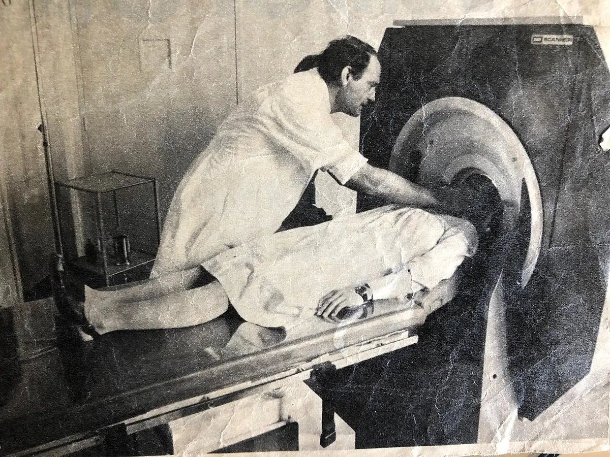
How a CT scanner works now
A modern scanner is a large ring, the gantry, with a hole for the table. Inside the ring, an X-ray tube and a curved row of hundreds of detectors face each other across the patient. They spin together around the body, two or three times a second, while the X-ray tube sends out a thin, fan-shaped beam. The detectors measure how strong the beam is after crossing the body, from every angle.
The computer turns these measurements into a picture of the slice, in gray tones: dense bone white, air black, and soft parts in many shades of gray between. The table slides slowly through the ring, so slice after slice is scanned. A whole body can be scanned in a few seconds, and the slices can be stacked into a 3D picture that a doctor can turn round on the screen.
The numbers in the squares are measured on a scale named after Hounsfield. Water is 0 and air is −1,000; fat is below zero, and bone is several hundred to more than a thousand. Because a CT scanner is far more sensitive than film, it can tell apart soft parts that differ only a little: fresh blood is a little denser than brain, so bleeding inside the head shows up as a pale patch.
In a CT scan of the head, which shows up whitest?
Pick an answer to see why.
In your life today
- Head injuries. After an accident, a CT scan shows within minutes whether there is bleeding inside the skull, and where. Big hospitals in Nepal have CT scanners, and road accidents are one of the main reasons they are used.
- Strokes. A stroke happens when part of the brain loses its blood supply, either because a blood vessel is blocked or because one bursts. The two look alike from outside, but they need opposite treatments. A CT scan shows which it is.
- Cancer. CT finds tumors, shows how big they are and lets doctors aim radiotherapy at them precisely, the very problem that first made Cormack think.
- Beyond medicine. CT is used to look inside fossils still in their rock, Egyptian mummies, luggage at airports and engine parts, without opening anything.
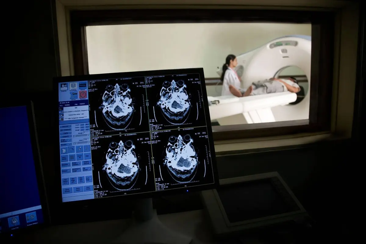
What came next
- Faster and finer. The first scanner took several minutes for each slice. Scanners now spin continuously while the table moves, taking hundreds of thin slices in a few seconds, fast enough to catch the beating heart.
- Other ways to make slices. The same mathematics, adding up along lines from many directions, is used in MRI, which uses magnets and radio waves instead of X-rays (the 2003 prize), in PET scans, and in electron microscopes that show the shapes of molecules (the 2017 Chemistry prize).
- Open question: less radiation. Engineers are building scanners with detectors that count each X-ray particle one by one, and computer programs that make clear pictures from fewer measurements. How low can the dose go before the picture is no longer good enough to trust?
In your science books
- Grade 9: X-rays, X-ray imaging and the CT scan, and the electromagnetic spectrum.
- How X-rays were found: the first Nobel Prize in Physics.
Learn more
- The Nobel Prize’s speed read, press release and the story of the first scanner, With a little help from my friends.
- The laureates’ Nobel lectures: Cormack and Hounsfield.
The official reason
The prize was given “for the development of computer assisted tomography.”