
Two brain halves, and how we see
Nobel Prize in Physiology or Medicine, 1981: Roger W. Sperry, David H. Hubel, and Torsten N. Wiesel
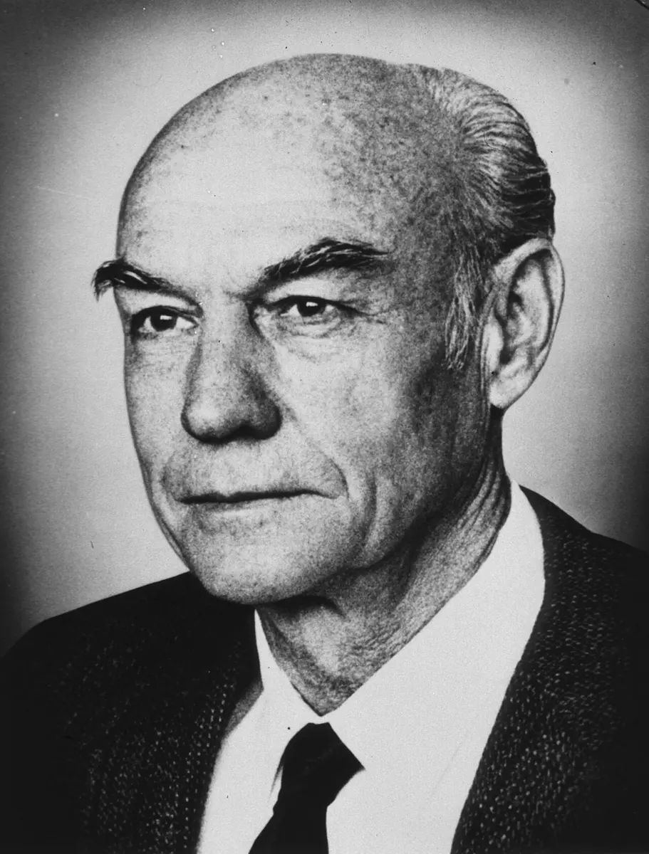
Roger W. Sperry
1913–1994, born in the United States
He studied English at college before switching to psychology and then to biology. Away from the laboratory he made sculptures and collected fossils.
Photo: Unknown author, Public domain
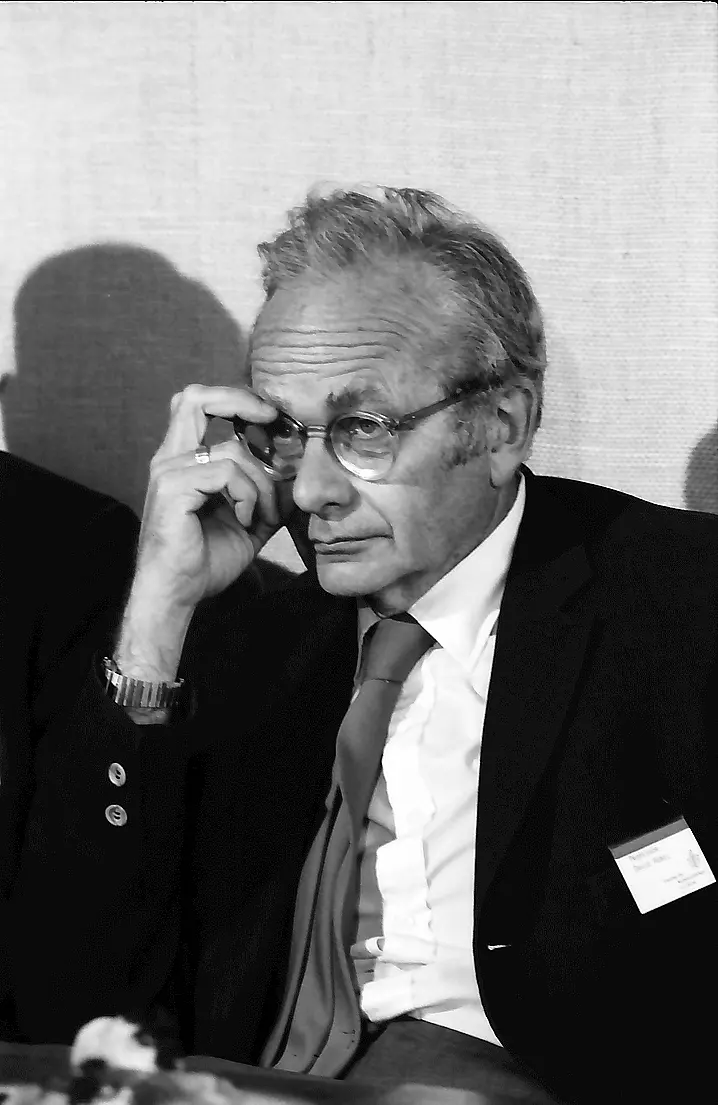
David H. Hubel
1926–2013, born in Canada
As a boy in Montreal he set off a homemade cannon and sent a hydrogen balloon all the way to the city of Sherbrooke. He played the piano and the flute.
Photo: Willy Pragher, CC BY 4.0
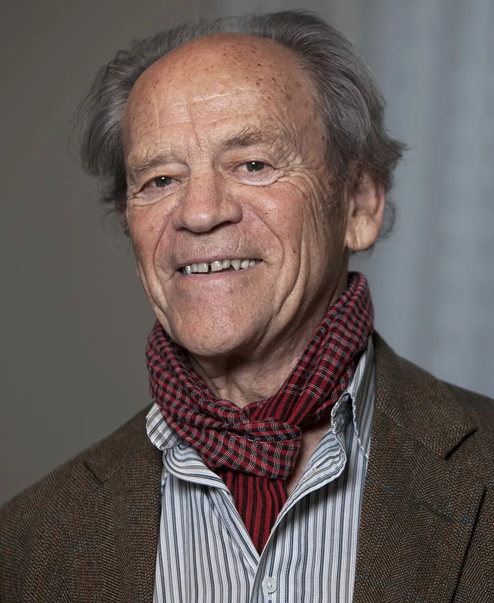
Torsten N. Wiesel
born 1924 in Sweden
The son of a psychiatrist, he grew up in a mental hospital where his father worked. He later led a university and worked to help scientists who were imprisoned for their views.
Photo: Suawikicommons, CC BY-SA 4.0
Look at this word: NEPAL. It took you less than a second to read it. But your eyes didn’t send your brain a word. They sent millions of tiny signals, one from each light-sensitive cell at the back of your eye. Somewhere inside your head, those signals became edges, then letters, then a word you know.
How does the brain do that? And why is it built in two halves? The 1981 Nobel Prize in Physiology or Medicine was shared between two discoveries about the brain. Half went to Roger Sperry, who found what each half of the brain is especially good at. The other half went to David Hubel and Torsten Wiesel, who found how the brain builds a picture from what the eyes see.
The problem: a brain nobody could look inside
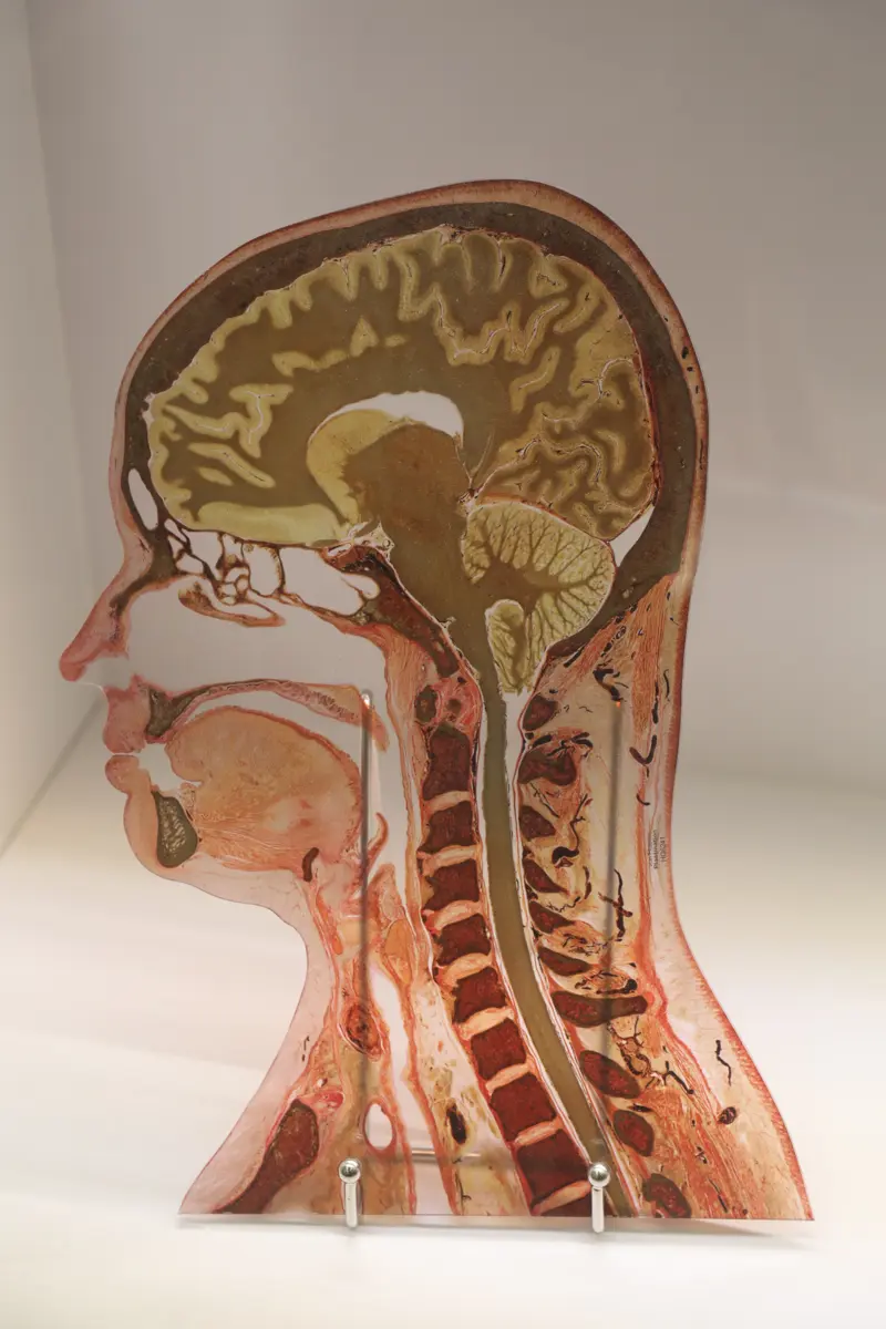
The biggest part of your brain, the cerebrum, is split by a deep groove into a left and a right half, called hemispheres Grade 9: cerebrum. The two halves look almost the same. They are joined by a thick bridge of about 200 million nerve fibers, the corpus callosum, so each half always knows what the other is doing.
By the 1950s, doctors knew two things. Each half controls the opposite side of the body: the left half moves your right hand. And in most people, speech comes from the left half. Doctors had learned this from people whose brains were damaged by a stroke or an injury. But what the right half did was a mystery. It couldn’t speak, so it couldn’t tell anyone.
Seeing was a mystery too. Scientists knew that signals from the eyes travel along the optic nerves to the visual cortex, at the back of the brain. Many imagined the cortex as a kind of cinema screen, with the picture from the eye simply projected onto it. But a picture on a screen still needs someone to look at it. What were the brain cells there actually doing?
What they did, step by step
Sperry: two minds in one head
Roger Sperry spent his life asking how the brain is wired.
- 1940s, eyes turned upside down. Sperry cut the optic nerve of newts and frogs and turned the eye upside down. The nerve grew back, but the animals now saw the world upside down: they struck downwards at a fly held above them, and no training ever cured it. Each nerve fiber had grown back to its old place in the brain. Sperry suggested that every nerve cell carries a kind of chemical label that guides it to the right partner. Much later, such guiding chemicals were found.
- 1950s, split-brain cats and monkeys. With his student Ronald Myers, Sperry cut the corpus callosum of cats, and later monkeys. They seemed completely normal. But when one half of the brain was trained to do a task, the other half had no idea how to do it. Each half was learning and remembering on its own.
- 1960s, the patients. Some people suffer from very severe epilepsy, in which storms of electrical activity spread through the brain and cause fits. In 1962, surgeons in Los Angeles began cutting the corpus callosum of a few such patients as a last resort, to stop the storms spreading from one half to the other. It worked: the fits became fewer. And the patients seemed just as before, in how they talked, behaved and felt.
- The clever tests. Sperry, with his student Michael Gazzaniga, found ways to talk to each half separately. A patient looked at a dot in the middle of a screen, and a word was flashed for a split second to one side of it. Anything to the left of where you are looking goes to the right half of your brain, and anything to the right goes to the left half.
The results were startling. When a word was flashed on the right side, the patient read it out easily. When it was flashed on the left, the patient said they had seen nothing. Yet if asked to feel among some hidden objects with the left hand, they picked out the very object the word named. The right half had read the word, and it knew the answer, but it couldn’t speak. The speaking left half truly hadn’t seen it.
From many such tests, Sperry showed that both halves think, each with its own memories and its own ways of working. The left half is better at speech, writing and sums. The right half is better at shapes and space: one right-handed patient could hardly copy a drawing of a cube with his right hand, but managed it quite well with his left. The right half is also better at recognizing faces and following a tune.
A split-brain patient sees a picture of an apple only on the left side of the screen. What happens?
Pick an answer to see why.
Hubel and Wiesel: listening to one cell at a time
David Hubel was born in Canada and trained as a doctor. In the army in the 1950s he made a new kind of microelectrode, a wire of tungsten so fine that its tip could pick up the tiny electrical pulses of a single nerve cell Grade 9: neuron. Torsten Wiesel, a Swedish doctor, had come to the United States in 1955. In 1958 they met in Stephen Kuffler’s laboratory at Johns Hopkins University in Baltimore, and began working together. They kept at it for more than twenty years.
- 1958, the method. In an anesthetized cat, they placed the electrode next to one cell in the visual cortex. The cat’s eyes faced a screen, and they shone patterns of light on it. Each time the cell fired, a loudspeaker gave a click, so they could hear the cell answer.
- Spots did nothing. Cells in the eye had been found to answer best to small spots of light. So Hubel and Wiesel tried spots, light ones and dark ones, for weeks. The cortex cells hardly answered.
- The lucky edge. One day they held a cell for nine hours. After three or four hours of nothing, they were sliding a glass slide with a black spot on it into their projector. Suddenly the loudspeaker crackled “like a machine gun”. It wasn’t the spot. The edge of the glass slide was throwing a faint, straight shadow line across the screen. The cell fired for that line, but only when it lay at one particular angle.
- 1959–1960s, a map of the cortex. Cell after cell, they found the same. Each cell watches one small patch of what the eye sees, and fires for a line or edge at its own favorite angle. Some fire for a line in one exact place; others fire for a line at their angle anywhere in a small patch, or only when it moves. Cells with the same favorite angle are stacked in columns, and the angle shifts a little from one column to the next. Other columns take turns between the left eye and the right eye.
What did Hubel and Wiesel find that a typical cell in the visual cortex answers best to?
Pick an answer to see why.
Seeing has to be learned in time
Next, Hubel and Wiesel asked whether these cells are ready at birth. They found that kittens are born with the wiring mostly in place. But it needs use to stay that way.
- 1963, one eye closed. When one eye of a kitten was kept closed during its first three months, the kitten was blind in that eye for good, even though the eye itself was perfectly healthy. The cells in its cortex had all been taken over by the open eye. Closing an adult cat’s eye for even longer did no harm.
- The critical period. There is a time early in life, the critical period, when the brain’s seeing cells are shaped by what the eyes see. The eyes must send sharp, patterned pictures, not just light. A blurred eye in that time can leave the brain unable to use it later. (Today, rules on experiments with animals are much stricter, and scientists must show that a question can’t be answered any other way.)
How the brain builds a picture
Hubel and Wiesel’s discoveries showed that seeing is built up in steps. The committee compared it with reading: some cells read the letters, and pass them on to cells that put letters into syllables, then others into words and sentences.
- In the eye, each cell answers to a spot of light in one tiny place.
- In the visual cortex, a cell that collects from a row of spot-cells answers to a line at one angle.
- Cells further along collect from many line-cells, and answer to corners, curves and shapes.
- Further still, in other parts of the brain, are cells that answer to whole objects, and even to faces.
In your life today
- Lazy eye. If one eye is blurred or turned (a squint) in early childhood, the brain can learn to ignore it. This is called amblyopia, or lazy eye. The treatment comes straight from this discovery: glasses to make the picture sharp, and a patch over the good eye for a few hours a day, so that the brain has to use the weak one. It works best when started young, while the brain can still change. That is why children’s eyes should be checked early, before school.
- Cataracts in babies. A few babies are born with a cataract, a cloudy lens Grade 10: cataract. Because of the critical period, it is now operated on in the first months of life, not years later. Nepal’s eye hospitals carry out a great many cataract operations, and children’s eye camps look for such problems early.
- Computers that see. In 1980, the Japanese engineer Kunihiko Fukushima built a computer program in layers, copied from Hubel and Wiesel’s cells: first lines, then shapes. Later programs built the same way are what recognize faces in your phone’s camera (see the Physics prize of 2024).
- Epilepsy. Most epilepsy today is controlled by medicines. Cutting the corpus callosum is still done, rarely, for some severe cases.
What came next
- More maps. Scientists have since found more than thirty areas of the brain that deal with seeing, some for color, some for movement, some for faces.
- Proof, sixty years on. Hubel and Wiesel guessed that a line-cell gets its angle by collecting from a row of spot-cells, as in Figure 4. In 2026 a team in Munich, Germany, watched single connections between cells in the brains of living mice and found that it does happen in the cortex itself, much as they had guessed.
- An open question. We know cells that answer to lines, and cells that answer to faces. But how does the brain put the color, the shape and the movement of a bus together, so that you see one red bus coming towards you, and not a jumble? And in a split-brain patient, are there two people in one head? Nobody yet knows for sure.
In your science books
- Grade 9: the human nervous system, the cerebrum and its two hemispheres, and the neuron.
- Grade 10: the human eye, defects of vision and cataract.
- Going further: messages along nerves.
Learn more
- The Nobel Prize’s summary of the 1981 prize, with the press release and the speech at the ceremony (nobelprize.org).
- The Nobel lectures: Sperry, Hubel and Wiesel.
The official reason
The prize was given “for his discoveries concerning the functional specialization of the cerebral hemispheres.”
The prize was given “for their discoveries concerning information processing in the visual system.”