
Microscopes that see past the limit of light
Nobel Prize in Chemistry, 2014: Eric Betzig, Stefan W. Hell, and W. E. Moerner

Eric Betzig
born 1960 in Ann Arbor, Michigan, USA
He left science, worked at his father’s machine-tool company, and came back to build his first super-sharp microscope in a friend’s living room.
Photo: Ecole polytechnique Université Paris-Saclay, CC BY-SA 2.0

Stefan W. Hell
born 1962 in Arad, Romania
His family moved to West Germany when he was fifteen. Senior scientists doubted his idea, so he went to Finland to work on it.
Photo: Bernd Schuller, Max-Planck-Institut für biophysikalische Chemie, CC BY-SA 3.0

W. E. Moerner
born 1953 in Pleasanton, California, USA
Always called by his initials, W. E., he was the first person to detect a single molecule by its light.
Photo: Kevin Lowder, CC BY-SA 3.0
In your school laboratory, a light microscope can show you the cells in a thin slice of onion, or the tiny creatures in a drop of pond water. Make the magnification bigger and bigger, and you might expect to see smaller and smaller things for ever. But you can’t. Below a certain size, everything turns to blur.
For more than a hundred years, scientists believed this blur was a law of nature that no light microscope could ever beat. The 2014 Nobel Prize in Chemistry went to three people who found ways round it, using molecules that glow. Now scientists can watch single molecules moving inside living cells.
The problem: the blur that would not go away
Abbe’s limit
In 1873 the German scientist Ernst Abbe, who designed microscopes, worked out why. Light is a wave Grade 9: Words used for waves, and waves spread out as they pass the edges of a lens. This spreading is called diffraction (Light bends round edges). Because of it, even a perfect microscope shows a tiny point of light not as a point but as a small blurred spot.
The size of the blur depends on the wavelength of the light. For visible light, it is about half the wavelength: about 0.2 micrometers (µm). A micrometer is a thousandth of a millimeter, so 0.2 µm is a fifth of a thousandth of a millimeter. Two things closer together than that blur into one, however much you magnify them. This is Abbe’s diffraction limit.
Too big to see the working parts
0.2 µm is small enough to see whole cells, and bigger parts inside them, like the mitochondria that release energy Grade 8: Parts of a cell. But many viruses are smaller than that, and a protein molecule is tens of times smaller. The real work of a cell is done by molecules: proteins meeting, joining and moving. Watching a cell with Abbe’s limit was, as the Nobel committee put it, like seeing the buildings of a city without seeing how the people live.
Electron microscopes can see far smaller things, but the sample has to be dried or frozen and put in a vacuum, so the cell is dead. Biologists wanted to see the molecules in living cells.
Two glowing molecules are 0.05 µm apart. What does an ordinary light microscope show?
Pick an answer to see why.
How it works: molecules that glow
All three laureates used fluorescence. A fluorescent molecule soaks up light of one color and, a moment later, gives out light of another color, usually redder. Highlighter pens look so bright because their ink is fluorescent. Biologists attach fluorescent molecules to the parts of a cell they want to see, such as a particular protein. Then, in a dark microscope, only those parts glow, like lamps in a dark city.
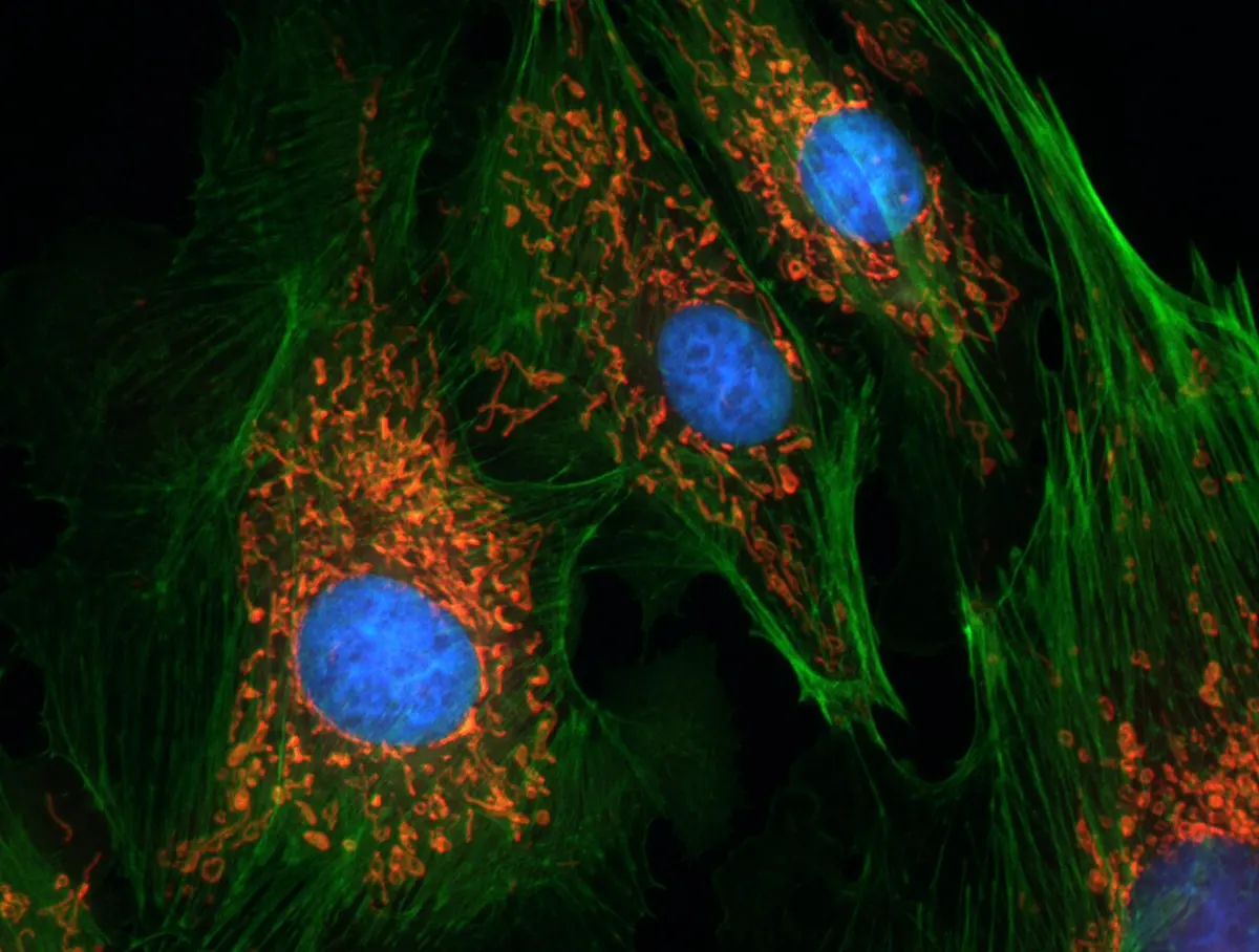
A special fluorescent protein helped a great deal: green fluorescent protein (GFP), from a glowing jellyfish, which won the 2008 Chemistry prize. A cell can be made to build GFP joined to any of its own proteins, so the protein glows wherever it goes.
But fluorescence alone does not beat Abbe’s limit. Two glowing molecules close together still blur into one. The laureates’ trick, in two different ways, was to make not all the molecules glow at the same time.
Why it was hard
- Abbe’s limit is real. It is not a fault of lenses that better lenses could fix. It comes from light being a wave. Most experts thought there was simply no way round it, and young scientists who tried were not taken seriously.
- One molecule gives very little light. To use single molecules, you must first be able to see one at all, against all the light and glow around it. Before 1989 no one had done it.
- Light can harm cells. Shining very bright light on living cells to see more can damage or kill them, and makes fluorescent molecules fade.
What they did, step by step
Hell: switch off the glow around the middle
Stefan Hell finished his PhD at Heidelberg, Germany, in 1990, determined to beat Abbe’s limit. Senior scientists in Germany were doubtful, so he took a job at the University of Turku in Finland.
- 1993, the idea. Leafing through a textbook on light, he read about stimulated emission: a molecule that is ready to glow can be made to give up its energy at once by another beam of light of the right color, so that it doesn’t glow in the usual way. “At that moment, it dawned on me,” he said later.
- 1994, the plan. He published how a microscope could use this. One laser spot makes the molecules glow. A second laser, shaped like a doughnut, switches off the glow everywhere except in the hole in the middle. The hole can be made far smaller than 0.2 µm. Sweep this tiny glowing spot across the sample, point by point, and you build a sharp picture. He called it STED (stimulated emission depletion).
- Göttingen, 2000. At the Max Planck Institute in Göttingen, Germany, he built the microscope. By 2000 he had used it to picture an E. coli bacterium more sharply than any light microscope ever had.
Moerner: seeing one molecule
W. E. Moerner worked at IBM’s research center in San Jose, California.
- 1989, a single molecule. Chemists had only ever measured millions of molecules at once, and seen their average. Moerner became the first to detect the light absorbed by one single molecule, in a crystal cooled with liquid helium.
- 1997, a lamp with a switch. At the University of California in San Diego, Moerner studied a variety of GFP. He found that blue light at 488 nm made it glow, until it faded and went dark. Then violet light at 405 nm brought it back, ready to glow again. Spread thinly in a gel, each molecule was like a tiny lamp that could be switched on and off.
Betzig: a few lamps at a time
Eric Betzig worked at Bell Laboratories in New Jersey, on another way of seeing small things: a light source on an extremely fine tip, held a few nanometers from the sample. A nanometer (nm) is a millionth of a millimeter.
- 1995, an idea, and a goodbye. Betzig decided this method could go no further, and left research. On a winter walk, he thought of another way. If neighboring molecules glowed in different colors, each color could be pictured on its own, with its molecules far enough apart to be seen separately. He published the idea in 1995, then joined his father’s machine-tool company.
- 2005, the missing piece. Years later he read about fluorescent proteins that can be switched on by light, like Moerner’s. They didn’t need different colors: they could glow at different times.
- A living-room laboratory. Betzig and his friend Harald Hess, both without jobs, built the microscope in Hess’s living room in California, each paying US$25,000 towards it. Then they took it to a laboratory near Washington to try it on cells.
- 2006, the result. A weak flash switches on only a few protein molecules, scattered far apart. Each shows as a blur, but the center of a blur can be found very exactly. When those molecules fade, another flash switches on a different few. Thousands of pictures later, all the centers are put together into one sharp picture. Betzig’s team published pictures of the membrane round a tiny part of a cell, the lysosome, far sharper than Abbe’s limit.
How it works now: from microscope to “nanoscope”
Both methods are now used in laboratories all over the world. STED and single-molecule methods can show details of about 20 to 30 nm, ten times smaller than Abbe’s limit, and some newer versions do better still.
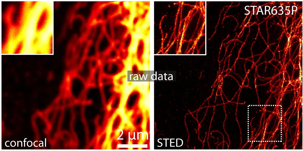
The laureates themselves turned their microscopes on life: Hell looked inside living nerve cells at the synapses, where nerve cells pass signals to each other; Moerner studied a protein linked to Huntington’s disease, a brain disease that runs in families; Betzig followed cells dividing inside developing embryos.
In your life today
- Understanding disease. Super-resolution microscopes show how proteins clump together in diseases such as Parkinson’s, Alzheimer’s and Huntington’s, how viruses enter cells, and how bacteria divide.
- Making medicines. Drug companies and hospitals’ research laboratories use them to see where a new medicine goes inside a cell.
- School microscopes have the same limit. The microscope in your school laboratory, with its glass lenses Grade 10: Lens, can never show you anything smaller than about 0.2 µm, however good it is. Your cheek cells and onion cells are hundreds of times bigger than that.
What do STED microscopes and single-molecule microscopes have in common?
Pick an answer to see why.
What came next
- Even sharper. In 2017 Hell’s team showed a method called MINFLUX, which combines both ideas and can locate molecules to within a few nanometers.
- Gentler, faster. In 2014 Betzig’s team showed the lattice light-sheet microscope, which lights a cell with a very thin sheet of light so it can be filmed for longer without harm.
- Open questions. Can we watch every kind of molecule in a living cell at once, quickly, without the light harming it or the glowing labels changing how the molecules behave? Each method still trades sharpness against speed and gentleness.
In your science books
- Grade 8: the cell and its parts, including mitochondria; bacteria.
- Grade 9: wavelength and other words used for waves, the electromagnetic spectrum and visible light.
- Grade 10: lenses and images formed by lenses.
- Going further: light bends round edges (diffraction) and Einstein’s packets of light.
Learn more
- The Nobel Prize’s own explanation: How the optical microscope became a nanoscope (nobelprize.org).
- The Nobel lectures: Betzig, Hell and Moerner.
The official reason
The prize was given “for the development of super-resolved fluorescence microscopy.”