
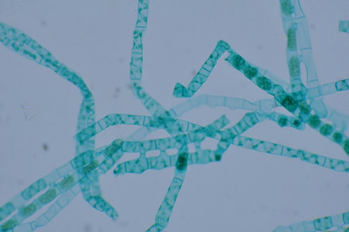
Pull a handful of the slimy green threads from the edge of a pond in the monsoon, the leu that makes the stones slippery, and look at one thread under a microscope. It is a row of glassy cells, each with a bright green ribbon wound round inside it like the stripe on a barber’s pole. That is Spirogyra. In autumn, two of its threads lying side by side may join by little bridges, and the whole of one thread’s living matter crawls across into the other.
Now think of a glass of water from an unsafe tap. It can carry, too small to see, the resting cysts of Entamoeba histolytica, a single-celled parasite that eats its way into the wall of the human gut and causes amoebic dysentery. One alga that feeds a pond, one protozoan that makes people ill: this unit follows both through their whole lives. Then it asks two questions about every living cell. How does water get into it and out of it, and up a tree with no pump (osmosis and transpiration)? And how does a cell get energy out of its food (intracellular respiration)?
2.1 Life cycle of Spirogyra and Entamoeba
A life cycle is the whole series of stages an organism passes through, from one stage back to the same stage in the next generation. Both organisms here are single eukaryotic cells or chains of them, and both can reproduce without a partner. But Spirogyra also reproduces sexually, while Entamoeba never does, and Spirogyra lives freely while Entamoeba can complete its life only inside a human.
Spirogyra
Spirogyra is a green alga of fresh water: ponds, ditches, rice fields, slow streams and water tanks. Its threads float in tangled green masses near the surface, buoyed up by bubbles of the oxygen they make in sunlight, and feel slimy to the touch, so in English it is called pond silk or water silk. There are about 400 species, all built the same way.
Its body. The plant body is an unbranched filament (thread), a single row of cylindrical cells joined end to end. Every cell is the same: there is no root, stem or leaf, no base and no tip, and any cell can divide. So the body is a simple thallus, and each cell feeds, grows and divides by itself. Every cell has:
- a cell wall of two layers: an inner layer of cellulose and an outer layer of pectin, which swells in water into a slimy mucilage sheath. The slime is why the threads feel slippery and why few animals can grip them. The cross walls between cells are called septa (one septum).
- a thin lining of cytoplasm pressed against the wall, round a large central vacuole full of cell sap.
- one or more chloroplasts, the feature that names the genus (speira, a coil): each is a long ribbon with wavy or toothed edges, wound in a spiral (a helix) just inside the wall, from one end of the cell to the other. A cell may have one ribbon or several, according to the species.
- on each ribbon, a row of pyrenoids, small round bodies where the cell gathers its food into starch.
- a single nucleus, hung in the middle of the vacuole by fine strands of cytoplasm that run out to the pyrenoids, like the spokes of a wheel.
All these nuclei are haploid (n): the filament has one set of chromosomes.
Reproduction in Spirogyra
Spirogyra reproduces in two ways: vegetatively, by breaking into pieces, and sexually, by conjugation.
Vegetative reproduction: fragmentation. A filament breaks easily, at a weak septum or where a cell dies, or when it is torn by moving water or animals. Every piece that has even one living cell grows into a new filament, its cells dividing by mitosis across the thread. This is how a pond fills with Spirogyra within weeks in warm, sunny weather.
Sexual reproduction: conjugation. At the end of the growing season, when the water cools or the nutrients run low, Spirogyra reproduces sexually. Two cells join by a tube and the contents of one pass into the other and fuse with them: this is conjugation (Latin conjugare, to yoke together). There are two kinds.
1. Scalariform conjugation (scalaris, of a ladder), between two filaments:
- Two filaments come to lie side by side, close and parallel, held together by their mucilage.
- Opposite cells of the two filaments each push out a small bulge, a papilla, towards the other.
- The papillae meet, push the filaments slightly apart, and the wall between their tips dissolves, making an open conjugation tube. With a tube between each pair of facing cells, the two filaments look like a ladder; the tubes are its rungs.
- In each joined cell, the whole protoplast (the living contents) loses water from its vacuole, shrinks away from the wall and rounds off. Each rounded protoplast is a gamete. Since it has no flagella and can’t swim, it is called an aplanogamete (a-planos, not wandering).
- The gametes of one filament move, by creeping like an amoeba, through the tubes into the cells of the other filament. The moving one is called the male gamete and the one that waits the female, but the two look exactly alike: they differ only in behavior. This is physiological anisogamy: unequal in what they do, equal in form.
- In the receiving cell the two gametes fuse, cytoplasm with cytoplasm and nucleus with nucleus, making a diploid (2n) zygote.
- The zygote secretes a thick wall of three layers and becomes a zygospore, oval and dark. At the end, one filament is a row of empty cells and the other a row of zygospores.
2. Lateral conjugation, between two neighboring cells of the same filament. It happens where one filament is alone. In one kind, a tube forms round the septum between two cells, on one side of the filament, and the contents of one cell pass through it into the other; the filament shows every second cell empty and every other cell with a zygospore. In another kind, a small hole opens in the septum itself and the gamete passes straight through.
The zygospore and its germination. Zygospores sink to the mud when the old filaments die and rot away. Their thick walls carry them through the cold or the dry season, for months if need be. When conditions improve:
- The diploid nucleus divides by meiosis into four haploid nuclei.
- Three of the four degenerate; one survives.
- The zygospore absorbs water and swells, its outer wall layers crack open, and the inner layer grows out as a short germ tube, holding the one haploid nucleus.
- The germ tube divides by mitosis, again and again, into a new filament.
So each zygospore gives just one new filament. The zygote is the only diploid cell in the whole life of Spirogyra; everything else is haploid. A life in which the adult is haploid and meiosis happens in the zygote is called haplontic (zygotic meiosis).
In scalariform conjugation, where are the zygospores formed?
Pick an answer to see why.
Entamoeba histolytica
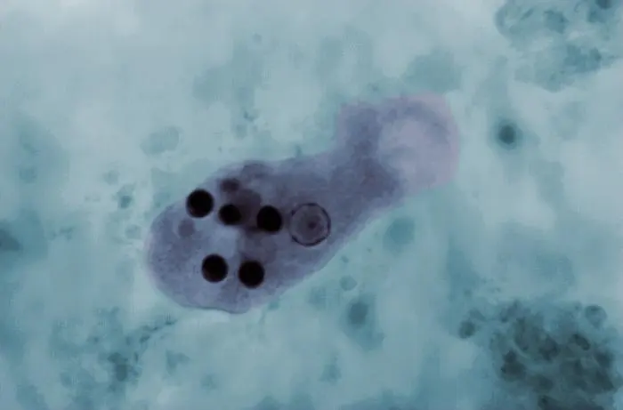
Entamoeba histolytica is a single-celled parasitic protozoan (kingdom Protista), a relative of the free-living Amoeba. It lives in the large intestine of humans, and humans are its only important host: its whole life cycle is completed in one host, with no insect or other animal in between. Its name says what it does: ent- (inside, in the gut), amoeba, and histolytica (histos, tissue; lysis, dissolving), the tissue-dissolver. In some of the people it infects, it eats into the wall of the large intestine and causes amoebic dysentery (amoebiasis): stomach pain and frequent stools with blood and mucus. From the gut it can travel in the blood to the liver and make an amoebic liver abscess, a pocket of dead tissue.
It has two main forms, each with a job:
| Trophozoite | Cyst | |
|---|---|---|
| What it is | the active, feeding, dividing form | the resting, infective form |
| Size | about 15–30 µm (up to 60 µm when it invades) | about 10–15 µm |
| Shape | irregular, changing; moves by one broad pseudopodium at a time | round, with a thick, smooth wall |
| Nuclei | one | one, then two, then four (mature) |
| Inside | food vacuoles, often with red blood cells it has eaten | a glycogen store and rod-shaped chromatoid bodies with rounded ends (food reserves) when young; both mostly used up as it matures |
| Survives outside the body? | no: dies within minutes to hours | yes: weeks in damp, cool places |
Each nucleus is round, with a small dot, the karyosome, at its center, and a fine ring of chromatin under its membrane. Unlike Amoeba, Entamoeba has no contractile vacuole: it lives in the gut, where it doesn’t need to pump out water.
The life cycle of Entamoeba histolytica
- Infection. A person swallows mature cysts, each with four nuclei, in water or food contaminated with human feces: unboiled water, raw vegetables washed in dirty water, food handled with unwashed hands, or food that flies have walked on. A few cysts are enough. Trophozoites, if swallowed, are killed by the stomach’s acid; the cyst’s wall survives it.
- Excystation. In the lower part of the small intestine, where the juices are alkaline, the cyst wall weakens and the four-nucleate amoeba squeezes out through a small opening. Its nuclei divide once more (4 → 8), and its cytoplasm divides round them, so each cyst gives eight small amoebae (metacystic trophozoites).
- Trophozoites in the large intestine. The young amoebae are carried on into the large intestine (the colon). There they grow into trophozoites, live on bacteria and food particles in the gut, and multiply by binary fission, the nucleus dividing by mitosis and the cell pinching in two. In most infected people, that is all they do: they live harmlessly in the gut, and the person has no illness.
- Invasion (in some people). In others, for reasons that depend on both the amoeba’s strain and the person’s gut, the trophozoites attack the gut lining. They kill the cells they touch, digest the tissue with enzymes, and burrow into the wall, making flask-shaped ulcers: a small opening that widens out underneath. They swallow red blood cells from the damaged vessels. This is amoebic dysentery. Some are carried by the blood of the portal vein to the liver, and rarely to the lungs or brain, where they form abscesses. These invading trophozoites can’t go on to the next stage: they are a dead end for the parasite.
- Encystment. As the contents of the gut move along the colon and dry out, many trophozoites in the gut’s cavity stop feeding, empty their food vacuoles and round up, becoming smaller precystic forms. Each secretes a tough wall round itself: it is now a cyst, with one nucleus, a glycogen store and chromatoid bodies.
- Maturing of the cyst. The nucleus divides twice by mitosis: one nucleus, then two, then four. The glycogen and chromatoid bodies are mostly used up. A four-nucleate cyst is mature and infective.
- Passing out. Cysts leave the body in the feces, sometimes in huge numbers. Outside, they can’t multiply, but they stay alive in damp soil, water and food for weeks, waiting to be swallowed by the next person.
Entamoeba: after Mariana Ruiz Villarreal (LadyofHats), Public domain, via Wikimedia Commons
So there is no sexual reproduction in the life of Entamoeba: it multiplies only asexually, by binary fission of the trophozoites and by the division of the cyst’s nuclei (one cyst giving eight amoebae). The trophozoite is the form that does harm; the cyst is the form that spreads.
Prevention. Since the cyst spreads the disease, the way to stop it is to keep human feces away from what people eat and drink:
- Drink boiled, filtered or treated water. Boiling kills cysts; the chlorine in ordinary tap water does not reliably kill them.
- Wash hands with soap after using the toilet and before cooking or eating.
- Use toilets, and keep them away from wells and water sources.
- Wash raw vegetables and fruit in clean water, or peel or cook them; cover food from flies and cockroaches.
- Don’t use untreated human waste (night soil) as manure on vegetables.
- Treat patients and carriers: doctors give medicines such as metronidazole, followed by one that clears the cysts from the gut.
How many trophozoites does one mature cyst of Entamoeba finally give?
Pick an answer to see why.
2.2 Osmosis and transpiration process
Water is about 70 to 90 percent of a living cell, and every cell is always taking water in or losing it. Two processes control this: osmosis, by which water moves into and out of cells, and transpiration, by which a plant loses water from its leaves and so draws more up from the soil.
Osmosis
Put a dried raisin in a cup of water overnight and it swells up plump again. Sprinkle salt on slices of cucumber for achar, and in a few minutes they sit in a puddle of their own water. In both, water has crossed the skins of living cells, in opposite directions. Why?
First, diffusion: the molecules of a liquid or gas are always moving at random, so any substance spreads from where it is more concentrated to where it is less concentrated, until it is evenly spread. A drop of ink in water spreads this way.
Now separate two solutions by a semipermeable membrane: a sheet with pores that let small water molecules through but not larger dissolved molecules such as sugar. Cellophane, the skin inside an egg’s shell and the membrane of every living cell behave like this (a cell membrane is better called selectively permeable, since it also lets some solutes through and controls which). The sugar can’t cross, so only the water can even things out. Water molecules keep crossing the membrane both ways, but the solution with more sugar dissolved in it has a lower concentration of water. More water molecules cross each second from the side with more water (less solute) to the side with less water (more solute) than come back, and the more concentrated solution gains water.
Osmosis is the movement of water (the solvent) through a semipermeable membrane, from a less concentrated solution (or pure water) to a more concentrated solution. In other words, water moves from where there is more water to where there is less.
Notice the two differences from ordinary diffusion: osmosis needs a membrane, and it is the water, not the solute, that moves.
The solution outside a cell is described by comparing it with the cell sap inside:
- A hypotonic solution is less concentrated than the cell sap (pure water is the extreme). Water enters the cell: endosmosis.
- An isotonic solution has the same concentration as the cell sap. Water enters and leaves equally, so there is no net movement.
- A hypertonic solution is more concentrated than the cell sap. Water leaves the cell: exosmosis.
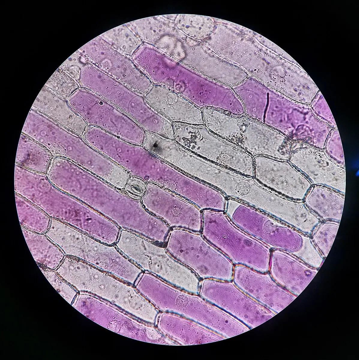
In a plant cell, water entering by endosmosis fills the vacuole and presses the cytoplasm hard against the cell wall, which, being strong, presses back, like a football’s leather round its bladder. The cell is firm, turgid; the pressure is the turgor pressure. In a hypertonic solution the cell loses water from its vacuole, which shrinks; the cytoplasm pulls away from the wall, first at the corners and then all round, and the cell is plasmolyzed. This shrinking of the protoplast away from the wall is plasmolysis. Put a plasmolyzed cell back into water soon enough and it swells back: deplasmolysis. Left too long, it dies.
In an animal cell, with no wall, there is nothing to stop it. A red blood cell in pure water swells until it bursts (hemolysis); in strong salt water it shrinks and wrinkles (crenation). That is why the blood plasma is kept isotonic with the cells, and why a drip given in a hospital is normal saline, 0.9% sodium chloride, isotonic with blood, never plain water.
A plant cell is placed in a solution more concentrated than its cell sap. What happens?
Pick an answer to see why.
Demonstration: the potato osmometer
An osmometer shows osmosis happening and measures it. A potato’s living cells make a good one.
Potato: ZooFari, Wikimedia Commons, public domain
How much water moves?
How fast and how much water moves depends on the difference in concentration across the membrane. A neat way to measure it is to weigh pieces of potato before and after they soak in solutions of different strengths. The percentage change in mass is
A gain means water went in (the solution was hypotonic to the potato’s cells); a loss means water came out (hypertonic). Where the line crosses zero, the solution is isotonic with the cell sap.
We use percentage change, not the change in grams, because the strips don’t start with exactly the same mass: a percentage compares them fairly.
Importance of osmosis
In plants:
- Absorption of water. The root hairs’ cell sap is more concentrated than the soil water, so water enters them by osmosis, and passes from cell to cell across the root to the xylem.
- Turgidity and support. Turgid cells keep soft stems, leaves and flowers firm and spread out; a herb such as spinach or mustard has little wood to hold it up. When a plant loses more water than it takes in, its cells lose turgor and it wilts.
- Opening and closing of stomata. The two guard cells of a stoma open it when they take in water by osmosis and swell, and close it when they lose water.
- Movements such as the drooping of a touch-me-not (lajjawati, Mimosa) leaf when it is touched: cells at the base of each leaf stalk lose water suddenly.
In animals, water is absorbed from the large intestine into the blood, and taken back from the kidney’s tubules, by osmosis, and the body’s fluids are kept isotonic with its cells.
In daily life:
- Salt in achar and salted fish, and sugar in jam and murabba, draw water out of any bacteria or molds that land on them by osmosis, so they can’t grow: the food keeps.
- Too much fertilizer near a plant’s roots makes the soil water hypertonic: water leaves the roots and the plant wilts (“fertilizer burn”). Salt sprinkled on weeds kills them the same way.
- Sea water can’t quench thirst: it is more concentrated than our body fluids, so drinking it draws water out of the cells.
Transpiration
A sunflower or a maize plant takes in a great deal of water through its roots, but uses only a small part of it: about 97% of the water a plant absorbs is lost into the air as vapor, mostly through its leaves. Transpiration is the loss of water as water vapor from the aerial parts of a living plant, mainly through the stomata of its leaves.
A leaf is built to let gases in and out. Inside it, the spongy mesophyll cells are loosely packed, with large air spaces between them, which open to the outside air through the stomata (one stoma), pores each between two kidney-shaped guard cells. The walls of the mesophyll cells are always wet. Water evaporates from them into the air spaces, so the air inside the leaf is nearly saturated with water vapor; when the stomata are open, the vapor diffuses out into the drier air outside.
There are three kinds of transpiration, by where the water leaves:
| Kind | Through | Share of the water lost |
|---|---|---|
| Stomatal transpiration | the stomata of leaves and young stems | about 90% |
| Cuticular transpiration | the waxy cuticle covering the leaves and stems | about 5–10% (more in shade plants with a thin cuticle) |
| Lenticular transpiration | the lenticels, small pores in the bark of woody stems and fruits | well under 1% |
How transpiration pulls water up. As water evaporates from a leaf’s cells, they draw water from the cells next to them, and those from the xylem in the leaf’s veins. The xylem is a set of continuous, very thin tubes from the root to the leaf, full of water, and water molecules cling to one another (cohesion) and to the walls of the tubes (adhesion). So the loss at the top pulls the whole thread of water up behind it, like drinking through a straw. This transpiration pull lifts water and the minerals dissolved in it to the top of the tallest trees: over 110 m in California’s coast redwoods. Root pressure, the push from osmosis in the roots, lifts water only a few meters at most.
Basil: F. M. Blanco, Flora de Filipinas (1880s), public domain
Mango leaf: traced from W. H. Fitch's plate in Curtis's Botanical Magazine (public domain)
Factors affecting transpiration
| Factor | Effect on transpiration | Why |
|---|---|---|
| Light | faster in light | stomata open in light (for photosynthesis) and mostly close in the dark; light also warms the leaf |
| Temperature | faster when warmer | water evaporates faster, and warm air can hold more vapor |
| Humidity | slower in humid air | the difference between the wet air inside the leaf and the air outside is smaller |
| Wind | faster in wind (up to a point) | moving air sweeps away the vapor around the leaf; a strong wind may close the stomata |
| Water in the soil | slower when the soil is dry | the plant loses turgor and its stomata close to save water |
| The leaf itself | slower with fewer or sunken stomata, a thick cuticle, hairs, small or rolled leaves, or spines | less surface and less open pore area |
The rate of transpiration is measured with a potometer (poton, drink): a leafy shoot is fixed, airtight, into a water-filled tube that ends in a thin glass tube with a scale. A small air bubble is let into the thin tube, and as the shoot takes up water, the bubble moves along the scale. Strictly, a potometer measures how fast the shoot takes up water, not how fast it loses it, but nearly all the water taken up is transpired, so the two are almost the same.
Importance of transpiration
- It lifts water and minerals. The transpiration pull draws water from the roots to the topmost leaves, and with it the minerals the roots absorb, without any pump.
- It cools the leaves. Evaporation takes heat: each gram of water evaporated carries away about 2,400 J. A leaf in full sun would overheat without it, and the shade of a tree is cooler than the shade of a tin roof partly for this reason.
- It keeps water moving through the plant, supplying every cell, and keeps the stream of water and minerals flowing to growing parts.
- It helps the open stomata take in carbon dioxide. The stomata must be open for carbon dioxide to enter for photosynthesis, and water vapor escapes through the same pores.
- It brings rain. Forests return huge amounts of water to the air: a large oak tree can transpire about 150,000 liters in a year. Much of the rain over a large forest, such as the Amazon, is water that the forest’s own trees transpired.
But transpiration also has a cost: in hot, dry weather a plant may lose water faster than its roots can take it in, and then it wilts, its stomata close, photosynthesis slows, and if the loss goes on, it dies. Farmers water crops, and gardeners transplant seedlings in the evening and shade them, to reduce the loss. Because it is both useful and harmful, transpiration has been called a necessary evil.
In which conditions does a plant transpire fastest?
Pick an answer to see why.
2.3 Intracellular respiration
You eat dal bhat at noon, and at three in the afternoon you are running a race. The energy for the race was in the rice’s starch, which your gut broke down into glucose and your blood carried to the muscles. But glucose is not energy you can use. Every cell has to break its food down to release the energy in it, and it does so inside itself, all the time.
Introduction
Intracellular respiration (also called cellular respiration or tissue respiration) is the process by which food, mainly glucose, is broken down inside living cells, step by step, by enzymes, to release the energy stored in it. The energy is caught in a molecule called ATP (adenosine triphosphate), which the cell then uses for its work.
It is “intracellular” (inside the cell) to tell it apart from external respiration, or breathing, which only exchanges gases between the body and the air in the lungs. Breathing supplies the oxygen and removes the carbon dioxide; the respiration itself happens in every living cell, of every plant, animal, fungus and microbe, day and night.
ATP, the energy currency. ATP is made from ADP (adenosine diphosphate) and phosphate, using energy released from food; when the cell needs energy, it splits ATP back into ADP and phosphate, and the energy released does work:
Each ATP carries a small, standard amount of energy, about 30.5 kJ per mole under the standard conditions chemists use, the right size for one job: one pull of a muscle protein, one link in a protein chain, one turn of a pump in the membrane. That is why ATP is called the cell’s energy currency: glucose is a large note, and ATP the small change the cell can spend anywhere.
Intracellular respiration is of two kinds: aerobic, which uses oxygen, and anaerobic, which doesn’t.
Aerobic respiration
In aerobic respiration, glucose is broken down completely, using oxygen, into carbon dioxide and water:
It happens in three stages, in two places in the cell:
- Glycolysis (glykys, sweet; lysis, splitting), in the cytoplasm. Each glucose molecule (6 carbon atoms) is split, in ten steps, into two molecules of pyruvic acid (pyruvate, 3 carbons each). This needs no oxygen. It gives a net gain of 2 ATP, and hydrogen atoms are taken off the food and carried away by a carrier molecule, NAD (as NADH).
- The Krebs cycle, in the matrix of the mitochondria. Each pyruvic acid enters a mitochondrion, loses one carbon as carbon dioxide (forming acetyl coenzyme A), and the rest goes round a cycle of reactions that takes it apart completely: all its carbon leaves as carbon dioxide, and its hydrogen is handed to the carriers NAD and FAD. It gives 2 ATP per glucose.
- The electron transport chain (oxidative phosphorylation), on the inner membrane of the mitochondria, its folds the cristae. The carriers hand over their hydrogen; its electrons pass along a chain of proteins in the membrane, and the energy they give up is used to make most of the ATP. At the end of the chain, oxygen takes the electrons and combines with hydrogen to form water. Without oxygen at the end, the whole chain stops.
So the carbon dioxide you breathe out comes from the Krebs cycle, and the oxygen you breathe in ends up in water. Because most of the ATP is made in the mitochondria, they are called the powerhouses of the cell; cells that work hard, such as heart muscle cells and sperm, are packed with them.
Cell drawings adapted from Servier Medical Art (smart.servier.com), CC BY 4.0
Anaerobic respiration
When there is no oxygen, or not enough, glycolysis can still go on: it doesn’t need oxygen. But the cell must get rid of the pyruvic acid and free its carriers for more glycolysis, so it turns the pyruvic acid into something else. This is anaerobic respiration (an-, without; aer, air), the breakdown of food without oxygen. Its products still hold most of the food’s energy, and it gives only the 2 ATP of glycolysis per glucose.
In yeast (and in plant roots in waterlogged soil), pyruvic acid becomes ethanol (alcohol) and carbon dioxide. This is alcoholic fermentation:
In our muscles during hard exercise, when the blood can’t bring oxygen fast enough, and in lactic acid bacteria, pyruvic acid becomes lactic acid. This is lactic acid fermentation:
Some microbes live only without oxygen, which is poison to them: those in the mud of a gobar gas plant, which turn dung into methane, are among them, and Entamoeba itself lives where there is very little oxygen.
Advantages of intracellular respiration
Respiration is not something a cell can do without: it is how every cell gets its energy. Its advantages, or importance, are:
- It releases energy for every life process. The ATP it makes powers muscle contraction, the beating of the heart, nerve impulses, the active transport of substances across membranes, cell division and growth, the building of proteins, fats and DNA, and the making of milk, hormones and enzymes.
- It releases energy in small, usable steps. Because the energy comes out a little at a time and is caught in ATP, the cell isn’t damaged by heat, and the energy can be carried to wherever it is needed and spent in small amounts.
- It keeps the body warm. The energy not caught in ATP is released as heat, which keeps the body of birds and mammals at a steady temperature, about 37 °C in humans.
- It provides building materials. Its intermediate products (pyruvic acid, the acids of the Krebs cycle) are the starting points for making amino acids, fats and other substances the cell needs.
- It makes carbon dioxide, which green plants use in photosynthesis, so respiration and photosynthesis together keep oxygen and carbon dioxide cycling through living things and the air.
- It can work with or without oxygen. Glycolysis works without oxygen, so cells can survive a short shortage (a sprint, a flooded root), and some organisms live where there is none.
- Its anaerobic form is useful to people. Yeast’s fermentation raises bread and makes alcohol and the starter cultures of jaand and chhyang; lactic acid bacteria turn milk into dahi and mustard leaves into gundruk; microbes in a gobar gas plant make methane for cooking.
And aerobic respiration has two advantages over anaerobic: it releases far more energy from the same food (about 15 to 19 times as much ATP), and its products, carbon dioxide and water, are harmless, while ethanol and lactic acid are harmful if they build up.
In which part of the cell is most ATP made in aerobic respiration?
Pick an answer to see why.
Exercise
1. Choose the correct option
a. The chloroplast of Spirogyra is:
Pick an answer to see why.
b. Conjugation between two adjacent cells of the same filament of Spirogyra is called:
Pick an answer to see why.
c. The infective stage of Entamoeba histolytica is the:
Pick an answer to see why.
d. A red blood cell placed in distilled water:
Pick an answer to see why.
e. Which of these shows that osmosis needs living cells?
Pick an answer to see why.
f. The end products of aerobic respiration are:
Pick an answer to see why.
2. Give reasons
Spirogyra feels slimy to touch.
Hint
Answer
Conjugation in Spirogyra is called physiological anisogamy.
Hint
Answer
Drinking unboiled water can cause amoebic dysentery.
Hint
Answer
The pot and soil are covered with polythene in the bell jar experiment on transpiration.
Hint
Answer
Transpiration is called a necessary evil.
Hint
Answer
Mitochondria are called the powerhouses of the cell.
Hint
Answer
3. Differentiate between
Scalariform conjugation and lateral conjugation
Hint
Answer
Osmosis and diffusion
Hint
Answer
Aerobic and anaerobic respiration
Hint
Answer
4. Answer the following questions
Describe scalariform conjugation in Spirogyra and the germination of its zygospore, with diagrams.
Hint
Answer
Describe the life cycle of Entamoeba histolytica with a diagram. How can amoebic dysentery be prevented?
Hint
Answer
Define osmosis. Describe an experiment to demonstrate osmosis using a potato.
Hint
Answer
What is transpiration? Describe an experiment to show that leaves lose water vapor, and that more is lost from the lower surface.
Hint
Answer
What is intracellular respiration? Explain aerobic respiration with its stages and equation, and write any four advantages of respiration.
Hint
Answer
5. Numerical problems
A potato strip weighing 3.20 g is left in a sugar solution for an hour and then weighs 2.88 g. Find the percentage change in its mass. Was the solution hypotonic, isotonic or hypertonic to the potato cells?
Hint
Answer
Beetroot discs of equal mass were left in salt solutions. Their percentage changes in mass were: 0.1 mol/L, +6%; 0.2 mol/L, +2%; 0.3 mol/L, −2%; 0.4 mol/L, −6%. Find the concentration isotonic with the beetroot cells.
Hint
Answer
In a potometer the air bubble moves 60 mm in 5 minutes along a tube whose cross-sectional area is 1.0 mm². When a fan blows on the shoot, it moves 60 mm in 3 minutes. Find the rate of water uptake in each case, and explain the difference.
Hint
Answer
A mango tree loses about 120 liters of water a day by transpiration. If 90% of this is lost through stomata, how much is lost through the stomata in a 30-day month? About how much water must the tree absorb each day, if 97% of what it absorbs is transpired?
Hint
Answer
One mole of glucose (180 g) gives about 2,870 kJ in aerobic respiration, and one mole of ATP stores 30.5 kJ. a. How much energy is stored in ATP from 360 g of glucose, at 38 ATP per glucose? b. How many moles of ATP would 360 g of glucose give in anaerobic respiration?
Hint
Answer
Printable PDFs
- The unit as a book (A5) 34 pages Answers as an A4 booklet, 9 sheets
- Study notes 3 pages
- Worksheet 5 pages Answer key
- Harder practice questions 3 pages Answer key
Practice every question in the book, and keep track of the ones you miss →
Found a mistake? Please tell us.