
You began as a single cell, a fertilized egg about 0.1 mm across, smaller than the full stop at the end of this sentence. Today you are somewhere between 30 and 37 trillion cells, depending on how they are counted, and nearly every one with a nucleus carries a copy of the same 46 chromosomes that were in that first cell. (Red blood cells, the most numerous of all, lose their nucleus as they mature; eggs and sperm carry 23.) Each day your body still makes hundreds of billions of new cells, to replace worn-out skin, blood and the lining of your gut. Each new cell must get a complete, exact copy of the instructions. How does a cell share out two meters of DNA without losing a single piece?
And there is a second puzzle. Your parents each have 46 chromosomes in their body cells, yet you have 46, not 92. So the egg and the sperm that made you must each have carried only 23, and not just any 23: exactly one of each pair. Making those cells needs a different kind of division, one that halves the number and, while it is at it, shuffles the genes so that no two eggs or sperm are the same. This unit is about both divisions, mitosis and meiosis, stage by stage; about why each matters; and about what they mean for the two ways living things reproduce, with and without a partner.
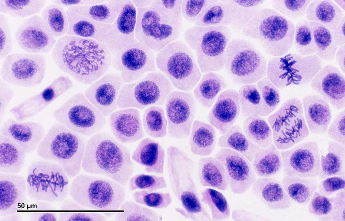
1.1 Mitosis cell division
Introduction
Cell division is the process by which one cell, the mother cell (or parent cell), divides to form new daughter cells. It always has two parts. First the nucleus divides: karyokinesis (Greek karyon, nucleus, and kinesis, movement). Then the cytoplasm divides: cytokinesis (kytos, a hollow vessel, a cell). Eukaryotic cells divide in two ways, mitosis and meiosis. (Bacteria, with no nucleus, divide more simply, by binary fission.)
Mitosis is the division in which one cell divides into two daughter cells that have the same number and kind of chromosomes as the mother cell, and as each other. A human body cell with 46 chromosomes gives two cells with 46 each, the same 46. Because the chromosome number stays equal, mitosis is also called equational division.
To follow mitosis you need four words from Grade 9:
- A body cell is diploid (2n): it has two sets of chromosomes, one set from the mother and one from the father. In humans 2n = 46, so n = 23. In an onion 2n = 16, in rice 24, in the garden pea 14, in a dog 78.
- The two chromosomes of a pair, one from each parent, of the same length and shape and carrying genes for the same characters at the same places, are homologous chromosomes (homologs).
- Before a cell divides, each chromosome is copied. The copy stays joined to the original at the centromere. The two copies are sister chromatids: identical, because one was copied from the other.
- So a chromosome may be single (one chromatid) or double (two sister chromatids). Either way it is one chromosome: chromosomes are counted by their centromeres.
Mitosis happens in the somatic (body) cells: wherever an organism grows or repairs itself. In plants it happens in the meristems, the tips of roots and shoots (which is why the onion root tip above is full of dividing cells) and the cambium that thickens stems. In animals it is spread through the body, fastest where cells wear out: the bottom layer of the skin, the bone marrow that makes blood cells, the lining of the gut. Some cells, such as most nerve cells and the muscle cells of the heart, rarely or never divide once they are mature.
The cell cycle
A cell that divides doesn’t spend most of its time dividing. It spends it growing and preparing. The whole life of a dividing cell, from one division to the next, is the cell cycle. It has two parts:
- Interphase, the long time between divisions.
- M phase (mitotic phase), the division itself: karyokinesis followed by cytokinesis.
In a typical human cell growing in a laboratory dish, one turn of the cycle takes about 24 hours, of which the division itself takes only about one hour (Figure 1.2).
Interphase
Interphase was once called the “resting stage”, because under a light microscope nothing seems to happen in the nucleus: the chromatin is a faint network and no chromosomes can be seen. In fact the cell is at its busiest. Interphase has three stages:
- G₁ phase (first gap, or growth): the new cell grows, makes proteins and new organelles, and does its everyday work. A cell that is going to divide again then passes a check: is it big enough, is its DNA undamaged, are the signals right?
- S phase (synthesis): the cell copies its DNA. By the end of S, each chromosome is made of two identical sister chromatids joined at the centromere. In an animal cell the centrosome is copied too, so the cell now has two.
- G₂ phase (second gap): the cell grows more, makes the proteins needed for division (including the tubulin for the spindle), and checks that all its DNA has been copied correctly.
Cells that stop dividing leave the cycle from G₁ into a state called G₀. Most nerve cells stay in G₀ for life; liver cells sit in G₀ but can return to the cycle and divide if part of the liver is removed.
In which stage of the cell cycle is the DNA copied?
Pick an answer to see why.
How much DNA is in a cell?
It helps to give the amount of DNA a name. Call the DNA in one set of chromosomes 1C. A diploid cell in G₁ has two sets: 2C. During S phase the DNA doubles to 4C, though the number of chromosomes stays the same (each is now double). Mitosis shares that 4C equally, so each daughter cell is back to 2C. You will see this pattern again in meiosis, with one more halving (Figure 1.6, further on).
Karyokinesis: the division of the nucleus
Mitosis is one smooth process, but it is described in four stages. Step through them in the widget, then read what happens in each.
1. Prophase
Prophase (pro, before) is usually the longest stage of mitosis.
- The long, thin chromatin threads coil and condense into short, thick chromosomes that can be seen with a light microscope. Each chromosome can be seen to be double: two sister chromatids joined at the centromere.
- The nucleolus gets smaller and disappears.
- In an animal cell the two centrosomes move apart, towards opposite ends (poles) of the cell. Short microtubules radiate from each like the rays of a star: the aster.
- Between them the spindle begins to form: a set of fibers (bundles of microtubules) that will move the chromosomes.
- At the end of prophase the nuclear envelope breaks up into small pieces. Some books call this last part, when the spindle fibers reach into the old nucleus and catch the chromosomes, prometaphase.
2. Metaphase
In metaphase (meta, after or between):
- Each chromosome is caught by spindle fibers at its centromere. On the centromere are two small discs of protein, the kinetochores, one on each sister chromatid, facing opposite poles. Fibers from one pole attach to one kinetochore, and fibers from the other pole to the other.
- Pulled from both sides, the chromosomes move to the middle of the spindle and line up in one plane across the cell: the equator, or metaphase plate. The centromeres lie on the plate; the arms may point in any direction.
- The chromosomes are now at their shortest and thickest, which is why they are counted and photographed at metaphase.
The cell does not go on until every chromosome is attached to both poles. This check (the spindle checkpoint) stops a cell from giving one daughter an extra chromosome and the other one too few.
3. Anaphase
Anaphase (ana, up or back) is the shortest stage, only a few minutes long.
- The sister chromatids, held together until now by rings of a protein called cohesin, are suddenly released: the centromere of each chromosome splits.
- The spindle fibers attached to the kinetochores shorten and pull the two chromatids of every chromosome to opposite poles, at about 1 µm a minute. Each chromatid moves centromere first, its arms trailing behind, so it looks like a V, J or rod depending on where its centromere is (Grade 9’s V, L, J and I shapes).
- At the same time the poles move farther apart, and the cell gets longer.
Once separated, each chromatid is called a daughter chromosome. So for a few minutes, a human cell in anaphase has 92 chromosomes, 46 moving to each pole.
4. Telophase
Telophase (telos, end) is prophase in reverse:
- The daughter chromosomes reach the poles, and the spindle breaks down.
- A new nuclear envelope forms round each group of chromosomes, from the pieces of the old one.
- The chromosomes uncoil back into thin chromatin threads.
- A nucleolus appears again in each new nucleus.
Now the cell has two nuclei, each with the same chromosomes as the original: karyokinesis is over.
Spindle fibers attach to a chromosome at its:
Pick an answer to see why.
In which stage of mitosis does a human cell briefly have 92 chromosomes?
Pick an answer to see why.
Cytokinesis: the division of the cytoplasm
Cytokinesis usually begins in late anaphase or telophase and divides the cytoplasm, with its organelles, between the two new nuclei. Animal and plant cells do it in opposite ways (Figure 1.3):
- In an animal cell, a ring of protein filaments (actin and myosin, the same proteins that make muscles contract) forms just under the cell membrane round the middle of the cell. As it tightens, it pulls the membrane in, making a groove, the cleavage furrow, which deepens from the outside in until the cell is pinched in two.
- A plant cell can’t pinch in: its firm wall is in the way. Instead, vesicles from the Golgi bodies, carrying the materials of the wall, gather on a framework of microtubules (the phragmoplast) across the middle of the cell. They join to form a disc, the cell plate, which grows from the center outwards until it meets the old wall. The cell plate becomes the middle lamella, and each daughter cell lays down its own new wall on either side of it.
The result is two daughter cells, each with a nucleus holding the same chromosomes as the mother cell, and a share of the cytoplasm. Each then enters G₁ and grows.
Mitosis in plant and animal cells
| Mitosis in an animal cell | Mitosis in a plant cell |
|---|---|
| Centrioles present; the centrosomes move to the poles. | No centrioles in flowering plants; the spindle forms without them. |
| Asters form round each pole (an astral spindle). | No asters (an anastral spindle). |
| Cytokinesis by a cleavage furrow, from the outside in. | Cytokinesis by a cell plate, from the center out. |
| Happens in tissues throughout the body. | Happens mainly in meristems (root and shoot tips, cambium). |
| The cell rounds up before dividing. | The cell keeps its shape inside its wall. |
1.2 Meiosis cell division
Introduction
Meiosis (Greek meiosis, making smaller) is the division in which one diploid cell divides twice to form four haploid daughter cells, each with half the number of chromosomes of the mother cell: one chromosome of every pair. Because it halves the number, meiosis is called reduction division (reductional division).
Meiosis happens only in the cells that will make gametes or spores, in the reproductive organs: in animals, in the testes (making sperm) and ovaries (making eggs); in flowering plants, in the anthers (making pollen) and ovules (making the egg). The diploid cell that undergoes meiosis is called a meiocyte: a primary spermatocyte in the testis, a primary oocyte in the ovary, a pollen mother cell in an anther.
Meiosis has two divisions, one after the other, with only one copying of DNA before the first:
- Meiosis I (the reduction division): the two chromosomes of each homologous pair separate. Two cells, each with n chromosomes, each chromosome still double.
- Meiosis II (an equational division, like mitosis): the sister chromatids separate. Four cells, each with n single chromosomes.
Meiosis I
Before meiosis begins, the meiocyte goes through an interphase just like the interphase before mitosis: G₁, then S, when every chromosome is copied into two sister chromatids, then G₂. This is the only copying of DNA in the whole of meiosis. Then the first division, karyokinesis I, runs through four stages.
Prophase I
Prophase I is long and complicated, much longer than the prophase of mitosis: in a man’s testes it takes about three weeks, and in a woman’s eggs it begins before she is born. It is where meiosis does its two special things: the homologous chromosomes pair up, and they swap pieces. It is described in five sub-stages (Figure 1.4):
- Leptotene (leptos, thin; tene, thread): the chromatin coils into long, thin threads. Each is already double, but the two chromatids can’t be told apart yet. The threads look beaded: the beads, chromomeres, are tightly coiled spots along them.
- Zygotene (zygon, yoke): the two homologous chromosomes of each pair find each other and come together side by side, zipping up gene for gene along their length. This pairing is synapsis. A ladder of protein, the synaptonemal complex, forms between them and holds them close. A paired couple of homologs is a bivalent: there are as many bivalents as there are pairs, n (23 in a human).
- Pachytene (pachys, thick): the pairs are tightly zipped, shorter and thicker. Each bivalent can now be seen to have four chromatids, so it is also called a tetrad. Now crossing over happens: a chromatid of one homolog and a chromatid of the other (non-sister chromatids) break at exactly matching points and join up the other way round, swapping corresponding pieces. The genes are exchanged, so each of these chromatids now carries a mix of the mother’s and the father’s genes. This is genetic recombination.
- Diplotene (diplos, double): the synaptonemal complex breaks down, and the two homologs of each pair begin to pull apart. They stay held together only at the points where crossing over happened, where two non-sister chromatids are seen to cross: each such point is a chiasma (plural chiasmata; Greek chiasma, a cross, like the letter χ). In human egg cells, prophase I stops at diplotene before birth and waits there, for 12 to 50 years, until that egg is released from the ovary.
- Diakinesis (dia, across): the chromosomes are coiled to their shortest and thickest. The chiasmata appear to slide towards the ends of the chromosomes (books call this terminalization). The nucleolus disappears, the nuclear envelope breaks up and the spindle forms.
A bivalent is:
Pick an answer to see why.
Metaphase I
The spindle catches the bivalents, and they line up on the equator, in pairs. The two homologs of each pair lie on either side of the plate: the kinetochores of one homolog face one pole, those of the other face the opposite pole. (In mitosis, by contrast, single chromosomes line up, each facing both poles.) The chiasmata hold the two homologs together.
Which homolog of a pair faces which pole is a matter of chance, and each pair lines up independently of every other pair. This is called independent assortment, and it is one of the reasons why every gamete is different (section 1.4).
Anaphase I
The chiasmata let go, and the spindle pulls the two homologous chromosomes of each pair to opposite poles. The centromeres do not split: each chromosome moves whole, still with its two sister chromatids. So each pole receives one chromosome of every pair, n chromosomes. This is the moment the chromosome number is halved. (Because of crossing over, the two sister chromatids of a chromosome are no longer quite identical.)
Telophase I
The chromosomes reach the poles. In most cells a nuclear envelope forms round each group and the chromosomes uncoil a little, and cytokinesis I divides the cell in two (a furrow in animal cells, a cell plate in many plant cells). The two daughter cells are haploid: each has n chromosomes, each chromosome made of two chromatids, and 2C of DNA.
Between the two divisions there may be a short pause, interkinesis. It is not a true interphase: no DNA is copied. (There is nothing to copy: each chromosome is already double.)
Meiosis II
Meiosis II happens in both cells made by meiosis I, and it is just like mitosis, except that each cell has only n chromosomes.
- Prophase II: the chromosomes coil up again (if they uncoiled), the nuclear envelope breaks up and a new spindle forms in each cell, often at right angles to the first.
- Metaphase II: the chromosomes line up singly on the equator of each cell, each held by fibers from both poles, one sister chromatid facing each pole.
- Anaphase II: the centromeres split at last, and the sister chromatids separate and move to opposite poles.
- Telophase II: nuclear envelopes form round the four groups of chromosomes, which uncoil; the nucleoli reappear.
- Cytokinesis: each of the two cells divides, giving four haploid cells, each with n single chromosomes and 1C of DNA. In animals these become gametes; in flowering plants they become pollen grains (in the anther) or, in the ovule, one becomes the embryo sac that holds the egg.
In which stage does the number of chromosomes per cell become halved?
Pick an answer to see why.
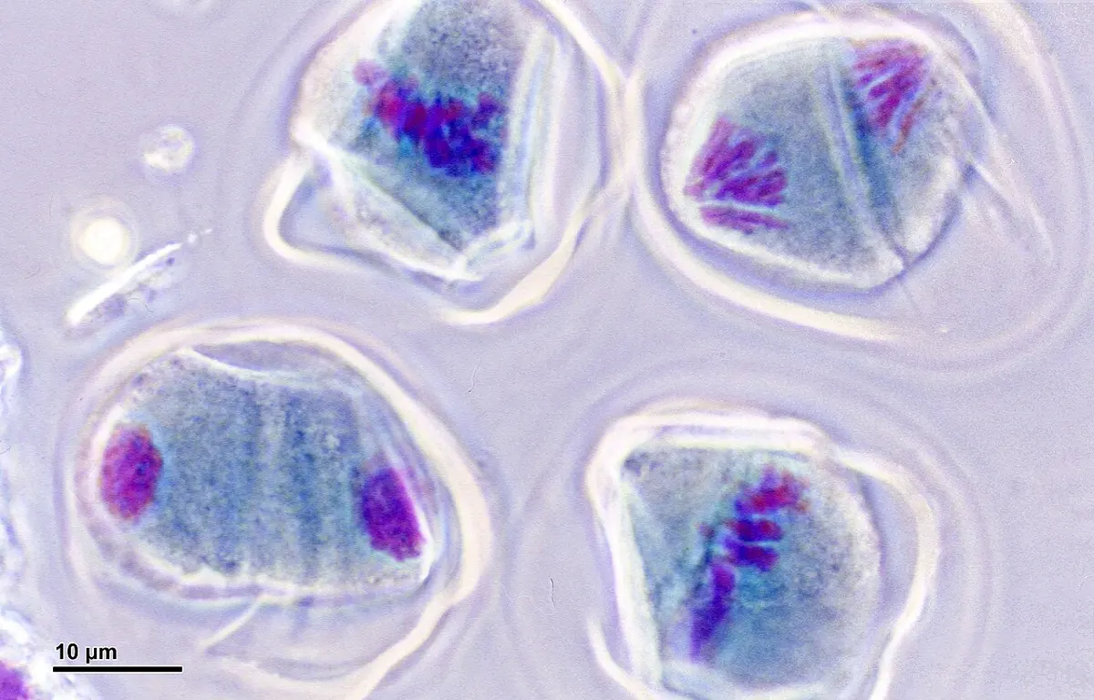
Sperm and eggs: four cells, or one?
In a man, each primary spermatocyte divides by meiosis into four equal cells, and all four become sperm. In a woman, each division of a primary oocyte is very unequal: almost all the cytoplasm, with its food store, goes to one cell, and the other gets a nucleus with very little cytoplasm, a tiny polar body that later dies. So one oocyte gives one egg (and two or three polar bodies). The egg keeps the food the embryo will need in its first days. In humans, meiosis II of the egg is completed only if a sperm enters it.
The chromosomes and the DNA, stage by stage
The amount of DNA in a cell rises and falls in the same pattern every time (Figure 1.6): it doubles once in interphase, then halves at every division of the cytoplasm. Mitosis has one division, so the DNA returns to where it started; meiosis has two, so it ends at half.
For a human cell (2n = 46), the numbers to know are:
| Stage | Chromosomes in each cell | Chromatids in each cell | DNA in each cell |
|---|---|---|---|
| G₁ | 46 | 46 | 2C |
| G₂, prophase, metaphase (mitosis) | 46 | 92 | 4C |
| Anaphase (mitosis) | 92 | 92 | 4C |
| Each daughter cell after mitosis | 46 | 46 | 2C |
| Prophase I, metaphase I (23 bivalents) | 46 | 92 | 4C |
| Each cell after meiosis I | 23 | 46 | 2C |
| Anaphase II | 46 | 46 | 2C |
| Each cell after meiosis II | 23 | 23 | 1C |
Mitosis and meiosis compared
Grade 10 Science lists the main differences. With the stages known, the full comparison is:
| Mitosis | Meiosis |
|---|---|
| In somatic cells (and in the cells that will make gametes, before they make them). | Only in meiocytes, in the testes and ovaries (anthers and ovules in plants). |
| One division. | Two divisions (meiosis I and II), with one copying of DNA. |
| Two daughter cells. | Four daughter cells. |
| Daughter cells diploid (2n), like the mother cell: equational. | Daughter cells haploid (n): reductional. |
| Daughter cells genetically identical to the mother cell and each other. | Daughter cells genetically different from the mother cell and each other. |
| Prophase short and simple. | Prophase I long, with five sub-stages. |
| No pairing of homologous chromosomes; no crossing over, no chiasmata. | Homologs pair (synapsis, bivalents); crossing over; chiasmata. |
| At metaphase single chromosomes line up on the equator. | At metaphase I pairs of homologs line up on the equator. |
| At anaphase the centromeres split and sister chromatids separate. | At anaphase I whole homologs separate (centromeres don’t split); at anaphase II the chromatids separate. |
| For growth, repair and asexual reproduction. | For making gametes or spores: sexual reproduction and variation. |
1.3 Significance of mitosis and meiosis cell division
Significance of mitosis
- Growth. A multicellular organism grows by increasing the number of its cells. From a single zygote, mitosis builds the whole body, every cell with the same chromosomes.
- Repair and replacement. Mitosis replaces cells that are worn out, damaged or lost: a healed wound, new skin (your outer skin is replaced every month or two), new red blood cells (about two million every second, made in the bone marrow), a regrown lizard’s tail.
- Asexual reproduction. Single-celled eukaryotes such as Amoeba and yeast reproduce by mitosis, and many plants and some animals reproduce by mitosis alone, by buds, runners, tubers and cuttings (section 1.4).
- Genetic stability. Each daughter cell gets exactly the same chromosomes as the mother cell, so all the cells of the body carry the same genetic information, generation after generation of cells.
- Keeping the right size and balance of cells. A cell that grows too large has too little surface for its volume (Grade 9 Optional Science: Why cells are small); dividing restores a workable size and the balance between nucleus and cytoplasm.
- Regeneration. Some animals rebuild lost parts by mitosis: a starfish regrows an arm, a planarian worm cut in pieces grows a whole worm from each piece.
When mitosis is not controlled, it is harmful. Cancer is a disease in which cells keep dividing when they should stop, because the genes that control the cell cycle (its checks) are damaged. The extra cells form a lump, a tumor, and some can spread through the body.
Significance of meiosis
- Making gametes and spores. Meiosis makes the sperm and eggs of animals, and the spores of plants, so sexual reproduction depends on it.
- Keeping the chromosome number constant. Meiosis halves the number in the gametes (n), and fertilization brings it back to 2n. Without meiosis the number would double every generation: 46, 92, 184…
- Variation. Crossing over makes new combinations of genes on each chromosome, and independent assortment makes new combinations of whole chromosomes in each gamete. So no two gametes are alike, and every child of the same parents is different (section 1.4). Variation lets a species adapt to change, and it is what evolution works on.
- Repairing DNA. While the homologs are paired in prophase I, some kinds of damage in one chromosome can be repaired using the matching stretch of its partner as a pattern.
When meiosis goes wrong, the result can be serious. If the two chromosomes of a pair fail to separate in anaphase I (or two chromatids in anaphase II), which is called non-disjunction, one gamete gets an extra chromosome and another gets one too few. A baby from an egg with two copies of chromosome 21 has three copies in every cell (47 chromosomes), and has Down syndrome, which affects about 1 in every 1,000 to 1,100 babies born worldwide. The chance rises as the mother gets older, most likely because her eggs have been waiting in diplotene of prophase I since before she was born.
Without meiosis, what would happen to the chromosome number of a species that reproduces sexually?
Pick an answer to see why.
1.4 Role of sexual and asexual reproduction
Living things make more of their kind in two ways. In asexual reproduction one parent produces offspring by itself, without gametes and without fertilization, through mitosis alone (or binary fission in bacteria). In sexual reproduction two parents each make gametes by meiosis, and a male and a female gamete fuse in fertilization to form a zygote, which grows into the new organism by mitosis. The difference between the two divisions decides the difference between the offspring.
Asexual reproduction and the formation of clones
Because mitosis gives daughter cells exactly like the mother cell, offspring made by asexual reproduction have exactly the same genes as their parent, and as each other. A group of organisms descended from one parent by asexual reproduction, all with the same genes, is a clone. (The word, from the Greek klon, a twig, was first used, spelled clon, in 1903 by the American plant scientist Herbert Webber, for plants grown from cuttings, buds or tubers.) A clone’s members differ only where the environment makes them differ, or where a rare mistake in copying the DNA (a mutation) has happened.
Clones are everywhere:
- Single-celled organisms: an Amoeba dividing into two, a yeast cell budding: every cell in the colony is a clone of the first.
- Plants, by vegetative propagation: every potato grown from a tuber, every sugarcane grown from a piece of stem, every banana grown from a sucker, every clump of bamboo grown from a rhizome and every garlic plant grown from a clove is a clone of its parent. So is every plant grown from a cutting or a graft: the fruit of a grafted mango tree is a clone of the tree the scion came from. A leaf of Bryophyllum grows tiny plantlets round its edge, each a clone.
- Animals: a Hydra budding, a planarian growing back from pieces, aphids and some lizards whose females produce young from unfertilized eggs.
- Ourselves: identical twins come from one zygote that split into two embryos early on, so they are natural clones of each other: the same genes, made by mitosis.
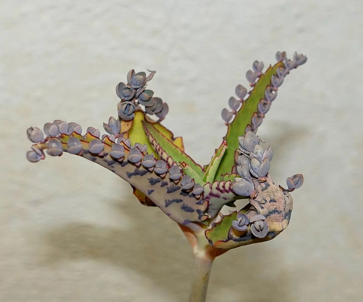
Making clones of an animal from one of its body cells, as with Dolly the sheep in 1996, is done in a laboratory; it is in Unit 5 of this book. Here the point is the natural one: mitosis makes clones.
Advantages and disadvantages of asexual reproduction
| Advantages | Disadvantages |
|---|---|
| One parent is enough: no need to find a mate. | No variation: all the offspring have the same genes. |
| Fast: many offspring in a short time (a bacterium can double every 20 minutes). | A disease or change that kills one can kill them all. |
| A successful combination of genes is kept exactly: a farmer knows what a cutting of a good variety will give. | Offspring often grow close to the parent and compete with it. |
| Uses less energy: no flowers, gametes or courtship. | Harmful mutations are not removed by mixing with other genes, and pile up. |
Sexual reproduction and genetic variation
Sexual reproduction makes offspring that are different from their parents and from each other: it makes genetic variation. The variation comes from meiosis and fertilization together, in three ways:
- Crossing over (prophase I): non-sister chromatids swap pieces, so each chromosome in a gamete is a new mixture of the grandmother’s and the grandfather’s genes. In humans each sperm carries, on average, about 27 crossovers and each egg about 42 (counted in parents’ and children’s DNA).
- Independent assortment (metaphase I): each pair of homologs lines up by chance, independently of the other pairs, so each gamete gets a random mixture of maternal and paternal chromosomes. With n pairs there are possible combinations. For a human, : more than eight million kinds of gamete from the line-up alone.
- Random fertilization: any one of a father’s millions of sperm may fertilize any one of a mother’s eggs. For humans that gives , about 70 trillion combinations for one couple’s children, before crossing over is counted. With it, the number is so large that no two children (except identical twins) have ever been genetically the same.
(A fourth source, new mutations, adds several dozen new changes to the DNA of every child; it happens in both kinds of reproduction, but in sexual reproduction they are mixed and sorted in every generation.)
Advantages and disadvantages of sexual reproduction
| Advantages | Disadvantages |
|---|---|
| Variation: every offspring is different. | Needs two parents (in most species) that must meet. |
| A population with variation is more likely to have some members that survive a new disease, a drought or a change of climate. | Slower: fewer offspring, more time and energy (flowers, pollen, courtship). |
| Variation lets a species adapt and evolve over many generations. | A very good combination of genes is broken up in the offspring. |
| Harmful mutations can be weeded out as genes are mixed. | Only half of each parent’s genes go to each child. |
| Asexual reproduction | Sexual reproduction |
|---|---|
| One parent. | Usually two parents. |
| No gametes and no fertilization. | Gametes made by meiosis, which fuse in fertilization. |
| Only mitosis (or fission). | Meiosis, fertilization, then mitosis. |
| Offspring genetically identical to the parent: a clone. | Offspring genetically different from the parents and each other. |
| No variation (except rare mutations). | Variation from crossing over, independent assortment and random fertilization. |
| Fast, many offspring. | Slower, fewer offspring. |
| Examples: Amoeba, yeast, Hydra, potato tubers, banana suckers. | Examples: humans, frogs, mustard, rice. |
Exercise
1. Choose the correct option
a. A cell is in G₂ phase. Compared with the same cell in G₁, it has:
Pick an answer to see why.
b. The nucleolus and the nuclear envelope disappear in:
Pick an answer to see why.
c. Sister chromatids separate during:
Pick an answer to see why.
d. Synapsis, the pairing of homologous chromosomes, takes place in:
Pick an answer to see why.
e. Crossing over takes place between:
Pick an answer to see why.
f. A cell with 2n = 24 completes meiosis I. Each daughter cell has:
Pick an answer to see why.
g. Cytokinesis in a plant cell takes place by:
Pick an answer to see why.
h. Which of these does not produce genetic variation?
Pick an answer to see why.
i. The fruit fly has 2n = 8. Ignoring crossing over, how many kinds of gamete can one fly make?
Pick an answer to see why.
j. A group of plants grown from the tubers of a single potato plant is called:
Pick an answer to see why.
2. Give reasons
Mitosis is called equational division.
Hint
Answer
Meiosis is called reduction division.
Hint
Answer
Meiosis II is similar to mitosis.
Hint
Answer
Interphase is not really a resting stage.
Hint
Answer
Meiosis is essential in organisms that reproduce sexually.
Hint
Answer
All the members of a clone are genetically identical.
Hint
Answer
Children of the same parents are not alike, except identical twins.
Hint
Answer
The tip of an onion root is used to study mitosis.
Hint
Answer
3. Differentiate between
Mitosis and meiosis
Hint
Answer
Meiosis I and meiosis II
Hint
Answer
Karyokinesis and cytokinesis
Hint
Answer
Cytokinesis in an animal cell and in a plant cell
Hint
Answer
Homologous chromosomes and sister chromatids
Hint
Answer
Asexual reproduction and sexual reproduction
Hint
Answer
4. Answer the following questions
What is the cell cycle? Describe its phases.
Hint
Answer
Describe the stages of mitosis with labeled diagrams.
Hint
Answer
Describe the sub-stages of prophase I of meiosis.
Hint
Answer
Describe meiosis I, with diagrams.
Hint
Answer
Describe meiosis II. How is it different from mitosis?
Hint
Answer
Write the significance of mitosis.
Hint
Answer
Write the significance of meiosis.
Hint
Answer
How does sexual reproduction produce genetic variation? Explain the three sources.
Hint
Answer
What is a clone? How are clones formed in nature? Give examples, and write two advantages and two disadvantages of asexual reproduction.
Hint
Answer
What is non-disjunction? How can it cause Down syndrome?
Hint
Answer
You are given a permanent slide of an onion root tip. How would you recognize the four stages of mitosis in it?
Hint
Answer
A disease is spreading through the potato fields of a district where almost every farmer grows the same variety, planted from tubers. Explain the danger, and suggest what farmers and plant breeders could do.
Hint
Answer
5. Numerical problems
A cell divides by mitosis every 30 minutes. Starting from one cell, how many cells will there be after 4 hours, if all go on dividing?
Hint
Answer
A cell of a plant has 2n = 20 chromosomes (maize). Write the number of chromosomes and chromatids in one cell at: a. G₁, b. metaphase of mitosis, c. anaphase of mitosis, d. metaphase I, e. each cell after meiosis I, f. each cell after meiosis II.
Hint
Answer
In a squash of an onion root tip, Sabina counted 400 cells: 352 in interphase, 28 in prophase, 6 in metaphase, 4 in anaphase and 10 in telophase. Find the mitotic index. If the whole cell cycle takes 20 hours, how long do interphase and prophase last?
Hint
Answer
How many sperm are formed from 25 primary spermatocytes, and how many eggs from 25 primary oocytes? How many meiotic divisions are needed to make 100 sperm?
Hint
Answer
Rice has 2n = 24. How many bivalents form in prophase I of a pollen mother cell? How many kinds of pollen can one rice plant make by independent assortment alone?
Hint
Answer
A cell in G₁ contains 12 pg of DNA. How much DNA is there in: a. the same cell in G₂, b. each cell after mitosis, c. each cell after meiosis I, d. a gamete?
Hint
Answer
A human has 2n = 46. Ignoring crossing over, find the number of kinds of sperm a man can make, and the number of different combinations possible in the children of one couple. Give the second in standard form.
Hint
Answer
A zygote divides by mitosis, and every cell divides again at each round. After how many rounds of division are there more than 1,000 cells? How many chromosomes does each of those cells have, if the zygote had 46?
Hint
Answer
Printable PDFs
- The unit as a book (A5) 34 pages Answers as an A4 booklet, 9 sheets
- Study notes 3 pages
- Worksheet 5 pages Answer key
- Harder practice questions 3 pages Answer key
Practice every question in the book, and keep track of the ones you miss →
Found a mistake? Please tell us.