
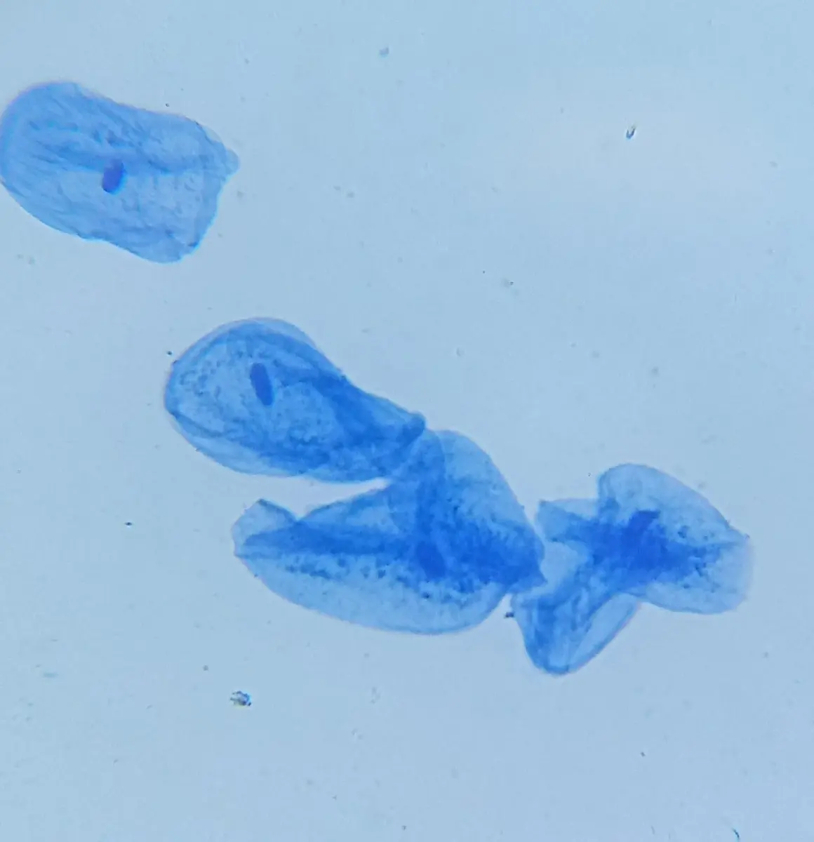
Stir a spoon of dahi into a drop of water, put the drop on a slide and look at it under a school microscope at its highest power. Among the fat droplets you will find tiny rods and beads, alone or in short chains: the bacteria that turn milk into dahi, such as Lactobacillus. Now gently scrape the inside of your cheek with a clean toothpick and look at that too. You will see flat, wide cells, each with a dark dot near its middle. Both are cells, and both are alive. But each of your cheek cells is about twenty times longer than one of those bacteria, and its dark dot, the nucleus, is something the bacterium doesn’t have at all.
Those are the two great kinds of cell: the small, simple prokaryotic cells of bacteria, and the larger eukaryotic cells of everything else, from yeast to rhododendrons to you. This unit is about both kinds, about the parts inside a eukaryotic cell, each with its own job, and about how those parts work together when a cell breathes, makes food, gets rid of its wastes and divides.
1.1 Prokaryotic and eukaryotic cell
Every cell, of every kind, has three things. A thin cell membrane encloses it. Inside is cytoplasm, a jelly of water, salts and proteins in which the cell’s chemistry happens. And somewhere in it is DNA, the instructions for making and running the cell, with ribosomes, the tiny machines that read those instructions and make proteins. What differs is how the inside is organized.
Prokaryotic cells (Greek pro, before, and karyon, kernel or nucleus) have no nucleus. Their DNA, usually one long, circular molecule, lies coiled in a region of the cytoplasm called the nucleoid, with no membrane round it. They have no other membrane-bound parts either: no mitochondria, no chloroplasts, no Golgi body, no endoplasmic reticulum. Bacteria, including the cyanobacteria (once called blue-green algae, such as Nostoc and Anabaena), are prokaryotes, and so are the archaea, microbes that look like bacteria but are a separate group. Most prokaryotic cells are 1–5 µm long.
Eukaryotic cells (Greek eu, true) have a true nucleus: their DNA, in several straight chromosomes, is kept inside a double membrane. Their cytoplasm is divided by more membranes into organelles (“little organs”), each a compartment with its own job: mitochondria, the endoplasmic reticulum, Golgi bodies, lysosomes, vacuoles and, in plants, plastids. Protists such as Amoeba and Paramecium, fungi, plants and animals are all eukaryotes. Their cells are usually 10–100 µm across, ten or more times longer than a bacterium and a thousand or more times bigger in volume.
The parts of a prokaryotic cell
Look at a bacterium such as Escherichia coli, which lives in our gut, from the outside in (Figure 1.2).
Capsule or slime layer. Many bacteria wrap themselves in a sticky layer of sugars (sometimes proteins) outside the wall. It stops them drying out, helps them stick to surfaces (the plaque on teeth is bacteria held together this way) and makes them harder for our white blood cells to swallow.
Cell wall. A firm wall outside the membrane gives the cell its shape and stops it bursting when water rushes in. In bacteria it is made of peptidoglycan (also called murein), chains of sugars cross-linked by short chains of amino acids, a material found nowhere else. Penicillin kills bacteria by stopping them building it, which is why it harms bacteria but not our cells, which have no wall.
Cell membrane. Inside the wall, the membrane controls what goes in and out. In a bacterium it also does the jobs that mitochondria do in our cells: the enzymes that release energy with oxygen sit in it.
Cytoplasm with ribosomes. The cytoplasm is packed with ribosomes, tens of thousands of them, which make the cell’s proteins. A bacterium’s ribosomes are smaller than ours: 70S, against the 80S ribosomes of a eukaryote’s cytoplasm (S is explained under ribosomes).
Nucleoid. The DNA, one circular chromosome, folded into a tangle in the middle. E. coli’s chromosome is about 1.5 mm long when stretched out, more than 700 times the length of the cell it fits in.
Plasmids. Many bacteria also carry one or more small extra rings of DNA. Their genes are not needed every day, but they can be lifesavers: genes to resist an antibiotic, for example. Bacteria pass plasmids to one another, which is one way resistance to antibiotics spreads.
Pili (singular pilus) and fimbriae. Short, thin, straight hairs of protein that let the bacterium cling to surfaces and to other cells; a special long pilus joins two bacteria while a plasmid passes from one to the other.
Flagella (singular flagellum). Long, thin, whip-like threads that some bacteria spin like a ship’s propeller to swim. A bacterial flagellum is a hollow tube of protein turned by a tiny rotary motor in the wall and membrane, quite unlike the flagellum of a sperm cell (which beats, and is built of microtubules).
Mariana Ruiz Villarreal, LadyofHats, Public domain, via Wikimedia Commons
Which of these is found in a prokaryotic cell?
Pick an answer to see why.
A drug that stops bacteria making peptidoglycan kills bacteria but doesn’t harm human cells, because human cells:
Pick an answer to see why.
Prokaryotic and eukaryotic cells compared
| Prokaryotic cell | Eukaryotic cell |
|---|---|
| No true nucleus: the DNA lies in a nucleoid, with no membrane round it. | A true nucleus, its DNA inside a double membrane (the nuclear envelope). |
| Usually one circular chromosome; in bacteria its DNA is not wound round the proteins called histones. Often plasmids too. | Several straight (linear) chromosomes, their DNA wound round histones. |
| No membrane-bound organelles: no mitochondria, plastids, ER, Golgi body or lysosomes. | Membrane-bound organelles: mitochondria, ER, Golgi bodies, lysosomes, vacuoles; plastids in plants. |
| Small ribosomes, 70S. | Larger ribosomes, 80S, in the cytoplasm (70S in its mitochondria and chloroplasts). |
| Cell wall (in bacteria) of peptidoglycan. | Cell wall of cellulose in plants and of chitin in fungi; none in animal cells. |
| Respiration on the cell membrane. | Respiration mostly in mitochondria. |
| Small, mostly 1–5 µm. | Larger, mostly 10–100 µm. |
| Divide by binary fission, with no spindle. | Divide by mitosis or meiosis, with a spindle. |
| Bacteria, cyanobacteria, archaea. | Protists, fungi, plants and animals. |
How small is small?
Cells and their parts are measured in micrometers (µm) and nanometers (nm):
A light microscope magnifies twice: the objective lens near the slide makes an enlarged image, and the eyepiece enlarges that image again. So
and if you know how big something looks in a picture or drawing, you can work out its real size:
A school light microscope magnifies about 1,000 times at most (the best, about 1,500 times), and however much it magnifies, it can’t show two points closer together than about 0.2 µm as two points. That is why ribosomes (about 25 nm) and the inside of a mitochondrion can’t be seen with it. An electron microscope, which uses a beam of electrons instead of light, can show detail down to well under 1 nm, and almost everything in section 1.2 was discovered with one.
Why cells are small
A cell takes in food and oxygen and lets out wastes through its surface, but it uses them in its whole volume. As a cell grows, its volume grows faster than its surface. Picture cells as cubes. A cube of side has a surface area of and a volume of , so for each unit of volume it has
A cube 1 µm across, the size of a bacterium, has 6 µm² of surface for every µm³ of inside. A cube 10 µm across has only 0.6 µm² for every µm³: ten times less surface to serve each part of its inside. A cell that grew too big would starve in its middle, because food and oxygen couldn’t come in through the surface fast enough.
Eukaryotic cells get round this in two ways. Their membranes fold inside them, the endoplasmic reticulum, the Golgi body and the inner membrane of the mitochondria, giving them far more membrane to work on than their outside surface alone. And they move things about inside actively, along tracks, instead of waiting for them to diffuse.
1.2 Structure and function of cell organelles
The parts of a eukaryotic cell are easiest to understand drawn in one cell. Figure 1.3 is an animal cell as an electron microscope shows it, cut open; a plant cell has most of the same parts, plus a cell wall, plastids and one large vacuole (Figure 1.4). The rest of this section takes each part in turn: what it is made of, and what it does.
Cell drawings adapted from Servier Medical Art (smart.servier.com), CC BY 4.0
Cell wall
The cell wall is a firm but usually porous layer outside the cell membrane of plant cells, fungi, most bacteria and many protists. Animal cells have none. In plants it is made mainly of cellulose, long chains of glucose lying side by side in bundles called fibrils, set in a jelly of other sugars (hemicellulose and pectin), like the steel rods in concrete. A plant cell’s wall has three layers:
- The middle lamella, shared by two neighboring cells, made of pectin (calcium and magnesium pectate). It glues the cells together. When a fruit ripens, enzymes break down its pectin and the fruit goes soft.
- The primary wall, laid down by a young, growing cell: thin and stretchy, so the cell can grow.
- The secondary wall, laid inside the primary wall by some cells once they stop growing, such as the cells of wood and fiber. It is thick and may be hardened with lignin, which makes wood strong. Many of these cells then die, leaving only their walls.
Fine channels called plasmodesmata (singular plasmodesma) cross the wall, joining the cytoplasm of neighboring cells, so water and food can pass from cell to cell.
Functions of the cell wall: it gives the cell its shape and strength; it protects it from damage and from germs; and it stops the cell bursting when water enters: the cell swells against its wall until it is firm (turgid), which keeps a plant’s soft parts, its leaves and young stems, upright. It lets water and dissolved substances pass freely: it is fully permeable. The cell membrane under it decides what really enters the cell.
Cell membrane
The cell membrane (or plasma membrane) is the thin, living skin round every cell, about 8 nm thick: you would need about 10,000 of them stacked to make the thickness of a page. It is far too thin to see with a light microscope, which shows only where it lies.
It is made mostly of lipids (fats) and proteins. In 1972 two Americans, S. J. Singer and Garth Nicolson, described its structure in the fluid mosaic model (Figure 1.5), which is still how we picture it:
- Its base is a double layer of phospholipids (the lipid bilayer). Each phospholipid has a head that is attracted to water and two fatty tails that avoid water. In water the molecules arrange themselves in two layers, heads outward on both faces (towards the water outside and the watery cytoplasm inside) and tails inward, meeting in the middle.
- Proteins sit in the bilayer like tiles in a mosaic. Some cross it from side to side (integral proteins), some forming channels or carriers through which particular substances pass. Others sit on one face only (peripheral proteins).
- Cholesterol (in animal cells) lies among the tails and keeps the membrane from getting too runny or too stiff.
- Chains of sugars stick out from some proteins and lipids on the outer face only. They act like name tags, by which cells recognize each other.
- It is fluid: the lipids and many of the proteins move about sideways within their layer, like people in a crowd. That lets a membrane bend, heal small holes by itself and join with other membranes.
Functions of the cell membrane:
- It encloses the cell and keeps its contents together.
- It is selectively permeable (semi-permeable): it lets some substances in and out and stops others, so it controls the cell’s traffic. Small molecules such as oxygen, carbon dioxide and water pass through it by diffusion and osmosis; others, such as glucose and ions, need its channel and carrier proteins, and some are pumped in or out using energy.
- It takes in large particles or drops by folding round them (endocytosis: white blood cells swallow bacteria this way, called phagocytosis), and sends substances out in the reverse way (exocytosis).
- Its sugar name tags let cells recognize one another, so the body can tell its own cells from invaders.
- Its receptor proteins receive signals, such as hormones, from outside the cell.
A cell wall is ____, and a cell membrane is ____.
Pick an answer to see why.
Mitochondria
Mitochondria (singular mitochondrion; Greek mitos, thread, and chondrion, little grain, after how they looked under a light microscope) are rods or grains about 0.5–1 µm wide and 1–10 µm long, in every eukaryotic cell except a few, such as our mature red blood cells. A cell may have one or two thousand of them. They were first seen in muscle cells in the 1850s and named by the German Carl Benda in 1898; what is inside them was seen only with the electron microscope, from the 1950s.
Structure (Figure 1.6):
- Two membranes. The outer membrane is smooth and lets small molecules through freely.
- The inner membrane is folded into shelves called cristae (singular crista) reaching into the middle. The folds give it several times the area of the outer one. Its inner face is covered in tiny knobs, the F₁ particles (oxysomes): these are the heads of the enzyme ATP synthase, which makes ATP.
- The narrow intermembrane space between the two membranes.
- The matrix, a dense fluid filling the inside, full of enzymes, with the mitochondrion’s own circular DNA and its own 70S ribosomes.
Cell drawings adapted from Servier Medical Art (smart.servier.com), CC BY 4.0
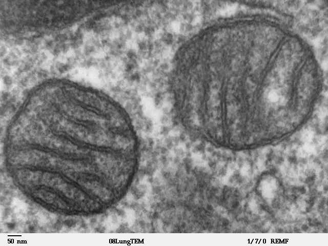
Functions: mitochondria are where most of a cell’s aerobic respiration happens. Glucose is first split in the cytoplasm; the pieces enter the mitochondrion, where the enzymes of the matrix break them down to carbon dioxide (the Krebs cycle), and the enzymes on the cristae use oxygen to make most of the cell’s ATP (adenosine triphosphate), the small molecule that carries energy to wherever the cell needs it. A mitochondrion is often called the powerhouse of the cell. Cells that use much energy have the most of them: muscle cells, the cells of the liver and kidneys, nerve cells, the tail of a sperm.
Because a mitochondrion has its own DNA and ribosomes, it makes some of its own proteins and grows and divides by itself, but it needs many proteins made by the cell’s nucleus as well. So it is called semi-autonomous. Its DNA is passed on only from mothers to their children, because a sperm’s mitochondria are destroyed in the egg after fertilization.
Plastids
Plastids are organelles found only in plants and algae. Each has two membranes, its own DNA and its own 70S ribosomes, and like mitochondria they can divide. There are three kinds, by color:
- Chloroplasts, green, containing the green pigment chlorophyll. They make food by photosynthesis.
- Chromoplasts (chroma, color), yellow, orange or red, containing carotenoids: carotene (orange) and xanthophyll (yellow), and in ripe tomatoes red lycopene. They color many flowers and fruits, and carrots, attracting the insects and birds that carry pollen and seeds.
- Leucoplasts (leukos, white), colorless, found in parts out of the light, such as roots and underground stems. They store food: amyloplasts store starch (a potato is packed with them), elaioplasts store oils and fats, and aleuroplasts store proteins. (Much of the oil and protein in a mustard seed is in fact kept outside plastids, in oil bodies and in protein-filled vacuoles of the cytoplasm; for the exam, the three kinds of leucoplast are as listed.)
One kind of plastid can turn into another. A green tomato is green because of chloroplasts; as it ripens they become chromoplasts and it turns red. A potato left in the light turns green on its surface, as its leucoplasts become chloroplasts (green potato skin also makes a poison, solanine, so cut it off before cooking).
The chloroplast
A chloroplast in a leaf cell is usually a lens-shaped disc about 5 µm long and 2–3 µm thick; a cell of a leaf’s palisade layer may have 20 to 100. Algae have other shapes: a cup in Chlamydomonas, a spiral ribbon in Spirogyra. Inside (Figure 1.8):
- A double envelope, an outer and an inner membrane.
- The stroma, a fluid filling it, with enzymes, starch grains, the chloroplast’s own DNA and 70S ribosomes.
- Thylakoids, flat sacs of membrane, stacked like coins into piles called grana (singular granum), with 10–20 thylakoids in a typical granum. The chlorophyll is in the thylakoid membranes.
- Stroma lamellae, single thylakoids joining one granum to the next.
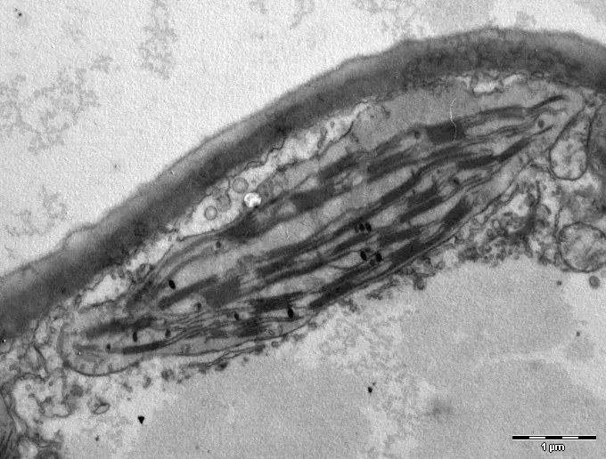
Functions: chloroplasts carry out photosynthesis. In the thylakoids, chlorophyll absorbs light, and its energy splits water, giving off oxygen, and makes ATP. In the stroma, that ATP and the hydrogen from water turn carbon dioxide into glucose. So the oxygen we breathe is made in thylakoids, and almost all the food anyone eats was first made in a stroma. Glucose can be stored in the chloroplast as starch grains.
Potato tubers store starch in:
Pick an answer to see why.
Nucleus
The nucleus is the largest organelle of most cells, a ball usually 5–10 µm across, seen by the Scottish botanist Robert Brown in orchid cells in 1831, who gave it its name. Most cells have one; some have many (the long fibers of our skeletal muscles, the threads of some fungi), and a few have none once mature, such as our red blood cells and the sieve tubes of phloem, which can’t then divide.
Structure (Figure 1.10):
- The nuclear envelope: two membranes with a narrow space between them. The outer one is joined to the endoplasmic reticulum and studded with ribosomes. The envelope is crossed by thousands of nuclear pores, each a ring of proteins that controls what passes between nucleus and cytoplasm: instructions (as RNA) and ribosome parts go out; proteins made in the cytoplasm come in.
- The nucleoplasm: the fluid inside.
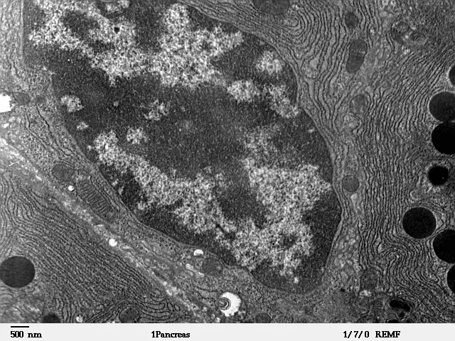
- Chromatin, threads of DNA wound round proteins (histones). Some of it is loosely packed and stains lightly (euchromatin): its genes are being read. Some is tightly packed and stains darkly (heterochromatin): its genes are mostly switched off. When the cell divides, the chromatin coils up tight into short, thick chromosomes, which can be seen with a light microscope.
- One or more nucleoli (singular nucleolus), dense, round bodies with no membrane, where the parts of ribosomes (ribosomal RNA and proteins) are made and put together.
Functions:
- It holds the cell’s DNA, its hereditary instructions, and passes a copy to each new cell when the cell divides; through the DNA it controls which proteins the cell makes, and so everything the cell does. It is the cell’s control center.
- It makes RNA: the copies of genes that go out to the ribosomes.
- Its nucleolus makes the parts of ribosomes.
- It starts cell division, with the copying of its DNA.
Golgi bodies
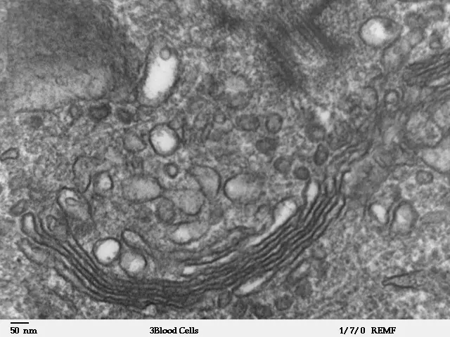
The Golgi body (Golgi apparatus, or Golgi complex) was first seen in nerve cells in 1898 by the Italian Camillo Golgi, using a silver stain he had invented. For half a century many scientists thought it was a mistake made by the stain, until the electron microscope showed it clearly. In plant cells there are many small ones, called dictyosomes.
Structure: a stack of 4–8 (sometimes more) flattened, curved sacs of membrane called cisternae (singular cisterna), with many small round vesicles budding off their edges. The stack has two faces: the cis face (receiving side), facing the endoplasmic reticulum, which receives vesicles from it, and the trans face (shipping side), on the other side, from which finished products leave in vesicles. Proteins and lipids pass through it from cis to trans, being changed on the way.
Functions: the Golgi body is the cell’s packing and shipping department.
- It modifies proteins and lipids made by the endoplasmic reticulum, often adding sugars to them.
- It sorts and packs them into vesicles, each labeled for where it is going.
- It secretes: its vesicles carry products such as digestive enzymes, hormones and mucus to the cell membrane, join it and empty outside the cell. So it is large in cells that secrete, such as those of the pancreas and of glands.
- It makes lysosomes.
- In plant cells it makes the materials of the cell wall, and when the cell divides, its vesicles build the cell plate that becomes the new wall between the two daughter cells.
- In a sperm it forms the acrosome, the cap of enzymes with which the sperm enters the egg.
Centrosome
The centrosome (“central body”), named by the German Theodor Boveri in 1888, lies near the nucleus of animal cells. It is made of two centrioles lying at right angles to each other, in a cloud of dense material (the centrosphere). Most flowering plants and conifers have no centrioles; algae and plants with swimming sperm (mosses, ferns) have them.
Structure of a centriole (Figure 1.12): a short, hollow cylinder about 0.25 µm wide and 0.5 µm long. Its wall is made of nine triplets of microtubules (nine groups of three tiny tubes of protein), set at a slant like the blades of a pinwheel. There is no tube in the middle, so the pattern is called 9 + 0. In a young centriole a hub and nine spokes, like a cartwheel, join the triplets.
Functions:
- When an animal cell divides, the centrosome is copied and the two centrosomes move to opposite ends of the cell. Between them grows the spindle, a set of microtubules that pulls the chromosomes apart, one copy to each end.
- It organizes the cell’s microtubules, its skeleton of tubes, along which vesicles and organelles are carried.
- Centrioles form the basal bodies at the base of cilia and flagella, from which these grow. A basal body has the centriole’s nine triplets; the cilium or flagellum growing from it has nine pairs of microtubules round two single ones in its middle (9 + 2). They beat to move the cell (a sperm, Paramecium) or to move things past it (the cilia that sweep dust out of our windpipe).
Lysosome
Lysosomes (Greek lysis, breaking up, and soma, body) were found by the Belgian Christian de Duve, who noticed in the early 1950s that a digestive enzyme from rat liver cells became fully active only after the tiny particles holding it had been damaged, as if it had been locked away in a bag. He named the bags lysosomes in 1955 and shared the 1974 Nobel Prize, partly for this discovery. They are found in animal cells; in plant cells the vacuole does most of their work.
Structure: a small round sac, usually 0.1–1 µm across, bounded by a single membrane and filled with about fifty kinds of digestive enzymes (hydrolases), which can break down proteins, fats, sugars and nucleic acids. Inside it is acidic, at a pH of about 4.5–5, and the enzymes work best in acid; the membrane pumps acid in and protects the rest of the cell. Lysosomes are made by the Golgi body. A new lysosome that hasn’t yet digested anything is a primary lysosome; once it has joined with something to digest, it is a secondary lysosome.
Functions:
- Digestion inside the cell. When a white blood cell swallows a bacterium, the bubble it is caught in joins with lysosomes, and their enzymes digest it. Amoeba digests its food the same way.
- Clearing and recycling (autophagy, “self-eating”): a worn-out mitochondrion or other part is wrapped in membrane and digested by lysosomes, and the useful molecules go back into the cytoplasm to be used again.
- Removing whole cells and tissues that are no longer needed, such as the tail of a tadpole as it becomes a frog.
- When a cell is badly damaged or dies, its lysosomes may burst and digest the cell itself (autolysis). For this reason lysosomes are sometimes called the suicide bags of the cell.
Vacuole
A vacuole is a sac bounded by a single membrane and filled with a watery fluid. In a mature plant cell there is usually one large central vacuole, which can take up 80–90% of the cell’s volume and push the cytoplasm and nucleus into a thin layer against the wall. Its membrane is the tonoplast, and its fluid, the cell sap, is water with sugars, salts, acids, pigments and wastes dissolved in it. Animal cells have only small, often short-lived vacuoles, if any. Some protists living in fresh water, such as Amoeba and Paramecium, have contractile vacuoles that fill with the water that keeps entering the cell and squirt it out, and food vacuoles in which food is digested.
Functions:
- It keeps a plant cell firm: water entering the cell sap by osmosis pushes the cytoplasm out against the wall, making the cell turgid. A plant wilts when its vacuoles lose water.
- It stores water, food (sugars, and proteins in seeds) and salts.
- It stores wastes and harmful substances away from the cytoplasm, such as the tannins of tea leaves and crystals of calcium oxalate (the needle-like crystals that make the raw stems of pindalu, taro, itch the throat).
- It holds pigments: the red, purple and blue of many flowers and fruits are anthocyanins in the cell sap of their vacuoles (a rhododendron’s red petals, purple cabbage, a red onion’s outer layers), unlike the yellow and orange carotenoids of chromoplasts.
- In a plant cell it does the work of lysosomes, breaking down old parts with its enzymes.
The red of a rhododendron petal is in its cells’:
Pick an answer to see why.
Ribosome
Ribosomes are the smallest organelles, about 20–30 nm across, and they have no membrane. They were first described clearly in 1955 by the Romanian-American George Palade, who shared the 1974 Nobel Prize with de Duve. Each is made of two parts, a large subunit and a small subunit, built of ribosomal RNA and proteins; the two parts come together only when the ribosome is making a protein. A busy cell has millions of them. They lie free in the cytoplasm, or bound to the endoplasmic reticulum and the outer nuclear membrane; several may read one strand of RNA at once, making a chain called a polysome.
Ribosomes are measured by how fast they sink in a fast-spinning centrifuge, in Svedberg units (S). The ribosomes in a eukaryote’s cytoplasm are 80S, made of 60S and 40S subunits; those of prokaryotes, and of mitochondria and chloroplasts, are 70S, made of 50S and 30S subunits. The numbers don’t add up (60 + 40 is not 80) because how fast something sinks depends on its shape as well as its mass.
Function: ribosomes make proteins (protein synthesis). A ribosome reads the copy of a gene (messenger RNA) that has come out from the nucleus and joins amino acids together in the order it gives. Free ribosomes make the proteins the cell uses in its own cytoplasm; ribosomes on the ER make proteins that are sent out of the cell, or into membranes and lysosomes.
Endoplasmic reticulum
The endoplasmic reticulum (ER; “network inside the cytoplasm”) is a network of membranes running through the cytoplasm of eukaryotic cells: flattened sacs (cisternae), tubes (tubules) and small sacs (vesicles), all enclosing one connected space, the lumen. It is joined to the outer membrane of the nucleus, and it was first seen in 1945 in some of the first electron micrographs of whole cells, by Keith Porter, Albert Claude and Ernest Fullam. It is of two kinds:
- Rough ER (RER), flattened cisternae studded with ribosomes on their outer face, which make them look rough. It makes proteins to be sent out of the cell or into membranes and lysosomes: the proteins pass into its lumen as they are made, are folded there, and are sent on in vesicles to the Golgi body. It is plentiful in cells that make and secrete proteins: the cells of the pancreas that make digestive enzymes, the cells that make antibodies.
- Smooth ER (SER), mostly tubules, with no ribosomes. It makes lipids: the phospholipids of the cell’s membranes and the steroid hormones (in the cells of the testes, ovaries and adrenal glands). In liver cells it detoxifies drugs, alcohol and poisons, making them easier to remove. In muscle cells a special smooth ER (the sarcoplasmic reticulum) stores calcium, which it lets out to make the muscle contract.
Functions of the ER as a whole: it is a system of channels that carries substances through the cell; it divides the cytoplasm into compartments, so different reactions can go on in different places; it gives a large surface for the enzymes that work on it; it makes new membrane for the cell; and it supports the cytoplasm.
How a cell makes and sends out a protein
The ER, Golgi body, vesicles and cell membrane work together as one system (often called the endomembrane system). Follow a digestive enzyme made by a cell of the pancreas:
- In the nucleus, the enzyme’s gene is copied into messenger RNA, which goes out through a nuclear pore.
- A ribosome on the rough ER reads it and makes the enzyme, threading it into the ER’s lumen.
- The enzyme is folded in the ER and carried in a vesicle to the cis face of the Golgi body.
- It passes through the Golgi’s cisternae, is finished (sugars may be added), and leaves the trans face in a vesicle.
- The vesicle joins the cell membrane and empties the enzyme out of the cell (exocytosis), into a duct that leads to the small intestine.
Every step needs energy, as ATP from the mitochondria. In 1967, by feeding cells of the pancreas radioactive amino acids for a few minutes and taking electron micrographs at intervals afterwards, George Palade’s team watched new protein travel exactly this way: rough ER first, then the Golgi body, then the vesicles, then out.
In which order does a protein to be secreted pass through these parts?
Pick an answer to see why.
1.3 Relation of cell functions with respiration, excretion, reproduction and photosynthesis
No organelle works alone. Each life process of a cell is a chain of jobs done by several parts, with materials passed from one to the next. Tap a process below to see which parts of an animal cell and a plant cell take part.
Respiration
Respiration releases the energy stored in food, as ATP. Every living cell respires, day and night, plant cells as well as animal cells. In aerobic respiration (with oxygen):
It happens in two places. In the cytoplasm, each glucose molecule is split into two smaller molecules (pyruvate), which gives a little ATP and needs no oxygen (glycolysis). These enter the mitochondria, where the Krebs cycle in the matrix breaks them down to carbon dioxide, and the enzymes on the cristae use oxygen to make most of the ATP. The cell membrane lets oxygen in and carbon dioxide out by diffusion. When oxygen runs short (in a muscle working very hard, or in yeast in dough), only the first step can happen: the cell gets far less energy, and makes lactic acid (muscle) or alcohol and carbon dioxide (yeast) instead.
Photosynthesis
Photosynthesis makes food from carbon dioxide and water, using the energy of light:
It happens only in the chloroplasts: light is absorbed by chlorophyll in the thylakoids, where water is split and oxygen given off, and glucose is made in the stroma. The cell wall and membrane let carbon dioxide in and oxygen out; the vacuole, by keeping the cells of the leaf turgid, keeps the leaf spread out to the light. Animal cells, and the cells of a plant’s roots and other parts away from the light, have no chloroplasts and can’t photosynthesize. The two processes fit together: the glucose and oxygen made in chloroplasts are what mitochondria use, and the carbon dioxide and water mitochondria give out are what chloroplasts take in.
Excretion
Excretion is the removal of the wastes made by the cell’s own chemistry: carbon dioxide from respiration, ammonia and urea from breaking down proteins, and others. In a cell:
- Lysosomes digest worn-out parts and whatever the cell has taken in; anything useful goes back to the cytoplasm, and what is left is thrown out.
- The smooth ER of liver cells changes drugs and poisons into harmless forms that can be removed.
- The Golgi body packs substances into vesicles, which carry them out of the cell.
- Vacuoles store wastes. Plants have no organs of excretion, and keep many wastes in the cell sap of their vacuoles (tannins, crystals, alkaloids such as the nicotine of tobacco), shedding them with old leaves and bark. The contractile vacuole of Amoeba pumps out extra water.
- The cell membrane lets carbon dioxide and urea out of the cell, into the blood (in an animal) or the air spaces of the leaf (in a plant).
Reproduction
A cell reproduces by dividing into two. In a eukaryotic cell:
- The nucleus comes first: its DNA is copied, its chromatin coils into chromosomes, and the two copies of each are shared equally between the two new cells. So each daughter cell gets the full set of instructions.
- In an animal cell the centrosome is copied, and the two centrosomes, at opposite ends of the cell, form the spindle that pulls the chromosomes apart. Most plant cells make a spindle without centrioles.
- Mitochondria (and chloroplasts) supply the ATP for division, and divide by themselves, so each new cell gets some; the cell can’t make new ones from nothing.
- At the end the cytoplasm divides: an animal cell’s cell membrane pinches in round the middle, while in a plant cell vesicles from the Golgi body gather across the middle and join into a cell plate, which becomes the new wall.
- The cell’s ribosomes make the many new proteins the two cells need as they grow.
A prokaryote divides more simply, by binary fission: it copies its circular DNA, the copies move apart as the cell grows, and a wall grows across the middle.
Exercise
1. Choose the correct option
a. Which of these organisms is made of prokaryotic cells?
Pick an answer to see why.
b. The cell wall of a bacterium is made of:
Pick an answer to see why.
c. The fluid mosaic model describes the structure of the:
Pick an answer to see why.
d. The F₁ particles (oxysomes) that make ATP are found on the:
Pick an answer to see why.
e. The part of the nucleus where ribosome parts are made is the:
Pick an answer to see why.
f. The cell plate in a dividing plant cell is formed by:
Pick an answer to see why.
g. A centriole is made of:
Pick an answer to see why.
h. Which organelle is called the “suicide bag” of the cell?
Pick an answer to see why.
i. Steroid hormones are made by the:
Pick an answer to see why.
2. Give reasons
A plant cell doesn’t burst when it is placed in pure water, but a red blood cell does.
Hint
Answer
Mitochondria are called the powerhouse of the cell.
Hint
Answer
Mitochondria and chloroplasts are called semi-autonomous organelles.
Hint
Answer
The inner membrane of a mitochondrion is folded.
Hint
Answer
Lysosomes are called the suicide bags of the cell.
Hint
Answer
Cells of the pancreas have a lot of rough ER and many Golgi bodies.
Hint
Answer
Root cells of a plant have no chloroplasts.
Hint
Answer
A ribosome can’t be seen with a school microscope.
Hint
Answer
3. Differentiate between
Prokaryotic cell and eukaryotic cell
Hint
Answer
Cell wall and cell membrane
Hint
Answer
Mitochondrion and chloroplast
Hint
Answer
Rough ER and smooth ER
Hint
Answer
Chloroplast and chromoplast
Hint
Answer
Lysosome and vacuole
Hint
Answer
70S ribosome and 80S ribosome
Hint
Answer
4. Answer the following questions
What are organelles? Name four organelles found in plant cells but not in animal cells, or the other way round.
Hint
Answer
Describe the structure of the cell wall of a plant cell, and give its functions.
Hint
Answer
Describe the fluid mosaic model of the cell membrane, with a labeled diagram.
Hint
Answer
Describe the structure of a mitochondrion with a labeled diagram, and state its functions.
Hint
Answer
What are plastids? Describe their kinds with examples.
Hint
Answer
Describe the structure of a chloroplast with a labeled diagram.
Hint
Answer
Describe the structure and functions of the nucleus.
Hint
Answer
Describe the structure of the Golgi body, and give four functions.
Hint
Answer
Describe the structure of a centrosome, and state its functions.
Hint
Answer
What are ribosomes? Why are they called the protein factories of the cell?
Hint
Answer
Trace the path of a digestive enzyme from the gene that codes for it to its secretion from a cell of the pancreas. Name each organelle and what it does.
Hint
Answer
How are the organelles of a cell related to respiration and photosynthesis? Explain with an equation for each.
Hint
Answer
Which organelles take part in excretion and in reproduction of a cell? Explain briefly.
Hint
Answer
What is the endosymbiotic theory? Give three pieces of evidence for it.
Hint
Answer
5. Numerical problems
A microscope has a 10× eyepiece and a 100× objective. What is its total magnification? An E. coli cell 2 µm long is seen with it: how long does its image appear?
Hint
Answer
In a photomicrograph at 400×, an onion peel cell is 120 mm long and 20 mm wide. Find its real length and width in micrometers.
Hint
Answer
A nucleus 8 µm across is drawn 4 cm across. Find the magnification of the drawing.
Hint
Answer
The field of view of a microscope at 400× is 0.45 mm across. Nine cheek cells lie side by side across it. Find the average width of one cell in micrometers.
Hint
Answer
Find the ratio of surface area to volume for cube-shaped cells of sides 1 µm, 5 µm and 10 µm. What happens to the ratio as a cell gets bigger, and why does this limit a cell’s size?
Hint
Answer
A model of an E. coli cell 2 µm long is made 30 cm long. At the same scale, how big should a ribosome 20 nm across be? Could you see it in the model?
Hint
Answer
Printable PDFs
- The unit as a book (A5) 34 pages Answers as an A4 booklet, 9 sheets
- Study notes 3 pages
- Worksheet 5 pages Answer key
- Harder practice questions 3 pages Answer key
Practice every question in the book, and keep track of the ones you miss →
Found a mistake? Please tell us.