
About one boy in every twelve in some countries can’t tell red from green the way his friends do. A red apple among green leaves looks to him like part of the tree, and a traffic light is read by its place, top or bottom, not its color. Yet among girls in the same countries, only about one in two hundred is color-blind in that way. The same is true of hemophilia, the disease in which blood doesn’t clot: almost everyone who has it is a boy.
Why should one sex suffer so much more than the other? The answer needs three ideas, and this unit builds all three: Mendel’s rules for following two characters at once, the chemistry of DNA and RNA in which every gene is written, and the X chromosome, of which a boy has only one.
3.1 Mendel’s law: law of independent assortment
In a monohybrid cross, two pure parents differ in one pair of characters, and the F₂ comes out 3 : 1. Mendel didn’t stop there. He wanted to know what happens when parents differ in two characters at once. Do the two travel together, as a package from each grandparent, or does each go its own way?
A cross between two pure parents that differ in two pairs of contrasting characters is a dihybrid cross. Mendel’s best-known one used two characters of the pea seed:
| Character | Dominant | Recessive |
|---|---|---|
| Seed shape | round (R) | wrinkled (r) |
| Seed color | yellow (Y) | green (y) |
Mendel’s dihybrid cross
Parents (P). Mendel crossed plants pure for round yellow seeds (RRYY) with plants pure for wrinkled green seeds (rryy). A gamete gets one allele of each gene, so every gamete of the first parent is RY and every gamete of the second is ry.
First filial generation (F₁). Every seed was RrYy, and every one was round and yellow: both dominant characters showed, just as each would in a monohybrid cross.
Gametes of F₁. Here is the key step. An RrYy plant puts R or r into a gamete, and Y or y, and the two choices don’t depend on each other. So it makes four kinds of gametes, RY, Ry, rY and ry, in equal numbers, a quarter each.
Second filial generation (F₂). Mendel let the F₁ plants self-pollinate. Each of the four kinds of pollen can meet each of the four kinds of egg, so there are 4 × 4 = 16 equally likely meetings. In his F₂, Mendel counted 556 seeds:
| Phenotype | Seeds counted | Expected out of 556 (9 : 3 : 3 : 1) |
|---|---|---|
| Round yellow | 315 | 312.75 |
| Round green | 108 | 104.25 |
| Wrinkled yellow | 101 | 104.25 |
| Wrinkled green | 32 | 34.75 |
| Total | 556 | 556 |
The expected numbers are 556 × 9/16, 556 × 3/16 and 556 × 1/16. Mendel’s counts are close to them: the F₂ phenotypic ratio is 9 : 3 : 3 : 1. Two of the four kinds, round green and wrinkled yellow, were in neither grandparent. They are new combinations (also called recombinants), and they could only appear if shape and color had separated.
Phenotypic chart of the dihybrid cross
The 16 meetings are set out in a checkerboard (a Punnett square): the F₁ male gametes across the top, the female gametes down the side, and in each box the genotype of the seed made where they meet. This chart, with the generations above it, is how the cross is drawn in an exam.
Read the checkerboard box by box and you find nine different genotypes:
| Phenotype | Genotypes (boxes out of 16) | Total |
|---|---|---|
| Round yellow | RRYY (1), RRYy (2), RrYY (2), RrYy (4) | 9 |
| Round green | RRyy (1), Rryy (2) | 3 |
| Wrinkled yellow | rrYY (1), rrYy (2) | 3 |
| Wrinkled green | rryy (1) | 1 |
So the F₂ genotypic ratio is RRYY : RRYy : RrYY : RrYy : RRyy : Rryy : rrYY : rrYy : rryy = 1 : 2 : 2 : 4 : 1 : 2 : 1 : 2 : 1, sixteen boxes in all. Notice the pattern: the four genotypes pure for both genes (RRYY, RRyy, rrYY, rryy) appear once each; the four pure for one gene and hybrid for the other appear twice each; and RrYy, hybrid for both, appears four times. The four pure ones lie on one diagonal of the checkerboard, and RrYy on the other.
Statement of the law
Separate the two characters and look at each alone. Of Mendel’s 556 seeds, 315 + 108 = 423 were round and 101 + 32 = 133 wrinkled: 423 ÷ 133 = 3.18, about 3 : 1. And 315 + 101 = 416 were yellow and 108 + 32 = 140 green: 416 ÷ 140 = 2.97, again about 3 : 1. Each character on its own behaves exactly as in a monohybrid cross, as if the other weren’t there. Two independent 3 : 1 ratios together make
since 3 × 3 = 9 round yellow, 3 × 1 = 3 round green, 1 × 3 = 3 wrinkled yellow and 1 × 1 = 1 wrinkled green. This is Mendel’s law of independent assortment:
When two pairs of contrasting characters are followed together, the alleles of one pair separate into the gametes independently of the alleles of the other pair. So an individual hybrid for two genes makes all four combinations of their alleles in equal numbers, and the characters can appear in new combinations in the offspring.
How many kinds of gametes does a plant of genotype RrYy make, and in what proportion?
Pick an answer to see why.
Why the law works: chromosomes in meiosis
Mendel had never seen a chromosome. We now know why his law holds. The gene for seed shape and the gene for seed color lie on different chromosomes of the pea. In meiosis I, each pair of homologous chromosomes lines up at the middle of the cell, and which member of a pair faces which pole is a matter of chance, decided separately for every pair. With two pairs there are two ways to line up, equally likely, and each gives two kinds of gamete. (To watch a whole meiosis stage by stage, see 1.2 Meiosis cell division in Unit 1.)
This is the link Sutton and Boveri saw in 1902 to 1903 (Grade 9’s chromosome theory of inheritance): Mendel’s factors assort independently because the chromosomes that carry them do.
Verification of the law of independent assortment
A law needs a test. If the F₁ (RrYy) really makes four kinds of gametes in equal numbers, there are two ways to see them.
1. The test cross
Cross the F₁ with a plant that is recessive for both genes (rryy). Every gamete of rryy is ry, carrying only recessive alleles, so it hides nothing: each offspring shows the alleles that came from the F₁ parent’s gamete.
| RY | Ry | rY | ry | |
|---|---|---|---|---|
| ry | RrYy, round yellow | Rryy, round green | rrYy, wrinkled yellow | rryy, wrinkled green |
If the F₁ makes the four gametes equally, the offspring come out 1 : 1 : 1 : 1. A cross of an individual with a parent recessive for the genes being studied is called a test cross; with two genes it is a dihybrid test cross.
Mendel did exactly this, both ways round. With the hybrid as the seed (female) parent and rryy as the pollen parent, he got 31 round yellow, 26 round green, 27 wrinkled yellow and 26 wrinkled green seeds. With rryy as the seed parent and the hybrid as the pollen parent, he got 24, 25, 22 and 27. Both are close to 1 : 1 : 1 : 1, so both the eggs and the pollen of the hybrid were of four kinds in equal numbers, as the law says.
2. Growing the F₂ for one more generation
Mendel also sowed his F₂ seeds and let each plant self-pollinate. A seed pure for both genes (RRYY, say) gives seeds all of one kind; a seed hybrid for one gene gives two kinds; RrYy gives all four again. The checkerboard predicts that the pure-for-both, hybrid-for-one and hybrid-for-both seeds come in the ratio 1 : 2 : 4. From the round yellow seeds alone, Mendel found 38 plants that bred true (RRYY), 65 and 60 hybrid for one gene (RRYy and RrYY), and 138 hybrid for both (RrYy). Over all nine genotypes, the pure ones averaged 33 plants each, the half-hybrid ones 65 and the double hybrid 138: close to 33 : 66 : 132, that is, 1 : 2 : 4. The genotypes were in the proportions the checkerboard gives.
Working out crosses quickly: the product rule
A checkerboard always works, but there is a faster way, which follows straight from the law. Because the two genes assort independently, the chance of a combination is the chance of one character multiplied by the chance of the other. This is the product rule of probability. Work out each gene on its own, as a monohybrid cross, then multiply.
3.2 RNA and DNA: their structure in detail
Grade 10 Science gave the outline: DNA is a double helix of nucleotides with the bases A, T, G and C, and RNA is a single strand with U in place of T. Now we look at what each part is made of and how the parts are joined.
The three parts of a nucleotide
Both DNA and RNA are polynucleotides: long chains of nucleotides. Every nucleotide has three parts.
1. Phosphoric acid. Phosphoric acid is H₃PO₄. In a nucleotide it is joined to the sugar, having lost water, and is present as a phosphate group. It carries a negative charge in the cell, which is why DNA and RNA are acids: nucleic acids.
2. A pentose sugar. The sugar has five carbon atoms, numbered 1′ to 5′ (read “one prime” to “five prime”; the primes keep the sugar’s numbers apart from the base’s). Four carbons and an oxygen make a five-sided ring; the 5′ carbon sticks out of it.
- In RNA the sugar is ribose, C₅H₁₀O₅, with an –OH group on its 2′ carbon.
- In DNA it is deoxyribose, C₅H₁₀O₄: the same, but with only H on the 2′ carbon. “Deoxy” means one oxygen less.
3. A nitrogen base. The bases are flat rings of carbon and nitrogen atoms, of two kinds.
- Purines have two rings, a six-sided ring joined to a five-sided ring: adenine (A) and guanine (G).
- Pyrimidines have one six-sided ring: cytosine (C), thymine (T) and uracil (U).
| Base | Kind | Formula | Found in |
|---|---|---|---|
| Adenine (A) | purine | C₅H₅N₅ | DNA and RNA |
| Guanine (G) | purine | C₅H₅N₅O | DNA and RNA |
| Cytosine (C) | pyrimidine | C₄H₅N₃O | DNA and RNA |
| Thymine (T) | pyrimidine | C₅H₆N₂O₂ | DNA only |
| Uracil (U) | pyrimidine | C₄H₄N₂O₂ | RNA only |
A sugar joined to a base is a nucleoside: adenine with ribose is adenosine, with deoxyribose deoxyadenosine. A nucleoside with a phosphate on its 5′ carbon is a nucleotide:
The bonds that hold them together
CDC’s course asks for the bonding among the parts. There are three kinds of bond, and each has its own job.
1. The N-glycosidic bond (sugar to base). The base joins the 1′ carbon of the sugar through one of its nitrogen atoms: nitrogen 9 of a purine, or nitrogen 1 of a pyrimidine. It is a strong covalent bond.
2. The phosphodiester bond (sugar to sugar, through a phosphate). The phosphate on the 5′ carbon of one nucleotide joins the 3′ carbon of the next. So the phosphate is joined to two sugars by two ester bonds, a phosphodiester bond. Sugar, phosphate, sugar, phosphate: these make the sugar–phosphate backbone of the strand, with the bases sticking out to one side. The bond is covalent and strong, so the order of the bases along a strand doesn’t change by itself.
A strand has two different ends. At one end a sugar’s 5′ carbon has a phosphate not joined to anything further: the 5′ end. At the other end a sugar’s 3′ carbon has a free –OH: the 3′ end. A strand has a direction, like a written word, and its sequence is always read from 5′ to 3′.
3. Hydrogen bonds (base to base, across the strands). In DNA the bases of one strand pair with the bases of the other: adenine with thymine by two hydrogen bonds, and guanine with cytosine by three. A hydrogen bond is a weak attraction between a hydrogen atom joined to a nitrogen and a nitrogen or oxygen atom of the other base. One alone is about twenty times weaker than a covalent bond, but millions together hold the two strands firmly, and they can still be opened a few at a time when the DNA is copied or read.
Why does A pair only with T, and G only with C? Two reasons, both visible in Figure 3.4. A purine (two rings) always pairs with a pyrimidine (one ring), so every rung of the ladder is three rings wide and the helix is the same width all along; two purines would bulge, two pyrimidines would leave a gap. And of the possible purine–pyrimidine pairs, only A–T and G–C put their hydrogen atoms opposite atoms that can attract them. Pairing that follows these rules is complementary base pairing: knowing one strand, you know the other.
The double helix of DNA
In 1953 James Watson and Francis Crick, using the X-ray photographs of Rosalind Franklin and Maurice Wilkins (the story is in Grade 10 Science), proposed the structure of DNA. Its main features:
- DNA has two polynucleotide strands twisted round a common axis into a right-handed double helix, like a spiral staircase that turns clockwise as it rises away from you.
- The sugar–phosphate backbones are on the outside; the bases are inside, stacked flat like the steps of the staircase.
- The two strands are antiparallel: one runs 5′ → 3′ upward, the other 5′ → 3′ downward.
- The strands are held together by hydrogen bonds between complementary bases, A with T (two) and G with C (three).
- The helix is about 2 nm wide. Neighboring base pairs are 0.34 nm apart, and the helix makes one full turn in about 10 base pairs, that is, about 3.4 nm. (Measurements of DNA in water give about 10.5 base pairs a turn; the round 10 is the figure most books use.)
- The backbones twist unevenly, leaving two grooves of different widths along the helix, the major groove and the minor groove, where proteins reach in to read the bases.
Most of these features can be seen in a real piece of DNA. Below are the atoms of the Dickerson–Drew dodecamer, twelve base pairs whose structure was found by X-rays in 1981; the measurements in the box are taken from its atoms.
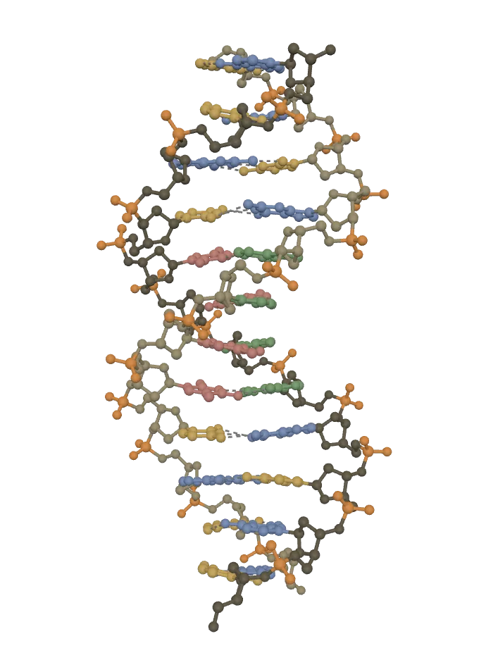
This browser can’t show the 3D model. The picture above is the same model.
Twelve base pairs of real DNA: a right-handed helix, the backbones outside, the bases stacked inside 0.34 nm apart, one turn in about 10 pairs. Turn it side-on for the wide major and narrow minor grooves. (X-rays don’t place hydrogen atoms, so none are shown.)
Sugar and phosphateThe sides of the ladder: each strand is a chain of deoxyribose sugars (brown; one strand darker) joined through phosphate groups (orange).
Adenine (A)Always paired with thymine (T) on the other strand, by two hydrogen bonds (dashed).
Thymine (T)Always paired with adenine (A) on the other strand, by two hydrogen bonds (dashed).
Guanine (G)Always paired with cytosine (C) on the other strand, by three hydrogen bonds (dashed).
Cytosine (C)Always paired with guanine (G) on the other strand, by three hydrogen bonds (dashed).
Drag to turn it. Tap a part, or a name below, to pick it out; tap it again to see the whole.
DNA: PDB 1BNA, Drew, Dickerson and others (1981), RCSB Protein Data Bank, CC0: www.rcsb.org/structure/1BNA
From Chargaff’s rules, A = T and G = C, so A + G = T + C: purines make up half the bases of DNA, and given one base’s share, you can find all four.
The structure of RNA
RNA is also a chain of nucleotides joined by phosphodiester bonds, with a 5′ end and a 3′ end, but:
- its sugar is ribose;
- its bases are A, G, C and U: uracil takes the place of thymine, and pairs with adenine by two hydrogen bonds;
- it is usually a single strand. A single strand can still fold back on itself where its bases happen to be complementary, making short double-stranded stretches (stems) and loops. Its folded shape gives each kind of RNA its job.
RNA is made by copying one strand of DNA (transcription), so its sequence is complementary to that DNA strand, with U wherever the DNA strand has A.
Which bond joins the phosphate of one nucleotide to the sugar of the next in a strand of DNA?
Pick an answer to see why.
3.3 RNA: types and function
A gene in DNA stays in the nucleus. To make the protein it codes for, the cell uses RNA, and three kinds of RNA share the work. How they work together, step by step, is told in How does a gene make a protein?; here we look at each kind.
Messenger RNA (mRNA)
Messenger RNA is a copy of one gene, made in the nucleus by transcription, which carries the gene’s message out to the ribosomes in the cytoplasm.
- Structure: a single, unfolded strand, as long as its gene: a few hundred to many thousands of nucleotides. Its bases are read three at a time; each group of three, a codon, stands for one amino acid. In our cells an mRNA gets a protective cap at its 5′ end and a tail of a hundred or more A’s at its 3′ end.
- Function: it carries the genetic code from DNA to the ribosome, where it is the template that sets the order of amino acids in the protein.
- It is short-lived: most mRNAs in a cell last minutes to hours before they are broken down, so the cell can change which proteins it makes. It makes up only a few percent of a cell’s RNA.
Transfer RNA (tRNA)
Transfer RNA is a small RNA that brings amino acids to the ribosome and matches each one to its codon.
- Structure: a single chain, typically 76 to 90 nucleotides, the smallest of the three kinds. Its complementary stretches pair up into four short stems, and it folds into the shape of a clover leaf, which in three dimensions is folded again into an L.
- The acceptor arm: the two ends of the chain, paired; the 3′ end always ends in the bases CCA, and the amino acid is attached to its last A.
- The anticodon arm: its loop carries the anticodon, three bases that pair with a codon on the mRNA.
- Two more arms with loops (the D arm and the T arm, named after unusual bases found in them) help the molecule fold and fit the ribosome.
- Function: each tRNA carries one particular amino acid; when its anticodon pairs with the matching codon on the mRNA, it leaves its amino acid in the right place in the growing protein. It is the adapter between the language of bases and the language of amino acids. Cells have dozens of kinds of tRNA, at least one for each of the 20 amino acids.
Ribosomal RNA (rRNA)
Ribosomal RNA is the RNA of the ribosomes.
- Structure: long RNA chains folded tightly together with dozens of proteins into the two subunits of a ribosome. In our cells there are four kinds (named by how fast they settle in a centrifuge: 28S, 18S, 5.8S and 5S); bacteria have three (23S, 16S and 5S). It is made in the nucleolus (Grade 9 Optional Science, Ribosome).
- Function: it forms the structure of the ribosome, holds the mRNA and the tRNAs in place, and, remarkably, it is the rRNA of the large subunit, not a protein, that joins each new amino acid to the chain. So the machine that makes every protein is itself mostly RNA.
- It is the most plentiful kind: about four-fifths of all the RNA in a cell.
| mRNA | tRNA | rRNA | |
|---|---|---|---|
| Full name | messenger RNA | transfer RNA | ribosomal RNA |
| Share of a cell’s RNA | a few percent | about a sixth | about four-fifths |
| Shape | a straight single strand | a folded clover leaf (an L) | folded with proteins into ribosomes |
| Size | hundreds to thousands of nucleotides | about 76 to 90 nucleotides | about 120 to 5,000 nucleotides |
| Job | carries the code of one gene to the ribosome | brings amino acids and matches them to codons | builds the ribosome and joins the amino acids |
| Special part | codons | anticodon, CCA end | – |
Which part of a tRNA pairs with the mRNA?
Pick an answer to see why.
3.4 Role of DNA in genetics
Everything genetics studies, from the color of a pea to a disease that runs in a family, comes back to DNA. Its roles are these.
1. It stores the genetic information
The order of the bases along DNA is the genetic information. A gene is a stretch of DNA whose base sequence codes for one protein or one RNA. With four bases, even a short stretch can be written in an enormous number of ways: a stretch of just ten base pairs can be arranged in 4¹⁰, about a million, different ways, and one set of your chromosomes holds about 3.1 billion base pairs and about 20,000 genes for proteins. DNA is also stable: its backbone is strong, and its two strands keep a copy of each other, so damage on one strand can be repaired from the other.
2. It copies itself: replication
Before a cell divides, its DNA is copied, so each daughter cell gets the full set of genes. This copying is replication.
- An enzyme breaks the hydrogen bonds and unzips the two strands, starting at many places along the DNA.
- Each old strand acts as a template: an enzyme, DNA polymerase, adds new nucleotides one by one, each pairing with the base it faces, A with T and G with C, and joins them into a new strand (always growing from its 5′ end toward its 3′ end).
- The result is two DNA molecules, exactly alike and exactly like the first. Each has one old strand and one new strand.
Because each new molecule keeps one of the old strands, replication is called semi-conservative (“half kept”).
3. It controls the characters: from gene to protein
DNA itself doesn’t make a pea round or an eye brown. It controls characters through the proteins it codes for: enzymes, pigments, hormones, the proteins of muscle and blood. The flow of information goes DNA → RNA → protein, which Crick called the central dogma of molecular biology.
- Transcription: in the nucleus, a gene’s DNA is copied into mRNA.
- Translation: at a ribosome, the mRNA is read codon by codon; tRNAs bring the amino acids, and rRNA joins them into a protein.
The code is read in codons of three bases. With four bases there are 4³ = 64 codons: 61 stand for the 20 amino acids (most amino acids have more than one codon), and 3 (UAA, UAG and UGA) mean “stop”. AUG both means methionine and marks where a protein starts. The same code is used by almost every living thing, from bacteria to people, which is why a human gene put into a bacterium can make a human protein: this is how insulin for diabetes is made.
Mendel’s round and wrinkled peas are an example. The R allele codes for a starch-branching enzyme, which the seed needs to build its branched starch. The r allele carries an extra stretch of about 800 base pairs in the middle of the gene, so the enzyme isn’t made. A wrinkled (rr) seed keeps more sugar, takes up more water while it grows, and wrinkles when it dries.
4. It brings variation
Children differ from their parents and from each other because of DNA.
- Mutation: a change in the base sequence (one base changed, added or lost, or a larger stretch). A mutation in a gene may change its protein; most mutations do little or harm, a few help. Mutations are the source of all new alleles; the r allele of the pea and the allele for color blindness both began as mutations.
- Recombination: in meiosis, independent assortment (3.1) and crossing over shuffle the alleles a person has into new combinations in each gamete, so every child of the same parents gets a different mix.
Over many generations, variation is what natural selection acts on, so DNA is also the basis of evolution.
5. It is the material passed from generation to generation
Through gametes, DNA carries the genes from parents to offspring: half from each. And its sequence can be read: DNA fingerprinting, in Unit 5, uses the differences in DNA between people to identify them.
How we know genes are made of DNA
Until the 1940s, most scientists thought genes were made of protein, which seemed more varied than a molecule of only four kinds of nucleotide. Three experiments changed their minds.
- Griffith, 1928. Frederick Griffith found that a harmless strain of the pneumonia bacterium became deadly when mixed with dead cells of a deadly strain: something from the dead cells transformed the living ones.
- Avery, MacLeod and McCarty, 1944. They purified the dead cells’ substances one at a time. Only DNA transformed the harmless bacteria; destroying the DNA with an enzyme stopped it, and destroying the proteins didn’t.
- Hershey and Chase, 1952. Alfred Hershey and Martha Chase labeled bacteriophages either in their protein (with radioactive sulphur) or in their DNA (with radioactive phosphorus). Only the DNA went into the bacteria, and new phages were made from it, as you saw in the lytic cycle in Grade 9 Optional Science.
In a few viruses, such as those of flu, measles and COVID-19, the genes are RNA, not DNA. Everywhere else, the genetic material is DNA.
How many codons are there, and how many of them stand for amino acids?
Pick an answer to see why.
3.5 Sex-linked inheritance
A human body cell has 22 pairs of autosomes and one pair of sex chromosomes: XX in a woman, XY in a man (see Grade 9 Optional Science). The sex chromosomes don’t only decide sex. The X chromosome is large, with about 800 genes for proteins, most of which have nothing to do with being male or female: genes for seeing colors, for clotting blood, for the muscles. The Y is small, with only a few dozen different genes for proteins, several of them in many copies.
Genes on the sex chromosomes are sex-linked genes, and the inheritance of the characters they control is sex-linked inheritance. Such characters are passed on differently to sons and daughters, because a son gets his X from his mother and his Y from his father, while a daughter gets one X from each.
Types of sex-linked genes on the basis of location
The X and Y share only small stretches at their tips; the rest of each is its own. Where a gene lies decides how it is inherited.
1. X-linked genes. These lie on the part of the X that has no partner on the Y (the non-homologous part of the X). A woman has two copies of each; a man has only one, and no second copy on his Y to hide a recessive allele. Examples: the genes for red–green color vision, for the clotting factors whose loss causes hemophilia, for the muscle protein dystrophin (Duchenne muscular dystrophy), and for the enzyme G6PD. Almost all sex-linked inheritance in humans is X-linked.
2. Y-linked genes (holandric genes). These lie on the part of the Y that has no partner on the X. They pass only from father to son, to every son, and never to a daughter: holandric means “wholly in males”. The best known is SRY, the gene that makes an embryo develop as a male. Most of the Y’s other genes are needed for making sperm.
3. XY-linked genes (incompletely sex-linked). These lie in the pseudoautosomal regions, the short stretches the X and Y share at their tips, where the two pair up and cross over in meiosis. A gene here has a copy on both the X and the Y, so it is inherited almost like a gene on an autosome. Example: SHOX, a gene needed for the growth of the bones of the arms and legs.
What is special about X-linked inheritance
Most X-linked disorders are caused by a recessive allele. Write the normal X as X and the X with the recessive allele as (for color blindness) or (for hemophilia). Then:
| Normal | Carrier | Affected | |
|---|---|---|---|
| Woman | XX | ||
| Man | XY | none |
A carrier is a person who has one recessive allele, shows no sign of the disorder, and can pass it on. A woman with one is a carrier: her other X hides it. A man with one has no other X, so he is affected. A man has only one copy of each X-linked gene; he is hemizygous for it. So:
- More men than women are affected. A man needs one recessive allele, a woman two. If one X in twelve carries the allele, about 1 man in 12 is affected, but only about 1 woman in 144 (1/12 × 1/12). (The real figure for women, about 1 in 200, is lower still, because red–green color blindness comes from two different genes: a woman with a faulty red gene on one X and a faulty green gene on the other sees normally.)
- A father never passes an X-linked character to his son, because he gives his son his Y, not his X.
- An affected father passes the allele to every daughter, who becomes a carrier (or affected, if her mother gave one too).
- Criss-cross inheritance: the character typically passes from an affected man, through his carrier daughter, to her son, his grandson, skipping a generation.
- An affected woman must have an affected father and a carrier or affected mother; all her sons are affected.
Color blindness
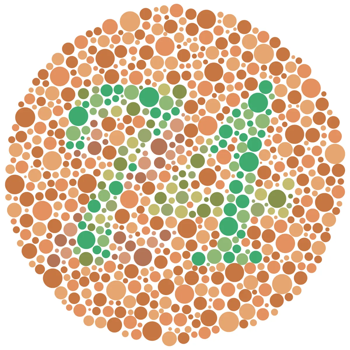
In the retina, light is sensed by cones of three kinds, each with a pigment most sensitive to red, green or blue light. The genes for the red and green pigments sit side by side near the tip of the long arm of the X chromosome. An allele that makes either pigment faulty or missing gives red–green color blindness: red and green, and colors mixed from them, look alike. (The blue pigment’s gene is on chromosome 7.) Among people of Northern European ancestry, red–green color blindness affects about 1 man in 12 and 1 woman in 200.
Here is how color blindness passes in the four kinds of marriage.
Cross 1: a normal woman and a color-blind man.
| Gametes | X (from mother) | X (from mother) |
|---|---|---|
| (from father) | , carrier daughter | , carrier daughter |
| Y (from father) | XY, normal son | XY, normal son |
All the daughters are carriers with normal vision; all the sons are normal.
Cross 2: a carrier woman and a normal man.
| Gametes | (from mother) | X (from mother) |
|---|---|---|
| X (from father) | , carrier daughter | XX, normal daughter |
| Y (from father) | , color-blind son | XY, normal son |
Half the sons are color-blind; half the daughters are carriers; no daughter is color-blind. Put Crosses 1 and 2 together and you see criss-cross inheritance: a color-blind grandfather, a carrier daughter, a color-blind grandson.
Cross 3: a carrier woman and a color-blind man. The daughters are (color-blind) or (carrier), half each; the sons are (color-blind) or XY (normal), half each. This is the only way a daughter can be color-blind: her father must be color-blind and her mother must carry the allele.
Cross 4: a color-blind woman and a normal man. Every daughter is , a carrier; every son is , color-blind.
Hemophilia
Hemophilia is a disorder in which the blood clots very slowly, because one of the proteins needed for clotting, a clotting factor, is missing or faulty. A cut bleeds for a long time, and bleeding inside joints and muscles, even from a bump, is painful and damaging. There are two main kinds, both X-linked recessive:
- Hemophilia A, the commoner, lacks clotting factor VIII; it affects about 1 in 4,000 to 5,000 boys born.
- Hemophilia B (also called Christmas disease, after Stephen Christmas, the first patient described with it, in 1952) lacks factor IX; it affects about 1 in 20,000 boys born.
It passes exactly as color blindness does. With for the hemophilia allele:
A carrier woman () and a normal man (XY) have the four kinds of child of Cross 2: a carrier daughter (), a normal daughter (XX), a hemophilic son () and a normal son (XY), each with a chance of 1 in 4.
A normal woman (XX) and a hemophilic man (): every daughter is a carrier, , and every son normal, XY.
A hemophilic woman, , needs a hemophilic father and a carrier mother, and both alleles are rare, so she is very rare; she is not, as older books say, impossible. Today hemophilia is treated by injecting the missing clotting factor, made from donated blood or by genetically engineered cells, and people with it can live a nearly normal life.
A man with hemophilia and a woman who is not a carrier have a son. What is the chance that the son has hemophilia?
Pick an answer to see why.
Exercise
1. Choose the correct option
a. In a dihybrid cross, the F₂ phenotypic ratio is:
Pick an answer to see why.
b. How many different genotypes appear in the F₂ of a dihybrid cross?
Pick an answer to see why.
c. Which of these is a pyrimidine?
Pick an answer to see why.
d. The RNA that carries amino acids to the ribosome is:
Pick an answer to see why.
e. A color-blind man marries a woman who is not a carrier. Their daughters will be:
Pick an answer to see why.
f. Genes found on the part of the Y chromosome that has no partner on the X are called:
Pick an answer to see why.
2. Give reasons
New combinations of characters appear in the F₂ of a dihybrid cross.
Hint
Answer
The two strands of DNA are called complementary.
Hint
Answer
DNA replication is called semi-conservative.
Hint
Answer
Color blindness and hemophilia are far commoner in men than in women.
Hint
Answer
3. Differentiate between
DNA and RNA
Hint
Answer
Purines and pyrimidines
Hint
Answer
X-linked genes and Y-linked genes
Hint
Answer
4. Answer the following questions
State Mendel’s law of independent assortment. Explain it with a dihybrid cross between pure round yellow and pure wrinkled green peas, drawing the checkerboard and giving the phenotypic and genotypic ratios.
Hint
Answer
How did Mendel verify the law of independent assortment? Show the test cross.
Hint
Answer
Describe the structure of DNA according to Watson and Crick, with a labeled diagram.
Hint
Answer
What is a nucleotide? Name the bonds that join a nucleotide’s parts and join nucleotides to each other.
Hint
Answer
Name the three types of RNA and give the function of each. Draw the clover-leaf structure of tRNA.
Hint
Answer
Explain the role of DNA in genetics.
Hint
Answer
What is sex-linked inheritance? Classify sex-linked genes on the basis of their location, with an example of each.
Hint
Answer
Show, with a checkerboard, the children of a woman who carries the allele for hemophilia and a normal man. What is criss-cross inheritance?
Hint
Answer
5. Numerical problems
In the F₂ of a dihybrid cross between pure tall pea plants with purple flowers (TTPP) and pure dwarf plants with white flowers (ttpp), 800 plants were raised. How many are expected to be a. tall with white flowers, b. dwarf with white flowers, c. pure for both characters?
Hint
Answer
A pea plant with round yellow seeds is test-crossed and gives 52 round yellow and 49 round green seeds. What is its genotype? Show why.
Hint
Answer
A DNA molecule is 6.8 µm long. How many base pairs and how many complete turns does it have?
Hint
Answer
A stretch of DNA has 500 base pairs and 1,300 hydrogen bonds between its strands. How many A–T pairs and how many G–C pairs does it have?
Hint
Answer
A protein is made of 250 amino acids. How many nucleotides of mRNA code for it, counting the stop codon? How many base pairs of DNA are needed for that much coding sequence?
Hint
Answer
A woman with normal vision, whose father was color-blind, marries a color-blind man. a. What are the genotypes of the woman and the man? b. What fraction of their sons, and of their daughters, are expected to be color-blind?
Hint
Answer
In a population, 1 man in 20 is color-blind. Estimate how many women in 10,000 are color-blind and how many are carriers.
Hint
Answer
Printable PDFs
- The unit as a book (A5) 37 pages Answers as an A4 booklet, 10 sheets
- Study notes 2 pages
- Worksheet 5 pages Answer key
- Harder practice questions 3 pages Answer key
Practice every question in the book, and keep track of the ones you miss →
Found a mistake? Please tell us.