
The parasite in the blood
Nobel Prize in Physiology or Medicine, 1907: Alphonse Laveran
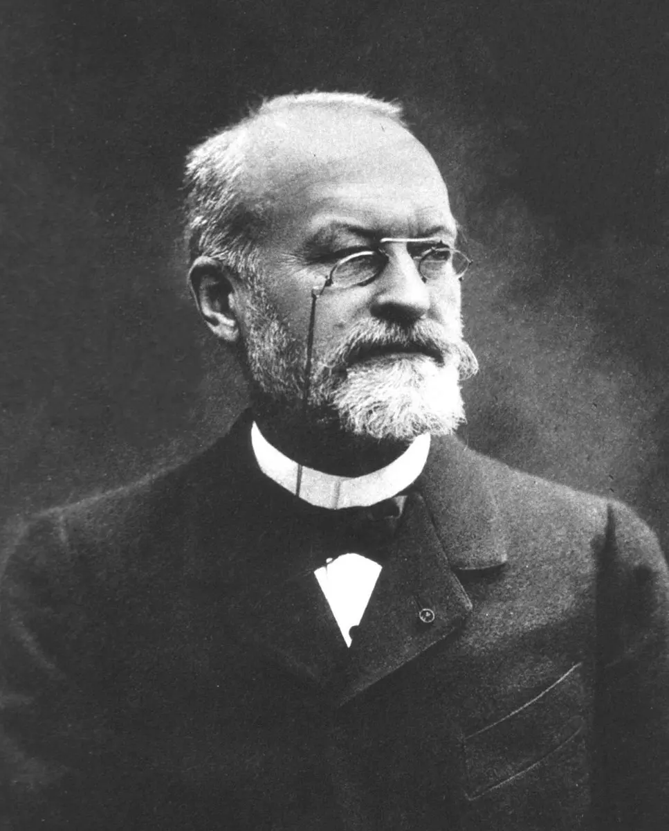
Alphonse Laveran
1845–1922, born in Paris, France
An army doctor, the son of an army doctor, who found the cause of malaria with a simple microscope and no stains.
Photo: Eugène Pirou (1841–1909), Public domain
If you go to a health post in the Terai with a high fever, the health worker may prick your finger and put a drop of blood on a glass slide. In the laboratory, the blood is stained purple and pink and looked at through a microscope. What they are searching for is a living thing small enough to hide inside one of your red blood cells, and thousands of red blood cells would fit on a full stop.
The first person to see it was a French army doctor, Alphonse Laveran, in Algeria in 1880. He showed that malaria is caused not by bad air or by a bacterium, but by a tiny single-celled animal-like creature: a protozoan. It was the first time anyone had shown that a protozoan causes a disease. For this, and for his later work on other protozoa, he received the Nobel Prize in Physiology or Medicine in 1907.
The problem: what causes a fever of the swamps?
Malaria was called “marsh fever”, because it struck people who lived near swamps. Its attacks of shaking chills and burning fever came back every two or three days. Since ancient times, people had blamed the bad-smelling air of the swamps; the very name comes from the Italian mal aria, “bad air”.
By 1880, though, doctors had a better idea: the germ theory, that many diseases are caused by tiny living things. Louis Pasteur and Robert Koch were finding the bacteria behind disease after disease: anthrax, then tuberculosis, cholera and diphtheria in the years that followed. So most experts were sure malaria must be a bacterium too. In 1879 two famous scientists, Edwin Klebs and Corrado Tommasi-Crudeli, announced that they had found it in the mud of the marshes near Rome, and named it Bacillus malariae. They were wrong.
Bacteria and protozoa: what is the difference?
Both are microorganisms, living things too small to see without a microscope Grade 8: Microorganisms. But they are very different.
- A bacterium is a very simple cell, usually one or two thousandths of a millimeter long. It has no nucleus: its DNA lies loose inside it. Bacteria belong to the kingdom Monera Grade 9: Kingdom Monera.
- A protozoan (plural protozoa, “first animals”) is a single cell built like our own cells, with a nucleus. Protozoa are often many times bigger than bacteria, and many of them move and feed like tiny animals. Amoeba is one Grade 8: Amoeba. They belong to the kingdom Protista Grade 9: Kingdom Protista.
The size of things this small is measured in micrometers (µm): one micrometer is a thousandth of a millimeter. A red blood cell is about 7.5 µm across.
Which of these tells you that a microbe is a protozoan and not a bacterium?
Pick an answer to see why.
Why it was so hard
- Everyone was looking for a bacterium. The great successes of the time were all bacteria, so scientists expected malaria to be one too, and looked in the wrong places: in swamp mud and water.
- No stains. Today, dyes color the parasite purple and the red blood cells pink, so it stands out. In 1880 those stains didn’t exist. Laveran looked at fresh, wet drops of blood, where the parasite is almost the same pale color as the red blood cell around it.
- A simple microscope. He used an ordinary microscope that magnified about 400 times, the kind a school laboratory has today.
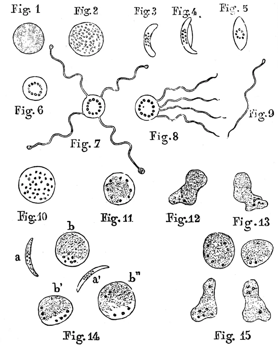
- It changes shape. The malaria parasite looks completely different at different stages of its life: a tiny spot, a blob, a crescent, a ball. It was hard to believe that all these were one creature.
What Laveran did, step by step
1. A clue in the black grains (1878–1880)
Laveran was born in Paris in 1845. Like his father, he became an army doctor, and served in the war of 1870 against Prussia. In 1878 the army sent him to Algeria, in North Africa, then ruled by France, where soldiers suffered badly from malaria. He worked in the military hospitals at Bône (now Annaba) and Constantine.
Doctors had known for 30 years that the blood of people with malaria holds tiny black grains of pigment, but nobody knew where they came from. Laveran set out to find out. Looking at fresh blood from patients, he noticed that the black grains were not loose: they sat inside small, pale bodies, some of them inside red blood cells. He began to suspect that these bodies were alive, and were parasites.
2. The threads that moved (late 1880)
His own report says that he first saw the strange bodies on 20 October 1880. A few weeks later, on 6 November 1880 by the usual account, he saw something that removed all doubt. On the edge of a round body full of black grains, in the blood of a patient, fine threads appeared and lashed about rapidly, pushing the red blood cells aside. Then they broke free and swam off among the cells.
Grains of dead pigment don’t whip about. This was a living thing. “From then on I had no more doubts,” he said in his Nobel lecture.
What convinced Laveran that the bodies in the blood were living parasites, not just lumps of pigment?
Pick an answer to see why.
3. Checking, again and again (1880–1884)
One sighting is not proof. Laveran had to show that the bodies are found in people with malaria and not in people with other illnesses. In one of his first reports he wrote that he had examined 44 patients with malaria and found the bodies in 26 of them, and had never found them in patients with other diseases. By 1884, when he published a big book on malaria, he had studied 480 cases.
He also looked for the parasite in the air, the water and the soil of the marshes, and tried to grow it outside the body. He never found it there. From this, he concluded in his 1884 book that the parasite must spend part of its life inside another animal, probably a mosquito, which would carry it from person to person.
4. Convincing the doubters (1882–1889)
Most experts didn’t believe him. Some said his bodies were just damaged red blood cells. In 1882 he traveled to Rome, where malaria was common in the marshes around the city, and found the same bodies in the blood of Italian patients. Slowly other scientists saw them too. The Italian Camillo Golgi showed that each attack of fever comes when a crop of parasites bursts out of the red blood cells together. In 1891 the Russian doctor Dmitri Romanowsky invented a stain that colored the parasites clearly, and then nobody could miss them. By about 1889 Laveran’s discovery was accepted.
His guess about the mosquito was proved by Ronald Ross in India in 1897–1898 (Ross’s story). When Ross sent his slides of infected mosquitoes to Paris in May 1898, Laveran checked them and confirmed that they held malaria parasites.
5. Other protozoa (1896–1907)
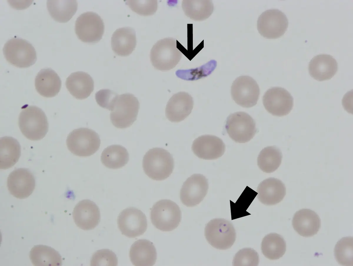
In 1896 Laveran joined the Pasteur Institute in Paris, to do research full time. There he studied other protozoa that live in blood, especially the trypanosomes: long, wriggling cells with a whip-like tail (a flagellum). They cause sleeping sickness in people in Africa, carried by the bite of the tsetse fly, and deadly diseases of cattle, horses and camels. Laveran described about thirty new kinds of trypanosome, more than anyone else.
In 1903 he received samples from a British doctor in Madras (now Chennai), Charles Donovan, of tiny oval bodies found in patients dying of a fever called kala-azar. Laveran and his colleague Félix Mesnil described them as a new parasite; it is now called Leishmania donovani.
How a malaria test works today
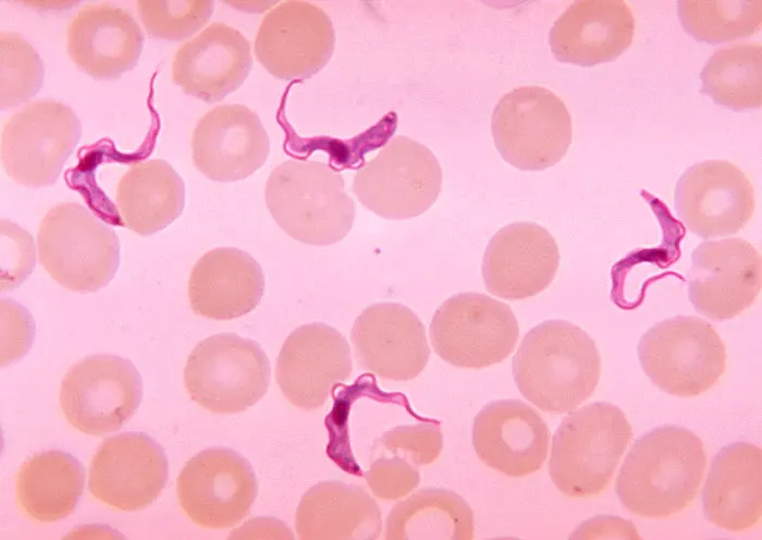
The test at a health post is still Laveran’s method, made easier by stains.
- A drop of blood is spread thinly on a glass slide (a blood smear), or a thicker drop is used to find parasites that are few.
- It is stained, usually with Giemsa stain, a mixture of dyes that colors the parasite’s nucleus dark purple.
- A trained laboratory worker looks at it through a microscope with an oil-immersion lens, magnifying about 1,000 times, and searches for rings, growing forms and crescents in the red blood cells. The shapes also show which kind of malaria parasite it is, which decides the medicine.
- Many health posts now also use rapid tests: a drop of blood on a strip that shows a colored line in about 15 minutes if proteins made by the parasite are present.
In your life today
Malaria in Nepal. Every case of malaria found in Nepal today is confirmed by a blood test, with a microscope or a rapid test, before treatment. Nepal now has very little malaria of its own, and aims to end local spread by 2030. How the parasite travels between people and mosquitoes is told on the page about Ronald Ross.
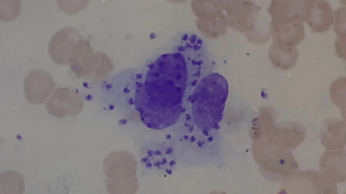
Kala-azar in the Terai. The Leishmania parasite that Laveran named is still found in Nepal. It is carried by the bite of a tiny sandfly, smaller than a mosquito, which breeds in the cracks of mud walls and damp soil near cattle sheds. The parasite lives inside white blood cells, and the disease, kala-azar (“black fever” in Hindi), brings months of fever, a huge spleen and wasting. Untreated, it is almost always fatal. Nepal, India and Bangladesh have worked together since 2005 to eliminate it, by finding and treating patients early and spraying houses. In Nepal the number of cases fell from more than 2,000 a year in the late 2000s to 218 in 2018 (WHO), and as of early 2026 Nepal hoped soon to be declared free of it as a public health problem.
Protozoa cause other illnesses common in Nepal too, such as amoebic dysentery, from Entamoeba in dirty water, and giardiasis, from Giardia: one reason to drink boiled or filtered water.
A village in the Terai has several cases of kala-azar. What does Laveran’s kind of work make possible?
Pick an answer to see why.
What came next
- A whole new science. Laveran’s discovery started medical protozoology, the study of protozoa that cause disease. He gave half of his Nobel Prize money to found a laboratory of tropical medicine at the Pasteur Institute.
- Medicines. Quinine, from the bark of the cinchona tree, had been used against malaria for centuries without anyone knowing what it killed. Once the parasite was known, chemists could test new drugs against it directly.
- Vaccines. Protozoa are much harder to vaccinate against than bacteria or viruses, because they are bigger, change shape and hide inside our cells. The first malaria vaccines were recommended by the World Health Organization only in 2021 and 2023, more than 140 years after Laveran saw the parasite.
- The open question: there is still no vaccine against kala-azar or sleeping sickness. Can we beat these diseases with medicines and by fighting the insects alone, or will we need vaccines?
In your science books
- Grade 8: microorganisms, including Amoeba and their harmful effects; fission, including the multiple fission of Plasmodium.
- Grade 9: Kingdom Monera and Kingdom Protista; parasitism.
- Grade 10: red blood cells.
- On this site: Ronald Ross and the mosquito (Medicine, 1902).
Learn more
- The prize at nobelprize.org: The Nobel Prize in Physiology or Medicine 1907, with the presentation speech.
- Laveran’s own Nobel lecture, “Protozoa as causes of diseases”.
The official reason
The prize was given “in recognition of his work on the role played by protozoa in causing diseases.”