
You began as a single cell, smaller than the dot on this i. That cell held a set of instructions from your mother and another from your father, and every cell of your body has carried a copy of them ever since. The instructions are written in DNA, and the DNA is packed into chromosomes: 46 of them in nearly every cell of your body.
Stretched out end to end, the DNA of the 46 chromosomes in one of your cells would be about 2 meters long. It fits inside a nucleus only a few thousandths of a millimeter across, wound and folded so neatly that the cell can still find and read any part of it. This unit is about those chromosomes, the kinds they come in, and how they carry the genes that pass your characters on from one generation to the next. That study is called genetics.
3.1 Chromosome and its types
Look at the nucleus of a cell that isn’t dividing, even under a good microscope, and you see only a fine, tangled network of threads. These threads are chromatin: long molecules of DNA wound around proteins called histones, like thread wound on many tiny spools. When the cell gets ready to divide, the chromatin coils and folds up tighter and tighter, until it forms short, thick bodies that can be seen clearly and counted. These are the chromosomes.
The name was given in 1888 by the German anatomist Wilhelm Waldeyer, from the Greek chroma, color, and soma, body: “colored bodies”, because they take up stains so strongly that they stand out darkly in a stained cell.
A chromosome is a thread-like body in the nucleus, made of DNA and histone proteins, that carries the genes. Each chromosome is one single, very long DNA molecule. Before a cell divides, it copies every chromosome, so a chromosome seen at the start of cell division is double: two identical copies lying side by side, joined at one point.
Bacteria are different. A bacterium (a prokaryote) has no nucleus: its DNA is usually a single ring lying in the cytoplasm, not wound on histones as ours is. In this unit, a chromosome means the chromosome of a eukaryote, a plant, animal or fungus.
Structure of a chromosome
A chromosome is easiest to study at metaphase of cell division, when it is most tightly coiled and lines up in the middle of the cell. Then it looks like this:
1. Chromatids. A chromosome at metaphase is made of two identical halves called chromatids. They are copies of each other, made when the DNA was copied, so they are called sister chromatids. When the cell divides, they are pulled apart, and each one becomes a chromosome of one of the two new cells.
2. Centromere. The sister chromatids are held together at a narrow point called the centromere. It is also called the primary constriction, because the chromosome is pinched in there. On each side of the centromere is a small disc of protein, the kinetochore, where the spindle fibers attach during cell division. The fibers pull on the kinetochores, so the centromere is the part of a chromosome that leads the way to the pole.
3. Arms. The centromere divides each chromatid into two arms. Unless the centromere is exactly in the middle, one arm is shorter than the other. The short arm is called p (said to be for petit, French for small) and the long arm q, the next letter.
4. Telomeres. The two ends of a chromosome are its telomeres. In humans, a telomere is the same six bases, TTAGGG, repeated hundreds to thousands of times. Telomeres protect the ends of the chromosome, as the plastic tips of a shoelace keep it from fraying, and stop the ends of different chromosomes from sticking to each other.
5. Secondary constriction and satellite. A few chromosomes have a second narrow point besides the centromere: a secondary constriction. Many secondary constrictions hold the genes for making ribosomes, and the nucleolus forms around them, so they are also called nucleolar organizers. The small piece of chromosome beyond a secondary constriction, at the end of an arm, looks like a knob on a stalk and is called a satellite. A chromosome with a satellite is a SAT chromosome. In humans, chromosomes 13, 14, 15, 21 and 22 carry satellites on their short arms.
At which part of a chromosome do the spindle fibers attach during cell division?
Pick an answer to see why.
Types of chromosome on the basis of structure
Chromosomes differ in size and shape, and the main way they differ in shape is where the centromere is. Its place decides how long the two arms are, and so how the chromosome looks when the spindle fibers drag it by its centromere at anaphase, its arms trailing behind. On this basis there are four types:
a. Metacentric chromosome. The centromere is in the middle, so the two arms are equal or nearly equal. At anaphase it is V-shaped. Examples: human chromosomes 1, 3, 16, 19 and 20.
b. Submetacentric chromosome. The centromere is a little to one side of the middle, so one arm is clearly longer, up to about three times the other. At anaphase it is L-shaped. Examples: human chromosomes 2, 4 to 12, 17, 18 and the X chromosome.
c. Acrocentric chromosome. The centromere is near one end, so the short arm is very short. At anaphase it is J-shaped. Examples: human chromosomes 13, 14, 15, 21, 22 and the Y chromosome.
d. Telocentric chromosome. The centromere is at the very end, so there is only one arm. At anaphase it is rod-shaped, like the letter I. Humans have none, but almost all of a house mouse’s 40 chromosomes have their centromere at, or almost at, one end.
The type is set by the arm ratio, the length of the long arm divided by the length of the short arm:
| Type | Centromere | Arm ratio, q ÷ p | Shape at anaphase |
|---|---|---|---|
| Metacentric | in the middle | 1 to 1.7 | V |
| Submetacentric | a little off the middle | 1.7 to 3 | L |
| Acrocentric | near one end | more than 3 | J |
| Telocentric | at the end | no short arm | I (rod) |
Some books also sort chromosomes by the number of centromeres: most have one (monocentric); a broken and wrongly joined chromosome may have two (dicentric) or none (acentric), and is torn or lost when the cell divides; and in a few living things, such as the worm Caenorhabditis elegans, the centromere runs along the whole length (holocentric).
At anaphase, a chromatid is pulled toward the pole and makes a J shape. Its chromosome is:
Pick an answer to see why.
Types of chromosome on the basis of function
By what they do, chromosomes are of two types.
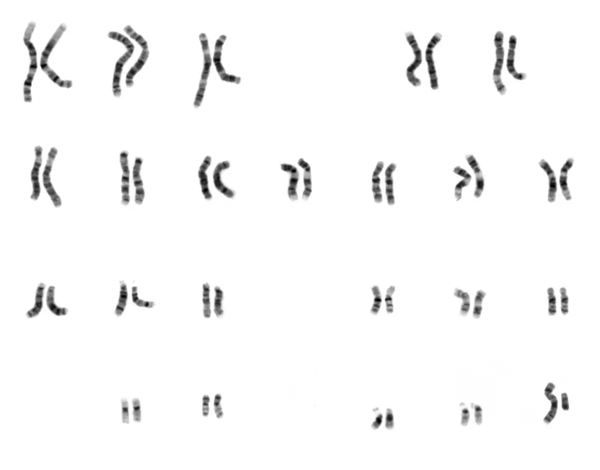
a. Autosomes. These carry the genes for the characters of the body (height, skin color, blood group and thousands of others), and have little to do with whether an individual is male or female. The two chromosomes of each pair are alike in size, shape and the genes they carry. Of our 46 chromosomes, 44 (22 pairs) are autosomes. They are also called somatic chromosomes.
b. Sex chromosomes. These decide the sex of an individual. In humans there is one pair: XX in a female and XY in a male. The X is a large submetacentric chromosome with about 800 genes for proteins; the Y is a small acrocentric one with only a few dozen. Because the two members of the pair can be different, sex chromosomes are also called allosomes or heterosomes (“different bodies”).
So a woman’s body cell has 44 + XX and a man’s 44 + XY. A picture of all the chromosomes of one cell, arranged in pairs by size, is called a karyotype.
| Autosomes | Sex chromosomes |
|---|---|
| Carry genes for the body’s characters. | Decide the sex of the individual. |
| 22 pairs (44) in a human body cell. | 1 pair (2) in a human body cell. |
| The two of a pair are alike in size and shape. | The two may differ (X and Y in a man). |
| The same in males and females. | XX in a human female, XY in a male. |
Not every living thing decides sex the same way. In birds, it is the female who has two different sex chromosomes, called Z and W (ZW), and the male who has two alike (ZZ). Most flowering plants, such as rice and the pea, have flowers that are both male and female, and have no sex chromosomes at all.
Every member of a species has the same number of chromosomes in its body cells, and it is usually even, because they come in pairs: one of each pair from the mother and one from the father. A body cell with two sets is diploid (2n); an egg or sperm, with one set, is haploid (n). A human body cell has 2n = 46, and a human sperm or egg n = 23: 22 autosomes and one sex chromosome.
How many autosomes are there in a human egg cell?
Pick an answer to see why.
3.2 Introduction to genetics
A calf looks like a cow, not a goat, and a child often has her mother’s eyes or her grandfather’s laugh. Characters pass from parents to their young: this is heredity. Yet no two children of the same parents are exactly alike, except identical twins, and even they differ a little. These differences between members of the same species are called variation.
Genetics is the branch of biology that studies heredity and variation: how characters pass from parents to offspring, and why offspring differ from their parents and from each other. The English biologist William Bateson coined the word, first in a letter in 1905 and then in public in 1906, from a Greek word meaning to give birth.
Some variation is hereditary: it is in the genes and can be passed on, like blood group or the shape of the earlobe. Some is environmental: it comes from how an individual lives, and isn’t passed on. A wrestler’s strong arms, a farmer’s tanned face and a scar are not inherited by their children. Most characters, such as height, depend on both: the genes set the range, and food and health decide where in it a person ends up.
Mendel, the father of genetics
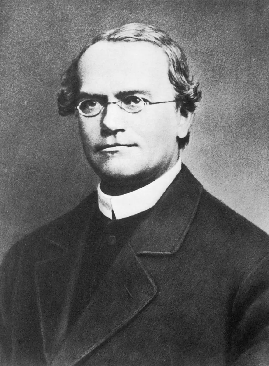
The rules of heredity were first worked out by Gregor Johann Mendel, a monk in the town of Brno (in today’s Czech Republic), who crossed thousands of pea plants in his monastery garden from 1856 to 1863. He showed that characters are passed on by particles that come in pairs, one from each parent, and that they don’t blend but stay separate, even when one is hidden for a generation. He published his results in 1866, but nobody understood how important they were. In 1900, sixteen years after his death, three botanists found the same rules on their own and then found his paper. Mendel is called the father of genetics.
The words you will need in genetics:
Gene: a part of a chromosome (a stretch of its DNA) that carries the instructions for one character, or more exactly for making one protein or one RNA. The Danish botanist Wilhelm Johannsen named it in 1909.
Allele: one of the different forms of the same gene. The gene for a pea plant’s height has two alleles: one for tall (written T) and one for dwarf (t).
Dominant and recessive: when both alleles are present, the one that shows is dominant (T), and the one hidden is recessive (t).
Homozygous and heterozygous: an individual with two alike alleles (TT or tt) is homozygous, or pure; one with two different alleles (Tt) is heterozygous, or hybrid.
Genotype and phenotype: the genotype is the set of alleles an individual has (TT, Tt or tt); the phenotype is the character we see (tall or dwarf). TT and Tt plants have different genotypes but the same phenotype: both are tall.
Why genetics matters
Farming. Plant breeders cross varieties to bring together the alleles they want, such as high yield, resistance to disease or drought, or a short stem that doesn’t fall over in the wind. The new rice, maize and wheat varieties grown in Nepal are made this way, and so are cows and buffaloes that give more milk.
Medicine. Some diseases, such as thalassemia, hemophilia and sickle-cell anemia, are inherited. Knowing how they are passed on lets doctors test for them and advise families.
Identifying people. Everyone’s DNA is different, except identical twins’, so DNA can show who a child’s parents are, or who was at the scene of a crime (DNA fingerprinting, in Grade 10 Optional Science).
Understanding life. Genetics explains why species vary, how they change over time, and how all living things are related.
A child of two tall parents grows up shorter than both because he was badly fed as a small child. This difference is:
Pick an answer to see why.
3.3 Relation between gene and chromosome
Mendel said characters are passed on by particles in pairs, but he had no idea where they were. Chromosomes were seen in the 1880s, but nobody knew what they did. The two discoveries came together in the early 1900s, and the link between them is the base of modern genetics: genes are parts of chromosomes.
A gene is a segment of the DNA in a chromosome. One chromosome is one very long DNA molecule, and it carries hundreds or thousands of genes, one after another along its length, like beads on a string. Each gene has its own fixed place on its own chromosome, called its locus (plural loci).
The chromosomes of a body cell come in pairs, and the two of a pair, called homologous chromosomes, carry the same genes at the same loci, one from the mother and one from the father. So every gene on an autosome is present twice in a body cell, once on each homologous chromosome. The two copies may be the same allele (TT) or different alleles (Tt). This is why genes come in pairs, as Mendel found.
The chromosome theory of inheritance
In 1902 and 1903, the American student Walter Sutton, watching chromosomes in grasshopper cells, and the German biologist Theodor Boveri, working with sea urchin eggs, saw that chromosomes behave exactly as Mendel’s particles should:
| Mendel’s factors (genes) | Chromosomes |
|---|---|
| Come in pairs in a body cell. | Come in pairs in a body cell. |
| The two of a pair separate when gametes form, so each gamete gets only one. | The two of a pair separate in meiosis, so each gamete gets only one. |
| Each pair is shared out independently of other pairs. | Each pair lines up and separates independently of other pairs. |
| One of each pair comes from each parent. | One of each pair comes from each parent. |
So, they said, the genes must be on the chromosomes. This idea is the chromosome theory of inheritance: genes are carried on chromosomes, and they pass from parents to offspring because the chromosomes do.
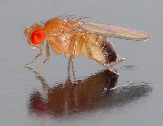
Morgan’s students found that genes on the same chromosome tend to be inherited together (linkage), and one of them, Alfred Sturtevant, while still an undergraduate, used how often linked genes were separated to draw the first map of genes along a chromosome (published in 1913): they lie in a line, each in its own place.
Gene and chromosome compared
| Gene | Chromosome |
|---|---|
| A segment of DNA. | A whole DNA molecule with its proteins. |
| Carries the code for one protein or RNA, and so for a character. | Carries hundreds or thousands of genes. |
| Too small to be seen under a light microscope. | Seen under a light microscope at cell division. |
| Lies at a fixed place (locus) on a chromosome. | Lies in the nucleus. |
| About 20,000 kinds (for proteins) in humans. | 23 kinds (46 in all) in a human body cell. |
Homologous chromosomes carry:
Pick an answer to see why.
Exercise
1. Choose the correct option
a. The part of a chromosome that holds the two sister chromatids together is the:
Pick an answer to see why.
b. Which type of chromosome is not found in a normal human cell?
Pick an answer to see why.
c. The term “genetics” was coined by:
Pick an answer to see why.
d. The fixed place of a gene on a chromosome is called its:
Pick an answer to see why.
e. The chromosome theory of inheritance was put forward by:
Pick an answer to see why.
2. Give reasons
Chromosomes are studied at the metaphase of cell division.
Hint
Answer
Chromosomes are found in pairs in a body cell.
Hint
Answer
3. Differentiate between
Metacentric and acrocentric chromosomes
Hint
Answer
Heredity and variation
Hint
Answer
4. Answer the following questions
What is a chromosome? Draw a labeled diagram of a chromosome at metaphase.
Hint
Answer
Write the functions of the centromere, the telomeres and the secondary constriction.
Hint
Answer
Classify chromosomes on the basis of the position of the centromere. Draw each type.
Hint
Answer
Explain the relation between gene and chromosome.
Hint
Answer
State the chromosome theory of inheritance. Give any three points in which genes and chromosomes behave alike.
Hint
Answer
5. Work it out
A chromosome 7 µm long has an arm ratio of 2.5. Find the length of each arm and name the type of chromosome.
Hint
Answer
In a chromosome 12 µm long, the centromere is 5.5 µm from one end. Find the arm ratio and the type.
Hint
Answer
A dog’s body cell has 78 chromosomes, and dogs decide sex by X and Y as humans do. How many autosomes are there in a dog’s body cell, and how many chromosomes, and which, in its sperm?
Hint
Answer
A human cell is at metaphase of mitosis. How many chromosomes, chromatids and centromeres does it have?
Hint
Answer
Printable PDFs
- The unit as a book (A5) 19 pages Answers as an A4 booklet, 5 sheets
- Study notes 2 pages
- Worksheet 5 pages Answer key
- Harder practice questions 3 pages Answer key
Practice every question in the book, and keep track of the ones you miss →
Found a mistake? Please tell us.