
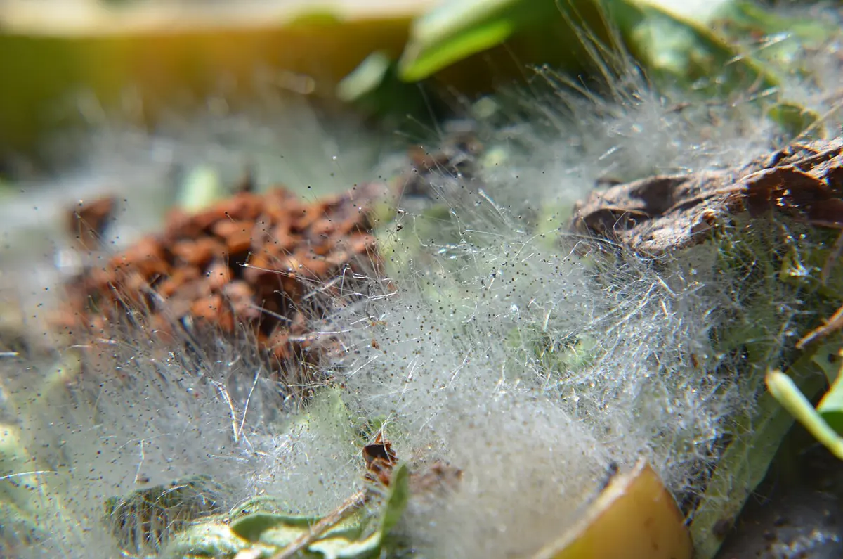
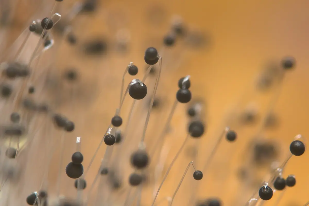
Leave a piece of roti in a closed tin in the rainy season, and after three or four days a gray fur grows over it, dotted with tiny black pins. Nobody put it there. It grew from spores far too small to see, which float in the air of every kitchen and land on everything. Those black pins are packed with more spores, millions of them, ready to start the same fur on the next piece of food.
The fur is a mold called Mucor, one of the living things too small to see one at a time that we call microorganisms. This unit is about them: what the science of microbes studies and why it matters, how a virus, a bacterium and a yeast make more of themselves, and the whole life of Mucor, the mold on the roti, with the good and the harm it does.
2.1 Microbiology
Microorganisms (also called microbes or germs) are living things so small that most of them can be seen only with a microscope. They include bacteria, fungi such as yeasts and molds, protozoa such as Amoeba, microscopic algae, and the viruses, which are not cells at all. The science that studies them, their structure, how they live and multiply, and what they do to us and to the world, is microbiology (Greek mikros, small; bios, life; logos, study).
Microbes come in very different sizes. A micrometer (µm) is a thousandth of a millimeter, and a nanometer (nm) is a thousandth of a micrometer.
| Microbe | Typical size | Seen with |
|---|---|---|
| A virus (bacteriophage T4) | about 200 nm (0.2 µm) long | an electron microscope only |
| A bacterium (E. coli) | about 2 µm long, 0.5–1 µm wide | a good light microscope |
| A yeast cell | about 5–10 µm across | a light microscope |
| Amoeba proteus | about 500 µm (half a millimeter) | a hand lens, just about |
| A colony of Mucor | its threads are about 10–20 µm wide, but millions together make a fur you can see | the naked eye |
A grain of rice is about 6 mm long, so 3,000 E. coli laid end to end would just reach along it, and about 30,000 phages would.
Branches of microbiology
Microbiology is now so large that it is split by the microbes studied:
- Bacteriology, the study of bacteria;
- Virology, the study of viruses;
- Mycology, the study of fungi (mykes, fungus);
- Protozoology, the study of protozoa, and phycology, the study of algae.
It is also split by where the knowledge is used: medical microbiology (germs that cause disease, and how to fight them), immunology (how the body defends itself), agricultural microbiology (microbes of soil, crops and animals), food microbiology, industrial microbiology (using microbes to make things) and environmental microbiology (microbes in water, soil, air and waste).
Importance of microbiology
Most microbes do no harm, and many are vital. Knowing them helps us use the useful ones and fight the harmful ones.
In medicine. Microbiology found the causes of diseases such as tuberculosis, cholera, typhoid and COVID-19, and so how each spreads and how to stop it: boiling water, washing hands, keeping flies off food. It gave us antibiotics (penicillin came from a mold, Penicillium) and vaccines. Bacteria now make medicines too: since 1982, human insulin for people with diabetes has been made by E. coli carrying the human insulin gene.
In agriculture. Rhizobium bacteria in the root nodules of beans, peas and lentils turn nitrogen from the air into a form plants can use, so legumes enrich the soil. Microbes rot dung and leaves into compost and humus. Microbiology also finds the fungi, bacteria and viruses that cause crop diseases, such as late blight of potato and rust of wheat, and ways to control them.
In food. Many of Nepal’s foods are made by microbes. Lactic acid bacteria turn milk into dahi and mustard leaves into gundruk; Bacillus bacteria ferment soybeans into kinema; yeasts and molds in marcha (murcha), the starter cake, turn cooked millet or rice into jaand and chhyang. Yeast makes bread and sel roti batter rise. Microbiology also explains why food spoils and how drying, salting, boiling, pickling and cold slow the microbes down.
In industry. Microbes grown in huge tanks make alcohol, vinegar, citric acid, vitamins, enzymes for washing powders and cheese, and many medicines.
In the environment. Bacteria and fungi are the great decomposers: they break down dead plants and animals and return their nutrients to the soil. Without them, the forest floor would be piled high with dead leaves. Microbes clean sewage in treatment plants, and in a gobar gas (biogas) plant, bacteria turn dung into methane for cooking.
In research. The bacterium E. coli and baker’s yeast are the two best-studied living things on Earth. Much of what we know about genes, how DNA is copied and how cells divide was first found in them.
Which of these is a use of microbes in agriculture?
Pick an answer to see why.
A scientist who studies how the mold Mucor reproduces works in which branch of microbiology?
Pick an answer to see why.
2.2 Mode of reproduction of virus, bacteria and fungi
Every living thing makes more of its own kind. A microbe usually does it asexually: one parent alone makes offspring that are copies of itself, so the offspring have the same genes. That is quick: when food is plentiful, a microbe can go from one cell to millions in a day. Here are three very different ways of doing it.
Reproduction in viruses: the lytic cycle of a bacteriophage
A virus has no cell, no food store and no machinery for making anything. Outside a living cell it does nothing at all. It can multiply only by getting inside a living host cell and making the cell build new viruses for it. So a virus doesn’t really reproduce the way a cell does: it is copied, by its host.
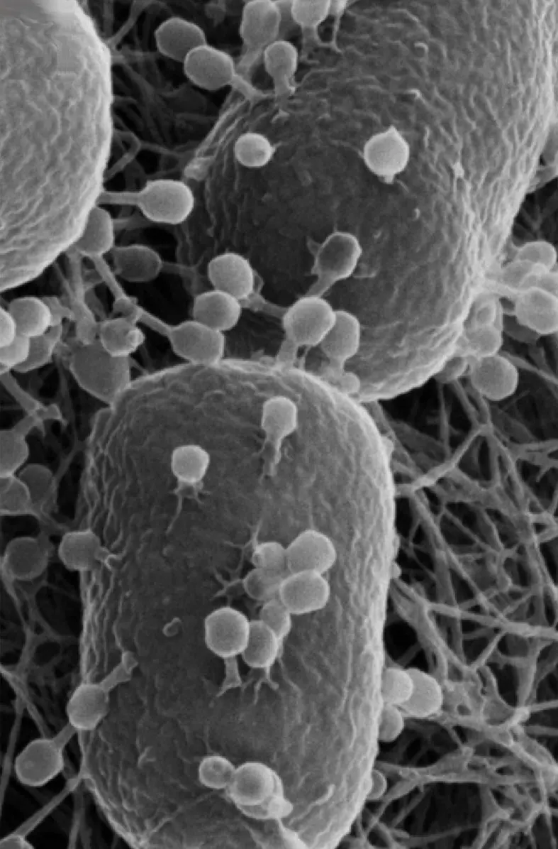
A bacteriophage, or phage (“bacteria eater”), is a virus that infects bacteria. The best-studied one, phage T4, infects E. coli, the bacterium that lives in our gut. It looks like a tiny landing craft: a six-sided head (the capsid, a coat of protein) holding its DNA, a collar, a hollow tail with a springy sheath around it, a baseplate, and six long, bent tail fibers, like legs.
ViralZone, SIB Swiss Institute of Bioinformatics, CC BY 4.0, via Wikimedia Commons
When a phage meets the right bacterium, it multiplies by the lytic cycle, named after the last step, when the bacterium bursts (lysis, breaking open). The cycle has five steps.
1. Attachment (adsorption). The phage drifts until it bumps into an E. coli cell. Its tail fibers grip particular molecules on the cell’s outer surface, its receptors, like a key fitting a lock. A phage can attach only to the kind of bacterium whose receptors fit its fibers, so phage T4 infects E. coli but not the bacteria in curd.
2. Penetration (injection). The baseplate settles on the cell wall, and the tail sheath contracts, driving the hollow inner tube through the wall like a syringe’s needle. An enzyme at the tube’s tip, lysozyme, digests a small hole in the wall. The phage’s DNA passes down the tail into the cell. The empty protein coat stays outside, a “ghost”.
3. Biosynthesis (replication). Within minutes, the phage’s genes take over the cell. The phage makes an enzyme that chops up the bacterium’s own DNA, so the cell stops making its own things. The cell’s ribosomes, enzymes and food store now make what the phage DNA orders: many copies of the phage DNA, and the proteins for heads, tails and fibers.
4. Maturation (assembly). The parts put themselves together. Heads are built and filled with a copy of the DNA; tails join the heads; fibers attach last. About 20 minutes after infection, the cell is crowded with complete new phages.
5. Release (lysis). Phage genes now make a protein that punches holes in the cell membrane and an enzyme (lysozyme) that digests the cell wall. Weakened, the cell bursts and dies, releasing all its new phages at once. Each is ready to attach to another E. coli and start the cycle again.
At 37 °C, the whole cycle takes phage T4 only about 25 to 30 minutes, and each burst cell releases roughly 100 to 150 new phages; the worked examples here mostly use 100, a round number. That is why phages spread so fast: one phage, one cell; then a hundred phages, a hundred cells; then ten thousand.
In which step of the lytic cycle is the bacterium’s own DNA broken down?
Pick an answer to see why.
What enters the bacterium during penetration?
Pick an answer to see why.
Reproduction in bacteria: binary fission in E. coli
A bacterium reproduces by binary fission (binary, two; fission, splitting): one cell grows and splits into two equal daughter cells. Each daughter gets a copy of the parent’s DNA, so both are genetically the same as the parent. Here is how E. coli does it.
1. The DNA is copied. E. coli has a single, circular chromosome, a loop of DNA about 4.6 million base pairs long, packed into a tangle called the nucleoid. It has no nucleus around it. The chromosome is copied, starting from one point on the loop, to make two identical loops.
2. The copies move apart. As the cell grows longer, the two loops move apart, towards opposite ends of the cell.
3. A septum forms. A ring of protein (made of FtsZ) forms round the middle of the cell, just inside the membrane. The membrane and the cell wall start to grow inward along this ring, like a drawstring being pulled tight. The new wall growing across the middle is the septum.
4. The cell divides. The septum closes across the cell, cutting it into two cells, each with its own DNA, its own share of ribosomes and cytoplasm, and a new rounded end. The two daughter cells separate.
Mariana Ruiz Villarreal, LadyofHats, Public domain, via Wikimedia Commons
Notice that there is no spindle and no stages like the mitosis of our own cells: a bacterium’s single chromosome needs only to be copied and pulled apart. That is one reason fission is so fast. In warm broth with plenty of food, at 37 °C, E. coli divides about every 20 minutes. The time between one division and the next is the generation time (or doubling time), because each time, the number of bacteria doubles.
So starting from bacteria, after generations the number is
A bacterium divides every 30 minutes. Starting from one cell, how many cells are there after 2 hours?
Pick an answer to see why.
Reproduction in fungi: budding in yeast
Yeast (baker’s and brewer’s yeast is Saccharomyces cerevisiae) is a fungus made of a single, egg-shaped cell about 5–10 µm long. Unlike a bacterium, it is a eukaryote: it has a nucleus, mitochondria and a large vacuole. When it has sugar to feed on, it reproduces asexually by budding:
1. A bud appears. A small bulge, the bud, pushes out from one point on the cell wall of the parent (mother) cell. Cytoplasm flows into it.
2. The nucleus divides. The nucleus moves to the narrow neck between mother and bud and divides by mitosis. In yeast, the nuclear envelope stays whole all the time: the nucleus stretches through the neck into an hourglass shape and pinches in two, leaving one nucleus in the mother and one in the bud.
3. The bud grows. The bud takes in cytoplasm, mitochondria and part of the vacuole, and grows almost as big as its mother.
4. The bud separates. A wall forms across the neck, and the bud breaks away as a new yeast cell. It leaves a ring-shaped mark, a bud scar, on the mother’s wall. A mother cell can bud again and again, each time from a new place, until it has produced about 20 to 30 daughters.
If the buds don’t separate, and each bud starts budding before it breaks away, the cells form a chain or a branched cluster. This chain of cells looks a little like a hypha and is called a pseudomycelium (a false mycelium).
Budding differs from fission in one way you can see: the two cells are unequal. The mother keeps her old wall and her scars; the bud starts small and new. With plenty of sugar, at about 30 °C, a yeast population doubles in about 1½ to 2 hours, much more slowly than E. coli, because a eukaryotic cell has far more to copy and sort.
| Binary fission (E. coli) | Budding (yeast) | |
|---|---|---|
| The parent | is a prokaryote: no nucleus | is a eukaryote: has a nucleus |
| How the DNA is shared | one circular chromosome copied and pulled apart; no spindle | the nucleus divides by mitosis |
| The two cells | equal in size | unequal: a big mother and a small bud |
| The parent | disappears into its two daughters | lives on and buds again, keeping a scar each time |
| Time to double (best conditions) | about 20 minutes | about 1½ to 2 hours |
Which feature shows that budding, not binary fission, is happening?
Pick an answer to see why.
2.3 Life cycle of Mucor
Mucor is a common mold, often called pin mold or black bread mold. It grows on damp bread and roti, rotting fruit and vegetables, dung, and soil: wherever there is dead, moist food rich in sugar and starch. It is a saprophyte: its threads release enzymes that digest the food outside them, and then absorb the dissolved nutrients. Mucor mucedo is the species most books describe.
The body of Mucor
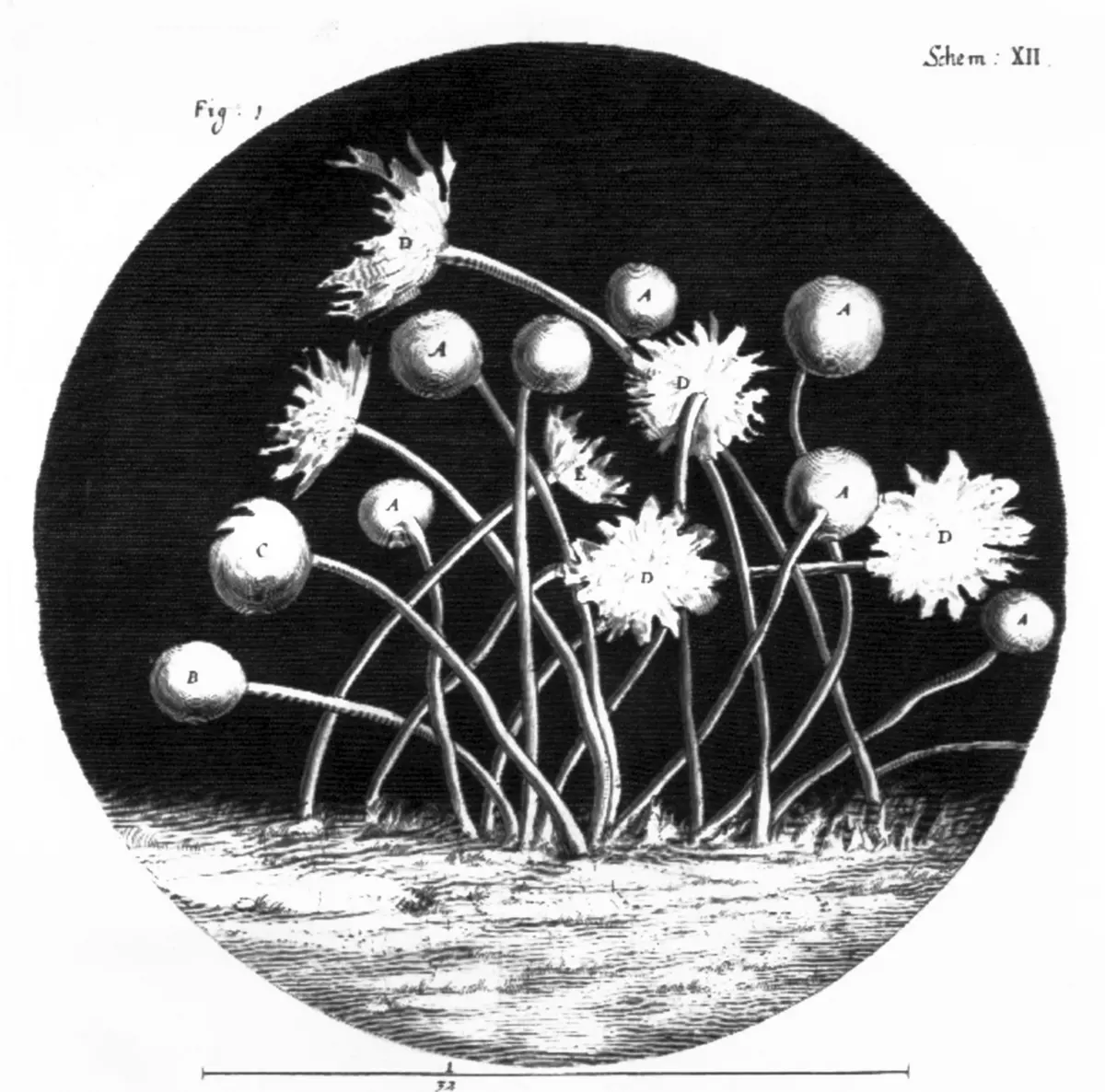
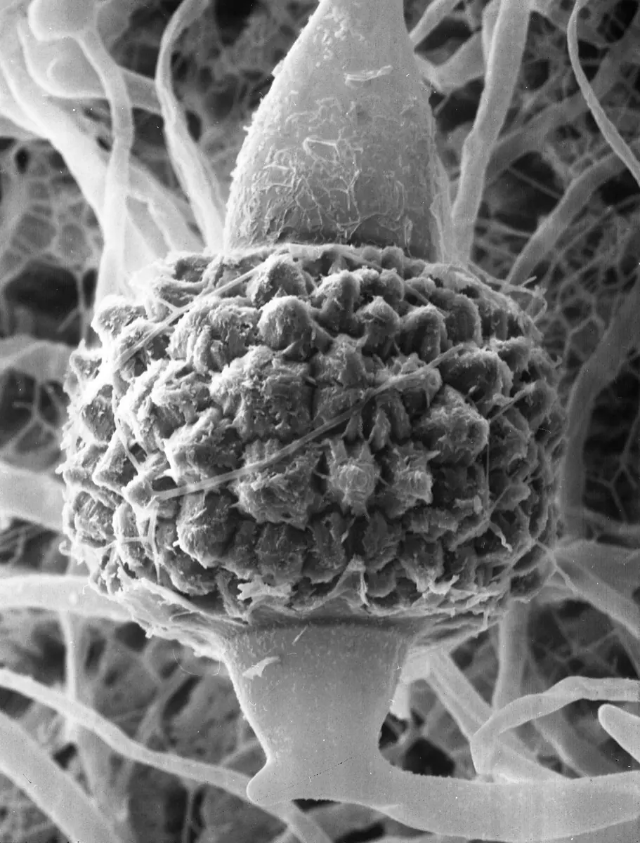
The body of Mucor is a mycelium, a cotton-like network of fine, branched, white threads called hyphae. Each hypha is a tube with a thin wall containing chitin, filled with cytoplasm and many small nuclei. Mucor’s hyphae have no cross walls (septa) dividing them into cells: each branched hypha is one long, continuous cell with hundreds of nuclei in it. Such a body is called coenocytic (koinos, shared; kytos, cell). Cross walls form only where reproductive parts are cut off from the rest. Every nucleus in the mycelium is haploid: it has one set of chromosomes (n).
Mucor drawn after Brefeld (Dodel-Port Atlas) and Meyers Konversations-Lexikon (public domain)
Mucor reproduces in three ways: vegetatively (fragmentation), asexually (spores) and sexually (conjugation).
Vegetative reproduction
If the mycelium is broken, each piece of hypha with some nuclei can grow into a new mycelium. This is fragmentation.
Asexual reproduction by sporangiospores
When the mycelium has fed well, it reproduces asexually, which is the way Mucor usually multiplies:
- Some hyphae grow straight up into the air. Each upright hypha is a sporangiophore (“spore-case bearer”).
- The tip of the sporangiophore swells into a round head, the sporangium. Cytoplasm and many nuclei stream up into it.
- A dome-shaped wall forms across the sporangium, separating a central sterile part, the columella, from the outer part. The outer cytoplasm divides into a great many small pieces, each with one or a few nuclei, and each piece grows a wall: these are the sporangiospores (or simply spores). They are haploid and are made by mitosis, so they have the same genes as the mycelium.
- As the spores ripen, the sporangium turns from white or yellow to gray-black. Its thin wall breaks, the spores are set free, and the wind carries them away. A small frill of the broken wall, the collar, is often left round the base of the columella.
- A spore that lands on damp food swells and puts out a germ tube, which grows and branches into a new mycelium.
Each sporangium can hold thousands of spores, and a single colony can carry many thousands of sporangia. That is why molds turn up everywhere: their spores are in almost every breath of air.
Some Mucor species have two more asexual tricks. When food or water runs short, parts of a hypha fill with cytoplasm, round off behind cross walls and grow thick walls: these chlamydospores survive bad times and grow into a new mycelium when things improve. And in some species growing in sugary liquid with little air, the hyphae break into round cells that multiply by budding, like yeast.
Sexual reproduction by conjugation
When food runs short or the colony meets another one, Mucor can reproduce sexually. Mucor mucedo is heterothallic: its mycelia come in two strains, called + (plus) and − (minus). They look exactly alike; there is no male or female. A mycelium can mate only with a mycelium of the other strain.
- Zygophores meet. When + and − hyphae grow near each other, each gives off chemical signals (hormones) that make the other grow short side branches, the zygophores, towards it. The tips touch and press together.
- Progametangia form. The tips of the touching zygophores swell into club-shaped progametangia, each filled with cytoplasm and many nuclei.
- Gametangia are cut off. A cross wall forms near the tip of each progametangium, cutting it into two parts: the end part, the gametangium, and the stalk behind it, the suspensor. Each gametangium is a multinucleate cell, one from the + strain and one from the − strain.
- Fusion. The wall between the two gametangia dissolves, and their contents mix. Nuclei from the + gametangium pair with nuclei from the − one and fuse, making diploid (2n) nuclei. This fusion of two gametangia is conjugation, and the fused cell is a zygote.
- The zygospore forms. The fused cell swells into a round zygospore (it forms inside a wall called the zygosporangium, and many books use the two names for the same thing). Its wall becomes thick, black and covered in warts. Held between the two empty suspensors, it can lie resting for months, through dryness and cold that would kill the mycelium.
- Germination. When conditions are good again, the diploid nuclei divide by meiosis, making haploid nuclei again, and the zygospore cracks open. Out grows a short hypha, the germ sporangiophore (promycelium), with a germ sporangium at its tip, full of haploid spores. Released, each spore grows into a new mycelium of the + or the − strain, and the cycle begins again.
So the zygospore is the only diploid stage in Mucor’s whole life; every other stage is haploid. And while asexual spores make copies, the zygospore mixes the genes of two different strains, so its offspring are new combinations.
Which part of Mucor is diploid?
Pick an answer to see why.
A gametangium and a suspensor are both cut from:
Pick an answer to see why.
2.4 Advantages and disadvantages of Mucor
Mucor is a nuisance in the kitchen, but in the wider world it does a great deal of good as well.
Advantages
- Decomposer. With bacteria and other fungi, Mucor breaks down dead plant matter, dung and spoiled food, returning carbon, nitrogen and minerals to the soil, where plants can use them again. It is one of the first molds to grow on fresh, sugary dead matter.
- Fermented foods and drinks. Molds of the Mucor group help make starter cultures for alcoholic drinks in Asia. A 2020 study of dry starter cakes from North East India, marcha from Sikkim among them, found Mucor circinelloides the most common mold, alongside Rhizopus and others; these molds break the starch of the grain into sugar, which the yeasts then ferment into alcohol. In China, Mucor species grow over cubes of tofu to make the cheese-like sufu.
- Enzymes. Molds of this group make useful enzymes: one, formerly called Mucor miehei, is grown in tanks to make a rennet that curdles milk into cheese without using calves’ stomachs, and enzymes for processing fats. Mucor species also make enzymes that break down starch and proteins.
- Alcohol and other products. Mucor indicus can ferment sugars from plant waste into alcohol (ethanol) as well as yeast does, so it is being studied for making fuel from straw and stalks. Some species store oils rich in useful fatty acids.
- Cleaning up. Mucor species can break down some pollutants in soil, such as certain oil residues, and their dead hyphae soak up metals from waste water.
- Research. Mucor has helped scientists study how fungi grow, how they switch between threads and yeast-like cells, and how + and − strains find each other: Blakeslee’s discovery of the two strains was made in this group.
Disadvantages
- Spoils food. Mucor rots bread, roti, fruits, vegetables, jam, cheese and stored grain. Its enzymes soften and sour the food, and it gives off off-flavors. Moldy food should be thrown away, not scraped clean, since the hyphae run deep inside.
- Damages crops after harvest. Mucor piriformis causes a soft rot of apples and pears in storage, and other species rot strawberries, citrus, mangoes and yams.
- Causes mucormycosis. A few Mucor species, with Rhizopus and other relatives, can infect people whose defenses are weak, especially people with poorly controlled diabetes, cancer patients, or patients on high doses of steroids. The disease, mucormycosis (sometimes called “black fungus” for the black, dead tissue it causes), usually starts in the nose and sinuses and can spread to the eyes and brain. It is rare but very serious: about half of the patients with the sinus form die. In 2021, during the COVID-19 pandemic, more than 40,000 cases were reported in India by the end of June, many in diabetic patients treated with steroids, and cases were reported in Nepal too. It doesn’t spread from person to person.
- Allergies. Its spores in the air of damp, moldy rooms can trigger sneezing and asthma in some people.
- Infects animals. Some species infect cattle, pigs and poultry.
| Advantages of Mucor | Disadvantages of Mucor |
|---|---|
| decomposes dead matter and recycles nutrients | spoils bread, fruit, vegetables and stored grain |
| helps ferment starters such as marcha, and foods such as sufu | rots fruit in storage |
| makes enzymes, such as microbial rennet for cheese | causes mucormycosis in people with weak immunity |
| ferments plant waste into alcohol | its spores cause allergies and asthma |
| breaks down some soil pollutants | infects some farm animals |
Which of these is NOT a disadvantage of Mucor?
Pick an answer to see why.
Exercise
1. Choose the correct option
a. The branch of microbiology that studies fungi is:
Pick an answer to see why.
b. The correct order of the steps of the lytic cycle is:
Pick an answer to see why.
c. In binary fission of E. coli, the wall that grows across the middle of the cell is called the:
Pick an answer to see why.
d. Yeast reproduces asexually by:
Pick an answer to see why.
e. The hyphae of Mucor are:
Pick an answer to see why.
f. The sterile, dome-shaped central part of a Mucor sporangium is the:
Pick an answer to see why.
g. A zygospore of Mucor is formed by the fusion of:
Pick an answer to see why.
h. One bacterium divides every 20 minutes. How many bacteria are there after 1 hour?
Pick an answer to see why.
2. Give reasons
A virus is called an obligate parasite.
Hint
Answer
A bacterium infected by a phage in the lytic cycle dies.
Hint
Answer
The two daughter cells formed by binary fission are genetically identical to the parent.
Hint
Answer
Mucor’s hyphae are called coenocytic.
Hint
Answer
Bread kept in a closed box in a damp, warm room grows mold faster than bread kept in a fridge.
Hint
Answer
The zygospore of Mucor helps it survive bad conditions.
Hint
Answer
3. Differentiate between
Binary fission and budding
Hint
Answer
Sporangiospore and zygospore
Hint
Answer
Gametangium and suspensor
Hint
Answer
Asexual and sexual reproduction in Mucor
Hint
Answer
4. Answer the following questions
What is microbiology? Name its branches based on the microbes studied.
Hint
Answer
Write any five points on the importance of microbiology.
Hint
Answer
Describe the lytic cycle of a bacteriophage with a labeled diagram.
Hint
Answer
Explain binary fission in E. coli with diagrams.
Hint
Answer
Describe budding in yeast with a diagram.
Hint
Answer
Describe the asexual reproduction of Mucor.
Hint
Answer
Describe the sexual reproduction of Mucor with labeled diagrams.
Hint
Answer
Draw a labeled diagram of the life cycle of Mucor, showing both its asexual and sexual cycles.
Hint
Answer
Write any three advantages and three disadvantages of Mucor.
Hint
Answer
Think: A student says, “A virus is the smallest living thing, so it must reproduce by binary fission, like bacteria.” What is wrong with this?
Hint
Answer
5. Numerical problems
An E. coli cell divides every 20 minutes. Starting with one cell, how many cells will there be after 4 hours?
Hint
Answer
A sample of milk has 500 bacteria in it. They divide every 30 minutes. How many bacteria will it have after 3 hours?
Hint
Answer
A culture of bacteria grew from 1,000 cells to 64,000 cells in 2 hours. Find its generation time.
Hint
Answer
A bacterium divides every 25 minutes. How long will one bacterium take to become 1,024 bacteria?
Hint
Answer
A phage infects a bacterium, and after 30 minutes the cell bursts, releasing 150 phages. If each of these infects a new bacterium and each of those cells releases 150 phages, how many phages are there after 1 hour, and how many bacteria have been killed?
Hint
Answer
Yeast in a bowl of sugar solution doubles every 2 hours. If there are 50,000 cells at 7 a.m., how many are there at 3 p.m. the same day?
Hint
Answer
In a lab, one E. coli (doubling every 20 minutes) and one yeast cell (doubling every 100 minutes) are each put in their own flask of food at the same time. How many of each will there be after 200 minutes? How many times more bacteria than yeast cells?
Hint
Answer
Think: A cook says, “If I leave rice out for 2 hours instead of 1 hour, there will be twice as many bacteria in it.” If the bacteria divide every 20 minutes, how many times more are there really after 2 hours than after 1 hour?
Hint
Answer
Printable PDFs
- The unit as a book (A5) 25 pages Answers as an A4 booklet, 7 sheets
- Study notes 2 pages
- Worksheet 5 pages Answer key
- Harder practice questions 3 pages Answer key
Practice every question in the book, and keep track of the ones you miss →
Found a mistake? Please tell us.