
Every year about 600,000 people die of malaria, most of them small children in Africa. In Nepal, for centuries the Terai’s malaria, which people called aul, kept the plains under forest, and travelers crossed them as fast as they could in the dry season. Today Nepal is close to getting rid of malaria for good. The enemy in this story is a single cell, Plasmodium, which spends half its life in a mosquito and half in a person.
8.1.4 Plasmodium vivax
Plasmodium is a protozoan parasite that causes the dangerous disease malaria in people and other animals. More than 200 species are known, and between them they infect people, other mammals, birds and lizards; they are often called malarial parasites. Four species are the common causes of malaria in people:
- Plasmodium vivax,
- Plasmodium malariae,
- Plasmodium ovale,
- Plasmodium falciparum.
(A fifth, P. knowlesi, a parasite of monkeys, also infects people in the forests of Southeast Asia.)
Classification
| Rank | Name |
|---|---|
| Kingdom | Protista |
| Phylum | Protozoa |
| Class | Sporozoa |
| Genus | Plasmodium |
| Species | vivax |
| Common name | Malarial parasite |
Habit and habitat
Plasmodium vivax is found in the warm temperate, subtropical and tropical parts of the world, including Nepal, India, Sri Lanka and Bangladesh. Most of Nepal’s malaria is caused by P. vivax. It is an intracellular parasite in people, living inside red blood cells (RBCs) and liver cells, but extracellular in the female Anopheles mosquito, where it lives in the gut and then the salivary glands.
The female Anopheles is called the vector of malaria: it carries the parasite from an infected person to a healthy one. (Only females bite: they need a meal of blood to make their eggs. Males drink nectar.)
P. vivax causes tertian malaria: the fever comes back every 48 hours. Counting the day of a fever as the first day, the next comes on the third, hence “tertian” (Latin tertius, third).
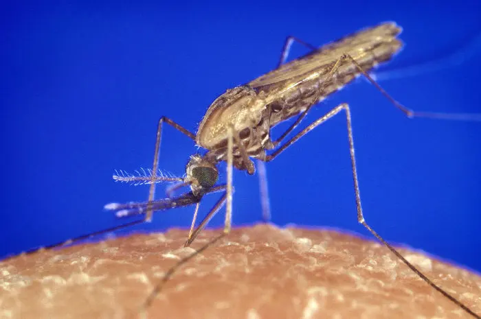
Who proved that malaria is spread by mosquitoes?
Pick an answer to see why.
Structure
Of the many stages of P. vivax, the trophozoite (Greek trophe, food: the feeding stage) is the grown, adult form that lives inside the RBCs of a person. It is a roughly spherical or amoeboid, uninucleate cell with a vacuole and granular cytoplasm. Under the electron microscope:
- i.It is covered by a membrane of two layers, the plasmalemma (the parasite also sits in a membrane sac made from the RBC’s own membrane, the parasitophorous vacuole).
- ii.Its nucleus is large and eccentric (off to one side), surrounded by a double nuclear membrane with ribosomes (RNA particles) on it; the nucleoplasm has a nucleolus, granules and fine fibrils.
- iii.Food vacuoles in the cytoplasm hold the dark granules of hemozoin (malaria pigment, below). It takes in the host’s cytoplasm through a mouth-like pore, the cytostome.
- iv.The endoplasmic reticulum is in the form of simple vesicles of varying shape, rough or smooth.
- v.There are several mitochondria, each bounded by a double membrane, with their cristae near the edge (peripheral cristae).
- vi.A concentric body, made of layers of membrane, seems to develop from the plasmalemma.
- vii.There are no locomotory organelles and no contractile vacuoles (inside a cell, there is no extra water to pump out).
Life cycle
An organism that harbors a parasite is its host. P. vivax is a digenetic parasite: it needs two hosts to complete its life cycle, the female Anopheles and a human. People suffer from malaria, but the mosquito is not harmed.
- The mosquito is the primary (definitive) host, because the sexual phase of the parasite’s life passes in it.
- The human is the secondary (intermediate) host, in which the parasite multiplies asexually.
The mosquito is also the vector, since it carries the parasite from one host to the next.
Why is the mosquito, not the human, called the primary host of Plasmodium?
Pick an answer to see why.
1. Asexual phase of P. vivax in a human
a. Infection, or inoculation of sporozoites
An infected female Anopheles carries the infective stage of the parasite, the sporozoites, in its salivary glands. When it bites a person to suck blood, it first injects some saliva, with dozens of sporozoites in it. The saliva holds anticoagulants, substances that stop the blood from clotting, so that the mosquito can keep sucking without its fine mouthparts getting blocked.
Structure of a sporozoite (under the electron microscope):
- i.It is a microscopic, uninucleate, sickle-shaped cell, tapering at both ends, about 12 µm long and 1 µm wide (a red blood cell is about 7.5 µm across).
- ii.It is covered by a thin, firm, elastic pellicle.
- iii.It moves by gliding and wriggling, using the contractile microtubules that run along its length under the pellicle.
- iv.At the front end is the apical cap, into which open the paired organelles (rhoptries). These release lytic juice, enzymes that help the sporozoite penetrate the cells it enters.
- v.In the cytoplasm are convoluted tubules (their function is unknown), a large central nucleus, a mitochondrion and a Golgi body. There is a tiny pore in the pellicle, the micropyle (cytostome).
Which stage of Plasmodium does a mosquito inject into a person?
Pick an answer to see why.
b. Schizogony in the liver
Within about half an hour of the bite, the sporozoites leave the blood and enter liver cells (hepatocytes), helped by the lytic juice of their paired organelles. There they multiply asexually by schizogony: the nucleus divides many times, and then the cytoplasm splits round each nucleus at once, so that one cell becomes many (multiple fission). A cell dividing like this is a schizont, and the daughter cells are merozoites.
Liver schizogony is usually described in two stages:
i. Pre-erythrocytic schizogony (the first schizogony, in liver cells). In the liver cell, the sporozoite grows and becomes round: a schizont. It divides by schizogony into thousands of small, uninucleate cryptomerozoites. The liver cell bursts and they are set free into the sinusoids (the liver’s fine blood channels).
ii. Exo-erythrocytic schizogony (the second, in fresh liver cells). The cryptomerozoites enter new liver cells, grow into round schizonts feeding on the cell’s contents, divide again, and release thousands of metacryptomerozoites when the cells burst. These are of two kinds: smaller and more numerous micro-metacryptomerozoites, which enter the blood and attack RBCs; and larger, fewer macro-metacryptomerozoites, which attack fresh liver cells again.
c. Schizogony in RBCs, or erythrocytic schizogony
The merozoites from the liver (the micro-metacryptomerozoites of the older account) invade red blood cells and pass through these stages:
- i.Young trophozoite. The merozoite feeds on the RBC’s contents, becomes rounded, and forms a vacuole.
- ii.Signet ring stage. As it grows, a large central non-contractile vacuole pushes the nucleus and cytoplasm to the edge, so the parasite looks like a ring with a gem: the signet ring. The parasite’s enzymes break down the RBC’s hemoglobin into its protein part, globin, and heme (hematin). The globin is its food. Free heme is poisonous to the parasite, so it packs it into harmless crystals, hemozoin, the yellowish brown malarial pigment, in fine granules.
- iii.Amoeboid stage. The ring grows into an active, amoeboid trophozoite with pseudopodia. At this stage small red-staining (eosinophilic) granules appear in the RBC’s cytoplasm, Schüffner’s dots, and the RBC becomes enlarged (up to twice its size) and pale.
- iv.Schizont and rosette stage. After feeding, the amoeboid stage becomes a large, round schizont. Its nucleus divides by mitosis into 12–24 nuclei, each of which moves to the edge, takes some cytoplasm, and becomes an oval merozoite. The merozoites round the edge, with the leftover cytoplasm and hemozoin in the middle, look like the petals of a rose: the rosette stage.
The RBC then bursts and releases its merozoites into the plasma. Each attacks a fresh RBC and repeats the cycle. In P. vivax, one erythrocytic cycle takes 48 hours, and the bursting of many RBCs at once gives the fever every 48 hours.
d. Post-erythrocytic schizogony
Older accounts add that some merozoites from the RBCs may enter liver cells again and multiply there: post-erythrocytic schizogony. This was once thought to explain relapses. It is now known that merozoites from the blood can’t infect liver cells; relapses of vivax malaria come from the hypnozoites (above).
Which event causes the regular attacks of fever in malaria?
Pick an answer to see why.
e. Formation of gametocytes
After several generations of schizogony in RBCs, some merozoites, instead of becoming schizonts, grow slowly inside their RBCs, with plenty of hemozoin, into gametocytes: cells that will make gametes. They are of two kinds:
- Macrogametocytes (female): larger, with a small compact nucleus and cytoplasm laden with food. They are usually the more numerous kind.
- Microgametocytes (male): smaller, with clear cytoplasm and a large central nucleus. One male is enough for several females, since each makes eight male gametes.
Gametocytes develop no further in the person. They circulate in the blood for some days or weeks, and either die there or are sucked up with blood by a female Anopheles, to continue the sexual phase.
2. Sexual phase of P. vivax in the female Anopheles
a. Sucking up blood
A female Anopheles sucks the blood of an infected person, with gametocytes and the other stages in the RBCs. In her stomach, all the other stages are digested, but the gametocytes survive, adapted to resist her digestive enzymes.
b. Gametogony (making gametes)
Gametogony is the making of male and female gametes from the gametocytes.
- Male gametes (microgametes). In the mosquito’s stomach, each microgametocyte undergoes exflagellation. It is triggered by the fall in temperature (from the warm-blooded human to the cold-blooded mosquito) and by a substance in the mosquito’s gut (xanthurenic acid). Its nucleus divides by mitosis into eight haploid nuclei (some books say 6–8), which move to the edge; each, with a little cytoplasm, is pushed out as a lashing, flagellum-like thread. The threads break free as motile male gametes. It is over in about 15 minutes.
- Female gamete (macrogamete). The macrogametocyte reorganizes itself into a non-motile female gamete, or ovum.
c. Fertilization and ookinete formation
The female gamete grows a small bulge, the receptive cone, to receive a male gamete. Many male gametes reach it, but only one enters through the cone. The two fuse and form a zygote; this fusion of two gametes is syngamy.
The zygote stays round and still for a while (about 24 hours, many books say; often less), then lengthens into a motile, worm-like (vermiform) cell, the ookinete (or vermicule), about 15–20 µm long and 3 µm wide.
d. Encystment and sporogony
The ookinete pushes through the wall of the mosquito’s stomach and comes to rest just beneath its outer layer. There it becomes round and is enclosed in a thin, elastic cyst wall, made partly by itself and partly from the stomach’s tissues: encystment. The encysted ookinete is now an oocyst. They stand out on the stomach like small warts: in nature usually one to a few on a mosquito, but in a heavy infection fifty or more.
Sporogony is the asexual multiplication inside the oocyst that makes sporozoites. Its nucleus has already been through meiosis (in the zygote and ookinete; many books place it at the first division in the oocyst), and now divides many times by mitosis, into many haploid nuclei. The cytoplasm meanwhile becomes spongy with large vacuoles and breaks into many masses, each taking a nucleus; each mass grows into a slender, spindle-shaped, haploid sporozoite. A mature oocyst may hold about 10,000 of them. The oocyst bursts, the sporozoites spill into the mosquito’s body cavity (hemocoel), and they make their way to the salivary glands. From there they pass down the duct in the hypopharynx (part of the mouthparts) with the saliva the next time the mosquito bites a person, and the asexual phase begins again.
From the mosquito’s meal of infected blood to sporozoites in its salivary glands takes about 9–18 days, longer in cool weather. That is why malaria spreads only where it is warm for long enough: in Nepal, in the Terai and the low valleys, not in the mountains.
Put the stages of the sexual cycle in order.
Pick an answer to see why.
8.1.5 Concept of Plasmodium falciparum
Plasmodium falciparum is found throughout the tropical and subtropical world; most of its cases and nearly all malaria deaths are in Africa south of the Sahara. It causes malignant tertian (or pernicious) malaria: the fever may be almost continuous or come at intervals of about two days. It is the most dangerous species:
- RBCs infected by it become sticky and cling to the walls of small blood vessels, especially in the brain, blocking them. This causes cerebral malaria: confusion, fits and coma.
- It can infect RBCs of every age, so very many may be infected at once, causing severe anemia.
- Blackwater fever: in some patients so many RBCs burst inside the blood vessels that hemoglobin leaks into the urine and turns it dark red-brown or black (hemoglobinuria).
- Without quick treatment it can kill within days, so its death rate is much higher than that of the other species.
Its incubation period is about 12 days on average (9–14). Its liver stage lasts about a week, and it makes no hypnozoites, so it doesn’t relapse.
In blood smears:
- the signet rings are small, often sit at the edge of the RBC, and often have two chromatin dots; there may be two or more rings in one RBC;
- trophozoites are small compared with those of P. vivax; the schizont is very small, about 5 µm across, and makes 8–24 merozoites; the older trophozoites and schizonts are rarely seen in blood taken from a finger, because they are stuck in the small vessels of the organs;
- the hemozoin is black;
- the RBC is not enlarged; instead of Schüffner’s dots it shows Maurer’s dots (clefts);
- the gametocytes are crescent-shaped (Latin falx, sickle: hence the name falciparum).
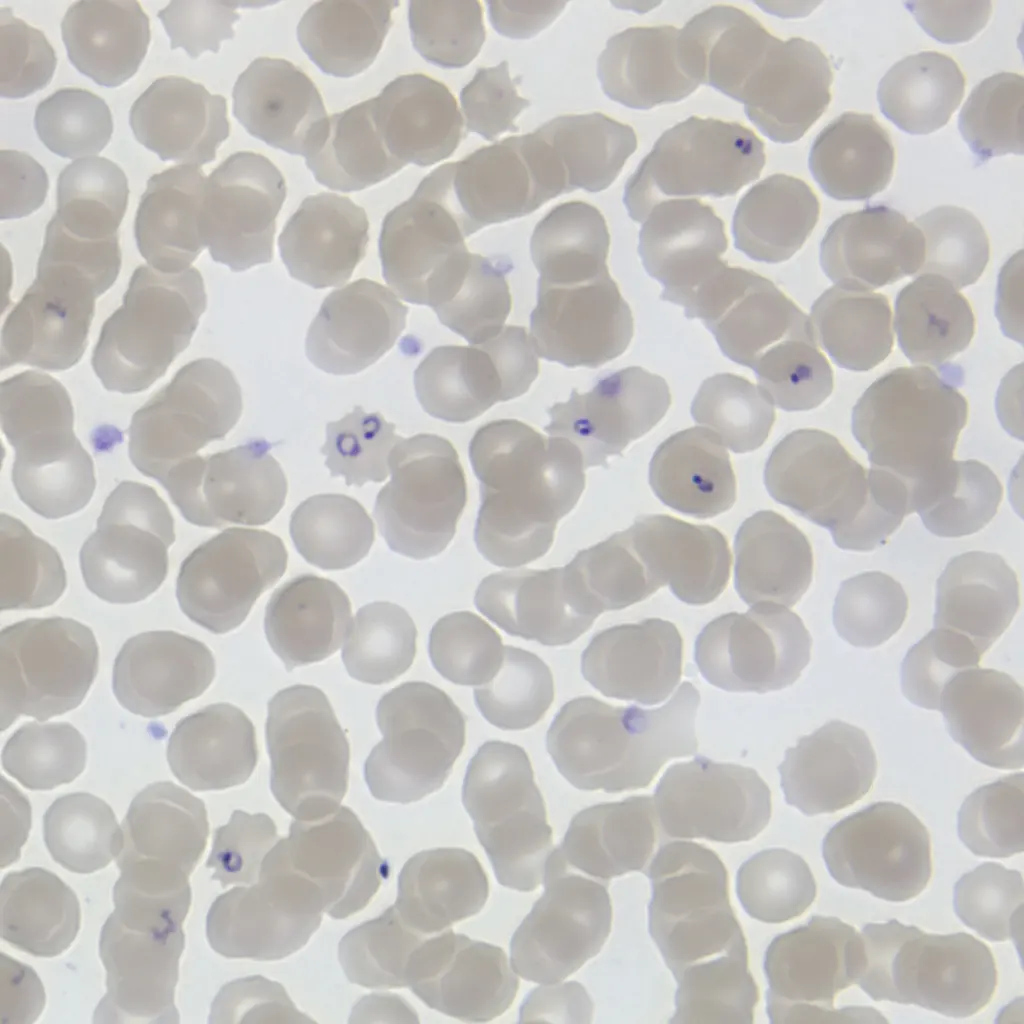
Comparison of the four species of Plasmodium that infect people
| P. vivax | P. malariae | P. ovale | P. falciparum | |
|---|---|---|---|---|
| Malaria | benign tertian; low death rate | quartan; usually mild, but can last for years | ovale (mild) tertian | malignant tertian; high death rate |
| Where | worldwide, mainly tropical, subtropical and warm temperate | worldwide, tropical and subtropical, patchy | mainly tropical Africa | worldwide in the tropics and subtropics |
| Incubation period | about 14 days | 18–40 days (often given as 18–24) | about 14 days | about 12 days |
| Erythrocytic cycle | 48 hours | 72 hours | 48 hours | 36–48 hours |
| Hypnozoites (relapses) | yes | no | yes | no |
| RBC | enlarged; Schüffner’s dots | not enlarged; Ziemann’s dots | slightly enlarged, often oval; Schüffner’s dots | not enlarged; Maurer’s dots |
| Merozoites per schizont | 12–24 | 6–12 | 6–14 | 8–24 |
Which species causes blackwater fever and cerebral malaria?
Pick an answer to see why.
8.1.6 Economic importance of Plasmodium
Plasmodium vivax does the mosquito no harm, but it causes benign tertian malaria in people. The symptoms appear about 14 days after the bite: the parasites use this time to multiply in the liver and then in the RBCs until there are enough of them. When a generation of schizonts bursts, merozoites and hemozoin spill into the plasma, and the body reacts with fever. Each attack has three stages:
- i.Cold stage. The patient feels very cold, with shivering and chattering teeth, even under blankets (about 15 minutes to an hour).
- ii.Hot stage. As the chill passes, the temperature rises, up to about 40–41 °C (104–106 °F), with a terrible headache (a few hours).
- iii.Sweating stage. The patient sweats heavily as the temperature falls back to normal, and feels tired but well, until the next attack (48 hours after the last, in vivax malaria).
Other effects of malaria are anemia (from the loss of RBCs), hemolysis (the bursting of RBCs), and splenomegaly, an enlarged spleen, which works hard to remove the damaged RBCs.
Control measures of mosquitoes
- Kill adult mosquitoes: by hand, with traps, by fumigation (smoke) or by spraying insecticides; above all, by spraying the inside walls of houses, where Anopheles rests after feeding.
- Remove breeding places near homes: swamps, marshes and stagnant water such as pools, ponds and ditches, where mosquitoes lay their eggs.
- Cover open drains.
- Remove tin cans, buckets, unused tyres, barrels and anything else that collects rainwater.
- Clear bushes and shrubs near houses, where adult mosquitoes rest.
- Spray oil, kerosene or other chemicals on water that can’t be drained: a film of oil stops the larvae from breathing at the surface.
- Biological control: put fish that eat mosquito larvae in ponds, pools, ditches and tanks, such as Gambusia (the mosquitofish), trout and sticklebacks. Gambusia is not native to Nepal, though, and where it spreads into wild waters it can harm native fish and frogs; native larva-eating fish are better where they will do.
And protect people:
- Sleep under a bed net treated with insecticide (it both blocks and kills mosquitoes; Anopheles bites mostly at night).
- Cover the arms and legs and use repellents in the evening.
- Get tested quickly for any fever in or after a visit to a malarial area, and take the full course of medicine.
Exercise
A. Tick the correct alternative.
- Which of the following is the carrier (vector) of Plasmodium?
Pick an answer to see why.
- Which of the following statements is correct for the life cycle of Plasmodium vivax?
Pick an answer to see why.
- Which of the following is the infective stage of Plasmodium (for people)?
Pick an answer to see why.
- What is the function of the part marked X in the diagram above?
Pick an answer to see why.
- In malaria, which event of erythrocytic schizogony is directly related to the periodic fever and chills?
Pick an answer to see why.
- Where does the pre-erythrocytic phase of Plasmodium take place?
Pick an answer to see why.
- Who discovered the relationship between malaria and the mosquito?
Pick an answer to see why.
- In which of the following does exflagellation take place?
Pick an answer to see why.
- In Plasmodium, the zygote changes after a while into a motile, worm-like structure called:
Pick an answer to see why.
- At which stage are Schüffner’s dots seen?
Pick an answer to see why.
- Which fish is commonly used to eat mosquito larvae?
Pick an answer to see why.
- Which of the following is the correct sequence in the sexual cycle of Plasmodium?
Pick an answer to see why.
- Which of the following is the diploid stage in the life cycle of Plasmodium?
Pick an answer to see why.
B. Answer the following questions.
Define schizogony. Explain the various types of schizogony in the life cycle of Plasmodium vivax with the necessary diagrams.
Answer
Plasmodium is a digenetic parasite that completes its life cycle in two hosts. The asexual phase passes in a human. Name the organism in which it completes its sexual cycle, and explain how.
Answer
Draw a neat and labeled diagram of a sporozoite.
Answer
Swarnim said that a spherical, mature, adult and vacuolated form of Plasmodium is a sporozoite. Is he right? Give a reason.
Answer
Describe liver schizogony with the required diagrams.
Answer
Malaria is a dangerous communicable disease found all over the world. Can it be prevented? Support your answer with some points.
Answer
Briefly describe the species of Plasmodium that causes blackwater fever.
Answer
How do encystment and sporogony take place on the wall of the stomach of Anopheles?
Answer
Compare and contrast the forms of Plasmodium vivax known as the feeding stage and the infective stage.
Answer
What is erythrocytic schizogony? Describe its stages in the life cycle of Plasmodium.
Answer
Study the diagram above and answer the following questions.
- i.Which phase is shown in the figure?
- ii.Name the stages A, B, C and D.
- iii.Copy the diagram and fill in the boxes.
Answer
Printable PDFs
- Study notes 3 pages
- Worksheet 5 pages Answer key
Quiz yourself: Classification of Living Beings, Grades 5–12 →
Practice every question in the book, and keep track of the ones you miss →
A companion to your textbook, not a replacement for it: study from the book your school uses, and where the two differ, follow your teacher. Found a mistake? Please tell us.