
In 1860 a young German doctor, unhappy in his job, went to the shore at Messina in Sicily with a microscope and a sketchbook. In a single winter he found and drew 144 new species of radiolarians, single cells smaller than a grain of sand that build skeletons of glass in the shape of stars, lanterns and helmets. His name was Ernst Haeckel, and he spent the rest of his life drawing such creatures. In 1866 he gave them a kingdom of their own, neither plant nor animal: the Protista.
This unit, the first of faunal diversity (the variety of animals and animal-like life), is about that kingdom: what protists are, how they are grouped, and one of them, Paramecium, in detail.
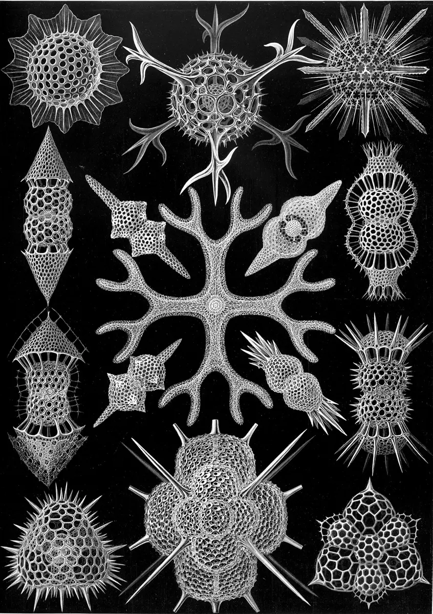
8.1.1 General introduction
Protists are eukaryotes (their cells have a true nucleus) that are not plants, animals or fungi. Most of them are single cells, too small to see without a microscope. Look at four of them:
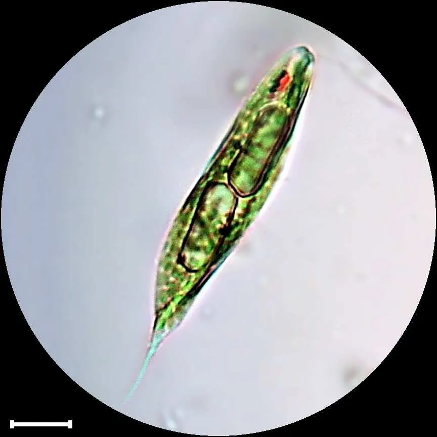
- Euglena is a microscopic, unicellular organism that swims with a flagellum. It lives mainly in fresh water. It has chloroplasts and makes its own food in light (it is autotrophic), but in the dark it can take in food from its surroundings (it is heterotrophic). Doing both is called mixotrophic nutrition.
- Paramecium is a microscopic, unicellular ciliate: it swims with thousands of short hairs, cilia. It lives in fresh water rich in decaying matter and is heterotrophic, eating bacteria and other tiny particles.
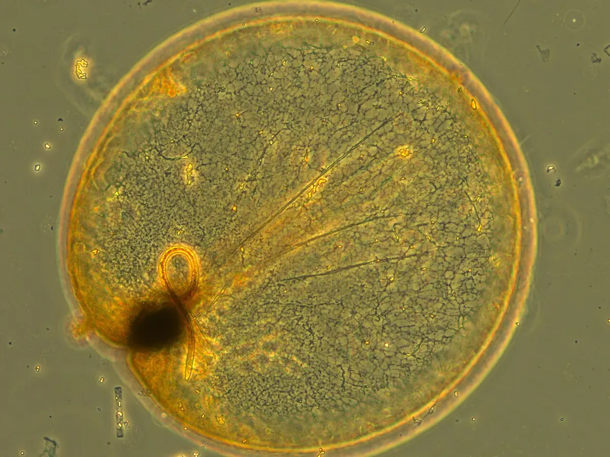
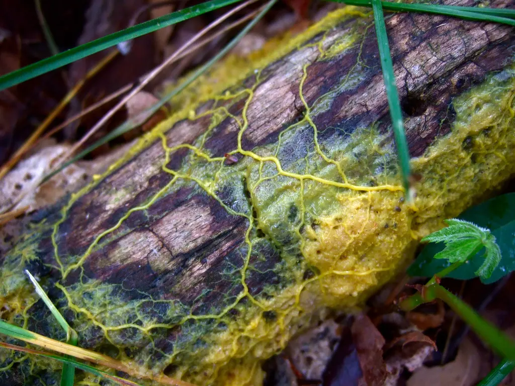
- Fuligo is a slime mold: a fungus-like protist that creeps over decaying wood as a slimy mass with many nuclei.
- Plasmodium is an intracellular blood parasite: it lives inside the red blood cells (and liver cells) of its host and causes malaria.
All four belong to the kingdom Protista.
8.1.2 Outline classification of Protista
The kingdom Protista (Greek protistos, the very first) was proposed by Ernst Haeckel in 1866. In 1969 the American ecologist Robert Harding Whittaker divided all living things into five kingdoms, by the kind of cell (prokaryotic or eukaryotic), the level of body organization (one cell or many), the mode of nutrition, and their evolutionary relationships Grade 11: Unit 4.
Protists are microscopic, single-celled, primitive eukaryotes. They are very varied:
- plant-like protists make their own food: golden algae, diatoms, dinoflagellates;
- animal-like protists (the protozoa) eat other things: Paramecium, Amoeba;
- some are both: Euglena photosynthesizes but can also feed (its colorless relative Astasia only feeds);
- some are fungus-like: the slime molds Fuligo and Protostelium.
Characteristic features of kingdom Protista
- They are usually aquatic (in fresh water or the sea); some live in moist soil, and many live inside other organisms.
- They are simple, microscopic, unicellular eukaryotes. A few live in colonies of similar cells joined together (Volvox, a ball of hundreds of flagellate cells, though most books now count it with the green algae).
- Their body is a single cell that does everything a whole animal does: this is the protoplasmic (or acellular) level of organization.
- The cell is covered by a cell membrane, a pellicle, a cell wall or a shell; some have no outer covering beyond the membrane.
- They have a single nucleus (most), two kinds of nucleus (Paramecium: nuclear dimorphism), or many nuclei (Opalina: the multinucleate or coenocytic condition).
- They move by pseudopodia, flagella or cilia; some (Plasmodium) have no organelles for locomotion.
- Their nutrition varies: holophytic (photosynthetic, like plants), holozoic (eating solid food, like animals), mixotrophic (both), saprozoic (absorbing dissolved food from dead matter) or parasitic.
- Food is digested inside the cell, in food vacuoles: intracellular digestion.
- Respiration and excretion happen by diffusion through the general body surface.
- Asexual reproduction is by binary fission, multiple fission, cyst formation, spore formation (sporulation) and so on; sexual reproduction by conjugation or by the fusion of gametes (syngamy).
Many protists are plankton: small organisms that drift in water, carried by currents because they are too small or too weak to swim against them. Plant-like plankton that photosynthesize (diatoms, dinoflagellates) are phytoplankton; animal-like ones that eat (foraminiferans, radiolarians, and many ciliates) are zooplankton.
Which feature is found in all protists?
Pick an answer to see why.
Classification of Protista
Kingdom Protista is divided into three groups, by how they feed:
- Group A: Photosynthetic protists (the plant-like ones);
- Group B: Consumer–decomposer protists (the slime molds, fungus-like);
- Group C: Protozoan protists (the animal-like ones).
Group A: Photosynthetic protists
They are divided into three phyla.
Phylum 1: Pyrrophyta (the dinoflagellates; Greek pyrrhos, fire-colored)
- i.They are aquatic, mostly in the sea.
- ii.They are unicellular, motile (with two flagella, one in a groove round the cell and one trailing behind) and photosynthetic.
- iii.Some are bioluminescent: they glow in the dark. The light is made by a chemical reaction in the cell (an enzyme, luciferase, acting on luciferin), set off when the water is disturbed. Example: Noctiluca.
- iv.The body is covered by a theca (some books call it a lorica), a coat of cellulose plates under the membrane. Some, the “naked” dinoflagellates such as Gymnodinium, have no plates.
- v.They reproduce mostly asexually, by binary fission; a few reproduce sexually too.
Examples: Noctiluca, Gymnodinium. (Noctiluca itself has lost its chloroplasts and eats other plankton, but it is a dinoflagellate all the same.) Some dinoflagellates multiply so fast that they color the sea red (red tides); their poisons kill fish and build up in shellfish, which can then poison the people who eat them.
Phylum 2: Chrysophyta (the diatoms and golden-brown algae; Greek chrysos, gold)
- i.They are unicellular, some forming colonies; found in the sea and in fresh water.
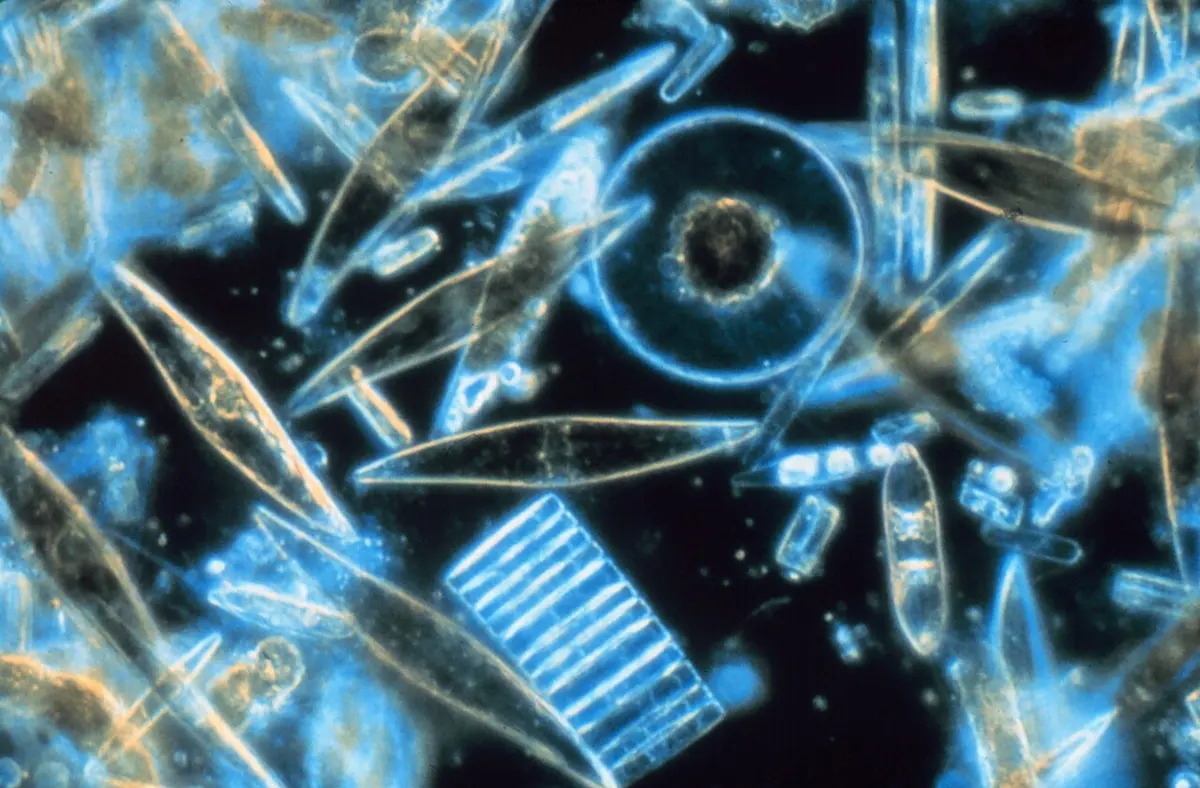
- ii.A diatom’s cell is enclosed in a hard shell of silica (glass), the frustule, in two halves that fit together like the lid and base of a box.
- iii.They reproduce both sexually and asexually; binary fission is commonest.
- iv.They include the diatoms and the golden-brown algae. Diatoms are an important food for animals in water: they are at the start of most food chains in the sea.
Examples: Navicula (a boat-shaped diatom), Triceratium (a triangular one).
Phylum 3: Euglenophyta (the euglenoids)
- i.They are motile, free-living flagellates of fresh water and damp soil.
- ii.They have no cell wall: their shape is kept by the plasma membrane and a flexible pellicle of protein strips under it, which lets them bend and squirm.
- iii.They swim with flagella (one long one that shows).
- iv.Some have chlorophyll and photosynthesize. Their reserve food is not starch but paramylon (paramylum), a different sugar chain.
- v.Near the base of the flagellum lies a red, light-sensitive eyespot (stigma), which helps the cell swim towards light.
- vi.They live like both plants and animals: mixotrophic nutrition.
- vii.They reproduce asexually by longitudinal binary fission (they split lengthwise).
Examples: Euglena, Peranema (colorless, eats other cells), Trachelomonas (in a brown shell).
A protist has chloroplasts, an eyespot and a flagellum, stores paramylon, and splits lengthwise. To which phylum does it belong?
Pick an answer to see why.
Group B: Consumer–decomposer protists, or slime molds
- i.They are also called protistan fungi, because they are like amoeboid protozoans at one stage (creeping and engulfing food) and like fungi at another (making spores in fruiting bodies).
- ii.They have no chlorophyll, so they can’t make their own food. They feed on decaying organic matter: bacteria, yeasts and bits of dead plants.
- iii.They reproduce both sexually and asexually (by spores).
They are of two types.
Type 1: Plasmodial (true, acellular) slime molds
- i.They are found on decaying leaves and wooden logs.
- ii.Their outer covering is the plasma membrane.
- iii.They are free-living, irregular masses of slimy protoplasm that creep slowly over their food.
- iv.The feeding stage, the plasmodium, is a large, multinucleate mass: one giant cell with thousands of nuclei and no walls between them, sometimes as wide as your hand.
Examples: Physarum, Tubifera, Fuligo.
Type 2: Cellular slime molds
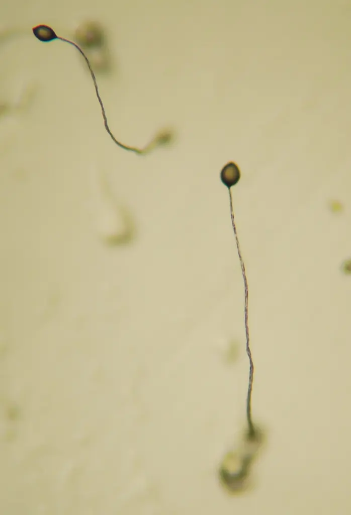
- i.They live in the upper layer of moist soil rich in decaying matter.
- ii.Their feeding stage is a single amoeboid cell, each living on its own and eating bacteria.
- iii.When food runs out, the cells aggregate: thousands crawl together to form a slug, but each keeps its own membrane (its individuality). The slug then makes a stalked fruiting body and reproduces by spores.
Examples: Dictyostelium, Acytostelium.
How does a cellular slime mold differ from a plasmodial one?
Pick an answer to see why.
Group C: Protozoan protists
- i.They live in water and on land; many are parasitic, and the rest free-living, commensal (living on another without harming it) or saprozoic.
- ii.They are the simplest, primitive, acellular, microscopic animalcules (“little animals”).
- iii.They are solitary or colonial, with a protoplasmic body organization.
- iv.The body is covered by a cell membrane, a pellicle or a shell.
- v.Their shape varies: spherical, oval, elongated or flattened.
- vi.The cytoplasm is divided into a clear outer ectoplasm and a granular inner endoplasm.
- vii.The number and shape of the nucleus vary.
- viii.Nutrition is holozoic and digestion is intracellular.
- ix.Respiration is by diffusion through the general body surface.
- x.Excretion is through the general body surface or the contractile vacuole; undigested food leaves by the cell anus (cytopyge).
- xi.In fresh-water forms, the amount of water and salt in the cell is kept steady by the contractile vacuole: osmoregulation.
- xii.Reproduction is asexual (fission, budding or sporulation) and sexual (by gametes, or by conjugation).
- xiii.In bad conditions many form cysts: encystment.
Protozoa are divided into four classes by their organelles of locomotion.
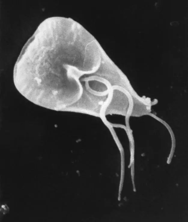
Class 1: Flagellata (Mastigophora)
- i.The body is covered by a pellicle.
- ii.They move by flagella.
- iii.They usually have a single nucleus (Giardia has two, side by side).
- iv.They are free-living or parasitic.
Examples: Leishmania (kala-azar), Trypanosoma (sleeping sickness), Giardia (diarrhea).
Class 2: Rhizopoda (Sarcodina)
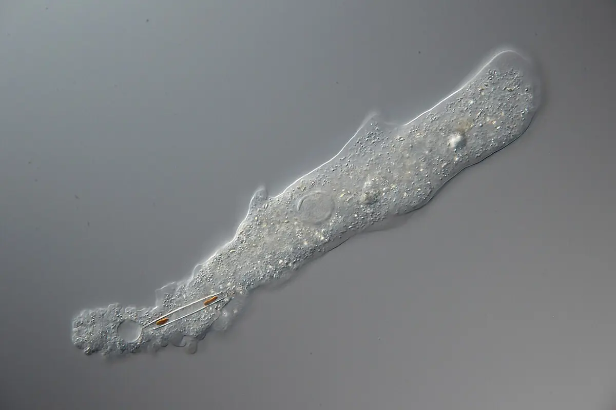
- i.The body has no fixed shape and is bounded only by the plasma membrane.
- ii.They move by pseudopodia (“false feet”), flowing extensions of the cell.
- iii.They are solitary, free-living or parasitic.
- iv.Asexual reproduction is by binary fission.
Examples: Amoeba (free-living), Entamoeba histolytica (amoebic dysentery).
Class 3: Sporozoa
- i.The body is covered by a pellicle.
- ii.They reproduce asexually (by multiple fission and spores) and sexually.
- iii.All are endoparasites (they live inside their host).
- iv.They have no organelles for locomotion (some glide slowly).
Examples: Plasmodium (malaria, Unit 23), Babesia (a blood parasite of cattle), Monocystis (in the earthworm’s seminal vesicles).
Class 4: Ciliata
- i.The shape is fixed, because of a firm pellicle on the outside.
- ii.They move by cilia.
- iii.They are free-living or parasitic.
- iv.They reproduce asexually and sexually (conjugation).
Examples: Paramecium, Balantidium coli (the largest protozoan that lives in people, caught from pigs; it causes dysentery), Vorticella.
A protozoan has no locomotory organelles, has a pellicle, and lives only as a parasite inside its host. In which class is it?
Pick an answer to see why.
8.1.3 Paramecium caudatum (slipper animalcule)
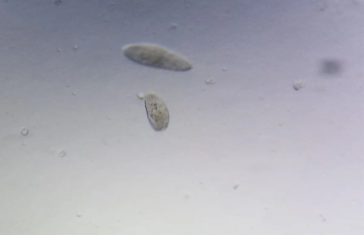
Paramecium is a unicellular, free-living, microscopic animalcule of fresh water rich in decaying organic matter. Its shape is like the sole of a slipper, so it is called the slipper animalcule. It moves by the beating of cilia, fine hair-like threads of cytoplasm all over its body.
Classification
| Rank | Name |
|---|---|
| Kingdom | Protista |
| Phylum | Protozoa |
| Class | Ciliata |
| Genus | Paramecium |
| Species | caudatum |
| Common name | Slipper animalcule |
Habit and habitat
Of the many species of Paramecium, Paramecium caudatum is the commonest. It lives in fresh water, in ponds, streams, ditches and lakes rich in decaying organic matter, and is most plentiful in stagnant water and slow streams, where bacteria (its food) are many. It is free-living and swims with its cilia. It is also called an infusorian animalcule, because it appears in infusions: water in which decaying plant or animal matter has soaked.
A. External structure (morphology)
a. Shape and size
Paramecium caudatum is slipper-shaped (some describe it as spindle-shaped), so it is called the slipper animalcule. It is asymmetrical, because of the oral groove that runs obliquely along one side. The body is long, blunt at the front (anterior) end, widest just behind the middle, and more pointed at the back (posterior) end. The side with the oral groove is the ventral or oral surface; the opposite side, the dorsal or aboral surface.
It is microscopic, but just visible to the naked eye as a whitish or grayish speck: P. caudatum is one of the largest species, about 170–300 µm long (0.17–0.3 mm; a µm, micrometer, is a thousandth of a millimeter).
Outline: unknown, CC0, via PhyloPic
b. Pellicle
The body is covered by a thin, firm but elastic protective layer, the pellicle. It is a membrane with flat sacs (alveoli) beneath it, so it is more than one layer thick. Being elastic, it lets the cell squeeze through narrow gaps between bits of debris and spring back into shape.
Under high power, the pellicle shows a regular pattern of hexagonal (six-sided) depressions. From the center of each hexagon rises a cilium, and at the front and back margins of the hexagons are small openings, through which the trichocysts are shot out.
c. Cilia
The whole body is covered with thousands of tiny, hair-like cilia (singular cilium). They are all about the same length, arranged in longitudinal rows, except at the posterior end, where a tuft of longer cilia forms the caudal tuft (Latin cauda, tail; hence the name caudatum). Each cilium grows from a small basal granule (basal body, or kinetosome) just below the pellicle, in the ectoplasm. Inside, a cilium has the 9 + 2 pattern of microtubules described in Unit 2.
Cilia are used for:
- locomotion: they beat like oars, not all together but one after another in waves that run along the rows (metachronal rhythm), like the wind passing over a field of wheat. The cell swims along a spiral path, turning on its long axis, at roughly a millimeter a second: several of its own lengths each second;
- feeding: the cilia of the oral groove sweep water and food into the mouth;
- sensing: they respond to touch, chemicals and temperature.
The caudal tuft helps the cell change direction while swimming.
What is the caudal tuft?
Pick an answer to see why.
B. Internal structure
a. Cytoplasm
The cytoplasm is clearly divided into two regions:
- i.Ectoplasm: a thin, clear, firm outer layer under the pellicle, which holds the trichocysts, the basal bodies of the cilia and the fine fibrils that link them.
- ii.Endoplasm: the large, central, granular, more fluid part, which holds the nuclei, contractile vacuoles, food vacuoles, mitochondria, Golgi bodies, ribosomes and other cell components.
b. Trichocysts
Trichocysts are small, rod-shaped or oval organelles in the ectoplasm, just under the pellicle, alternating with the basal bodies of the cilia. They lie at right angles to the body surface and open by small pores on the ridges of the hexagons of the pellicle.
An undischarged trichocyst is a sac, the shaft, packed with a dense protein that bends light (refractive), with a pointed tip (spike) at its outer end, covered by a cap.
A trichocyst is discharged when something stimulates the cell: a touch, a chemical, heat, an electric current, or the attack of a predator. Then:
- Calcium enters the cell, and the trichocyst’s membrane fuses with the membrane at its pore: its cap opens to the outside.
- The packed protein inside swells and unfolds, taking in water as it does. In a few thousandths of a second it stretches to about eight times its length.
- Out shoots a long, striated (cross-banded) thread with the opaque spike at its end, like an upside-down nail with a cap. The long striated shaft is seen only after discharge.
Functions: trichocysts are thought to help Paramecium anchor itself to a surface while feeding, and, as experiments with its predators have shown, in defense: a fired volley of threads can push the cell away from an attacker such as Dileptus. (They are of little use against another hunter, Didinium.) In some other ciliates they help in offense, to catch prey.
Paramecium in (c): unknown, CC0, via PhyloPic; parts placed from Kahl's drawing (public domain)
c. Nucleus
Paramecium caudatum has two kinds of nucleus: this is nuclear dimorphism.
- The macronucleus (meganucleus) is large and bean-shaped. It holds many copies of the genes the cell uses every day (hundreds of copies), and controls the metabolism (the daily activities) of the cell. It is also called the somatic or vegetative nucleus.
- The micronucleus is small and spherical, lying close to the macronucleus, often in its hollow (concavity). It holds two sets of chromosomes (it is diploid) and controls reproduction: it is the one that divides by mitosis in fission and by meiosis in conjugation. So it is also called the generative nucleus.
d. Contractile vacuoles
Unlike Amoeba, which has one, Paramecium has two large, fluid-filled contractile vacuoles, one near each end (anterior and posterior), in the endoplasm close to the dorsal (aboral) surface. Their places are fixed, and each opens to the outside by a permanent pore in the pellicle on the dorsal surface. Now and then a vacuole seems to disappear for a moment: just after it has emptied.
Each contractile vacuole is surrounded by about six to ten long, narrow tubes, the radiating (radial) canals, arranged like the rays of a star. They collect the extra water from the cytoplasm all round and pour it into the central vacuole. When the vacuole is full, it contracts and empties its contents to the outside through the pore. Then the canals fill again.
The posterior vacuole contracts more often than the anterior one, because it lies near the cytopharynx, where water comes in with each new food vacuole.
Function: osmoregulation, keeping the right amount of water and salts in the cell. It also removes some dissolved wastes.
Why does the posterior contractile vacuole contract more often than the anterior one?
Pick an answer to see why.
e. Food vacuoles
Food vacuoles (also called gastrioles) are roughly spherical, non-contractile sacs in the endoplasm, of varying number and size. Each holds swallowed food particles (mostly bacteria), a little water and digestive enzymes, inside a thin membrane.
Food vacuoles are carried round the cell by the streaming of the cytoplasm, along a fixed path: this movement is cyclosis. As they travel, the food in them is digested, and the digested food is absorbed into the cytoplasm and assimilated (used to build the cell). Whatever can’t be digested is thrown out at the cytopyge.
f. Oral groove
The oral groove is a large, oblique depression on the ventro-lateral side of the body, which makes Paramecium asymmetrical. At its hind end it leads into a short, conical, funnel-shaped depression, the vestibule. The vestibule leads into the cytopharynx (cell gullet) through a fixed oval opening, the cytostome (cell mouth). The cytopharynx runs obliquely back into the endoplasm, and at its inner end a new food vacuole forms.
Rows of cilia line the whole oral apparatus. Their beating sets up a current of water that collects food particles and passes them down to the cytostome, where they are trapped in a forming food vacuole. When the vacuole reaches a certain size, it pinches off and starts its journey.
g. Cytopyge (cell anus)
At the time of egestion (getting rid of undigested food), a tiny opening becomes visible in the pellicle on the ventral surface, just behind the cytostome: the cytopyge, also called the anal spot or cytoproct. The emptied food vacuole fuses with the pellicle here and throws out the undigested remains. The cytopyge is seen only while it is in use.
In which order does a food particle pass into Paramecium?
Pick an answer to see why.
Reproduction
Reproduction is the life process by which living things produce offspring of their own kind, so that their kind continues from generation to generation.
In Paramecium, asexual reproduction is by transverse binary fission, and sexual reproduction (more exactly, the exchange and renewal of nuclei) is by conjugation, with other processes such as autogamy.
Transverse binary fission
Binary fission happens in favorable conditions: plenty of food and a suitable temperature. P. caudatum divides two or three times a day this way, and each division takes about one to two hours. It is transverse: the cell divides across its middle, the plane of division at right angles to the long axis of the body. The division of the nuclei (karyokinesis) comes first, then the division of the cytoplasm (cytokinesis).
- i.Paramecium stops feeding, and its oral apparatus (the buccal structures) begins to disappear.
- ii.The micronucleus divides by mitosis into two daughter micronuclei, which move towards opposite ends.
- iii.At the same time, the macronucleus lengthens, is pinched in the middle, and divides by amitosis Grade 11: Unit 3; the daughter macronuclei also move apart.
- iv.Two oral grooves begin to form on the ventral side, one in the front half and one in the back half.
- v.Each half develops a new contractile vacuole, so each will have two; the oral structures are completed.
- vi.Meanwhile a transverse constriction (furrow) appears across the middle, deepens, and finally cuts the cytoplasm in two: two daughter Paramecia.
- vii.The front daughter is the proter; the back one, the opisthe.
The daughters are genetically identical to each other and to their parent: they form a clone. After many generations of fission, though, the rate of division slows and may stop: the line must be rejuvenated (renewed) by conjugation.
Outline: unknown, CC0, via PhyloPic
In the binary fission of Paramecium, how do its two nuclei divide?
Pick an answer to see why.
Conjugation
Conjugation is the simplest kind of sexual reproduction: a temporary union of two Paramecia of the same species but of different mating types (strains), in which they exchange micronuclei. It needs to happen now and then between long runs of binary fission, because after many generations (a few hundred) of fission alone, a line of Paramecium grows weak and can no longer divide.
The two partners look alike but differ in their chemistry, as two mating types. (The American biologist Tracy Sonneborn discovered mating types in Paramecium in 1937.) Partners ready to conjugate are pre-conjugants. Conjugation is brought on by:
- i.a shortage of food, or starvation;
- ii.being genetically and physiologically weak and small after a long run of binary fission;
- iii.sudden darkness or a fall in temperature, in some species;
- iv.mating-type substances: specific proteins on the cilia of partners of opposite mating types, which make them stick together.
Events in conjugation
- Two P. caudatum of different mating types meet and stick together by their ventral (oral) surfaces, at the oral grooves.
- Their oral structures disappear, and they stop feeding.
- At the point of contact the pellicle and ectoplasm dissolve, forming a protoplasmic bridge between them. They are now called conjugants.
- In each, the macronucleus breaks into pieces, which are later absorbed by the cytoplasm. Meanwhile the micronucleus grows and divides by meiosis into four haploid daughter micronuclei.
- Three of the four degenerate. The remaining one divides by mitosis into two pronuclei (gametic nuclei), of unequal size.
- The smaller, active pronucleus is the migratory (male) pronucleus; the larger, passive one is the stationary (female) pronucleus.
- The two migratory pronuclei cross the bridge, each into the other conjugant, and each fuses with the stationary pronucleus there.
- Each conjugant now has a zygote nucleus (synkaryon), diploid again, half from each partner. This complete fusion of two nuclei from different individuals is amphimixis.
- After about 12–48 hours together, the two separate. They are now called exconjugants.
- In each exconjugant, the zygote nucleus divides three times by mitosis, giving eight nuclei. Four of them grow into macronuclei; the other four stay micronuclei.
- Three of the four micronuclei degenerate.
- The remaining micronucleus divides by mitosis, and the cell divides by binary fission: two daughters, each with two macronuclei and one micronucleus.
- Each daughter divides again: the micronucleus divides by mitosis, and the two macronuclei are shared out, one to each. Four cells, each with one macronucleus and one micronucleus.
- So each exconjugant gives four, and the pair together eight, daughter Paramecia, which are genetically different from their parents.
Outline: unknown, CC0, via PhyloPic
Significance of conjugation
- i.It reorganizes the nuclei, which is essential if binary fission is to go on.
- ii.It rejuvenates the line: it restores vitality and genetic vigor in the offspring.
- iii.It replaces the old macronucleus with a new one made from the zygote nucleus.
- iv.It brings variation in the offspring, as genes from two parents are mixed.
- v.It multiplies the numbers too: eight offspring from one pair.
After conjugation, why are the eight daughter Paramecia genetically different from their parents?
Pick an answer to see why.
Exercise
A. Tick the correct alternative.
- Which of the following is a bioluminescent protist?
Pick an answer to see why.
- In which of the following is the mixotrophic mode of nutrition found?
Pick an answer to see why.
- Which of the following is a fungus-like protist?
Pick an answer to see why.
- What is the cell anus of the protozoans called?
Pick an answer to see why.
- In protozoans, which of the following performs osmoregulation?
Pick an answer to see why.
- Which of the following protozoans moves by flagella?
Pick an answer to see why.
- Paramecium lives in fresh water, and water keeps flowing into its body from outside, with the current that brings food particles. Which of the following structures is responsible for maintaining the osmotic balance in the body?
Pick an answer to see why.
Outline: unknown, CC0, via PhyloPic
- Which process is responsible for the movement of part A in the diagram above?
Pick an answer to see why.
- What may happen when the macronucleus of Paramecium is removed?
Pick an answer to see why.
- What is the function of the contractile vacuole in Paramecium?
Pick an answer to see why.
- How many radiating canals are found round each contractile vacuole of Paramecium?
Pick an answer to see why.
- How many daughter Paramecia are formed after each conjugation?
Pick an answer to see why.
- What will happen when Paramecium can’t perform conjugation?
Pick an answer to see why.
- Which type of digestion is found in Paramecium?
Pick an answer to see why.
- Which of the following is the external protective cover of Paramecium?
Pick an answer to see why.
- Which organelle is used for locomotion in Paramecium?
Pick an answer to see why.
- Which of the following parts helps in feeding in Paramecium?
Pick an answer to see why.
B. Answer the following questions.
A: Pearson Scott Foresman, public domain, via Wikimedia Commons
B: PhyloPic (CC0); parts placed from Leidy’s drawing (public domain)
To which groups of Protista do the organisms A and B above belong? Write any four differences between these groups.
Answer
What are slime molds? List their characters.
Answer
Outline: unknown, CC0, via PhyloPic
Observe the diagram above and answer the following questions.
- i.Identify the parts A, B, C and D.
- ii.What is the function of C and B?
- iii.B is long at the posterior end. Write its function.
Answer
Draw a neat and labeled diagram of Paramecium caudatum.
Answer
What are trichocysts? Discuss their working mechanism.
Answer
What is the significance of conjugation in Paramecium?
Answer
Osmoregulation is the process of maintaining salt and water in the body. Which organelle of Paramecium helps in this process? Describe it briefly.
Answer
Paramecium shows nuclear dimorphism: it has two types of nucleus, the macronucleus and the micronucleus. What are the differences between them?
Answer
Outline: unknown, CC0, via PhyloPic
Observe the diagram above and answer the following questions.
- i.Which method of asexual reproduction is shown in the diagram?
- ii.In which conditions does this process occur?
- iii.Briefly describe the process.
Answer
Why is Paramecium called the slipper animalcule? Describe the external features of Paramecium caudatum with a well-labeled diagram.
Answer
Which method of reproduction does Paramecium adopt in unfavorable conditions such as a shortage of food or a sudden fall in temperature? Describe it with the necessary diagram.
Answer
Printable PDFs
- Study notes 4 pages
- Worksheet 5 pages Answer key
Quiz yourself: Classification of Living Beings, Grades 5–12 →
Practice every question in the book, and keep track of the ones you miss →
A companion to your textbook, not a replacement for it: study from the book your school uses, and where the two differ, follow your teacher. Found a mistake? Please tell us.