
A single maize plant sheds millions of pollen grains from its tassel, a cloud of yellow dust that drifts over the field for a week. Each silk on a young cob below is the long style of one flower, and each one needs a single grain to land on it, grow a tube all the way down, and deliver its two male cells to one ovule. Every kernel on a cob of makai is the result of one such journey. Miss it, and that place on the cob stays empty: that is why cobs from the edge of a field, or from a lone plant, are often gappy.
A flower is a machine for making this meeting happen. In the last unit a plant copied itself; in this one two parents mix their genes. The unit follows the whole story in order: how the flower makes pollen and an egg, how pollen is carried from one flower to another, how the two male cells in a pollen tube do two different jobs (a feature found only in flowering plants), and how the fertilized ovule builds an embryo and its food store, the seed.
Flower: after Mariana Ruiz (LadyofHats), Mature flower diagram (public domain)
4.2.1 General introduction
The flower is the organ of a flowering plant that carries out sexual reproduction. It is a short shoot whose leaves have become four kinds of floral parts, in rings (whorls) on the swollen tip of the stalk, the receptacle (thalamus):
- Calyx, the outer whorl of sepals, usually green, which protects the flower in the bud.
- Corolla, the whorl of petals, often colored and scented, which attracts pollinators.
- Androecium, the whorl of stamens, the male reproductive organ. Each stamen is an anther on a stalk, the filament; the anther makes the pollen grains.
- Gynoecium, the pistil, the female reproductive organ, made of one or more carpels. A pistil has a sticky stigma that receives pollen, a style, and a swollen ovary holding the ovules.
Calyx and corolla are the accessory whorls; androecium and gynoecium are the essential whorls, because they make the gametes.
Flower: after Mariana Ruiz (LadyofHats), Mature flower diagram (public domain)
Sexual reproduction in plants is reproduction by the formation and fusion of male and female gametes. Because each gamete carries a half set of genes chosen by meiosis, and two parents usually contribute, every seed is a new combination of genes. In a flowering plant it happens in four steps:
- Gametogenesis: the stamens and the ovules make the gametes.
- Pollination: pollen is carried from an anther to a stigma.
- Fertilization: the male gametes reach the ovule and fuse with the egg and the central cell.
- Seed and fruit formation: the fertilized ovule grows into a seed (embryo, endosperm and coat), and the ovary into a fruit.
Which two whorls of a flower are called the essential whorls?
Pick an answer to see why.
4.2.2 Gametogenesis
Gametogenesis is the formation of gametes. In a flowering plant it happens in two steps. First, meiosis in the anther and in the ovule makes haploid spores: microspores in the anther (microsporogenesis) and a megaspore in the ovule (megasporogenesis). Then each spore grows, by mitosis, into a tiny haploid plant, a gametophyte, that makes the gametes: the pollen grain is the male gametophyte (microgametogenesis), the embryo sac the female gametophyte (megagametogenesis).
This is the plant life cycle you met in mosses, ferns and Pinus, the alternation of a diploid sporophyte and a haploid gametophyte, shrunk to its smallest: in a flowering plant the male gametophyte is just two or three cells and the female one seven cells, both living entirely inside the sporophyte’s flower.
1. Microsporogenesis
Microsporogenesis is the formation of haploid microspores from diploid microspore mother cells by meiosis, inside the microsporangia (pollen sacs) of the anther. The microspores then become pollen grains.
The anther
The anther is the head of the stamen. A typical anther is bilobed: two lobes, joined by a strip of sterile tissue, the connective, which carries a vascular bundle down from the filament. Each lobe is a theca (plural thecae), so a typical anther is dithecous. Each theca has two pollen sacs (microsporangia) running along its length, so the whole anther has four microsporangia: it is tetrasporangiate. In a cross-section the anther looks like a butterfly, with one pollen sac in each corner. When the anther is ripe, the wall between the two sacs of each theca breaks down, and the theca splits open along a line of thin cells (the stomium) to release the pollen.
The wall of the microsporangium
A cross-section of a young anther shows each pollen sac, nearly round, surrounded by a wall of four layers, from outside in:
- Epidermis: a single layer of flattened cells that protects the anther.
- Endothecium: a single layer of larger cells just below the epidermis. As the anther matures, their inner and side walls develop bands of fibrous thickening (mostly of cellulose). When the anther dries, the thin outer walls shrink more than the thickened ones, and the pull splits the anther open (dehiscence) at the stomium.
- Middle layers: one to three layers of thin-walled cells. They store food used by the developing pollen and are crushed and disappear as the anther matures.
- Tapetum: the innermost layer, in contact with the developing pollen. Its cells are large, with dense cytoplasm and often more than one nucleus (or a polyploid nucleus). The tapetum nourishes the developing pollen; it also makes the enzyme that frees the microspores from their tetrads, and it supplies material for the pollen’s outer wall and the sticky, colored coat (pollenkitt) on insect-pollinated pollen.
In the middle of the sac is the sporogenous tissue: compactly arranged cells, each a potential microspore mother cell.
A plant has a mutation that makes its tapetum break down early. What would you expect?
Pick an answer to see why.
Formation of microspores
The cells of the sporogenous tissue divide by mitosis and become microspore mother cells (MMC, pollen mother cells, 2n). Each one divides by meiosis into four haploid microspores, which stay together for a while as a microspore tetrad. As the anther matures and dries, the tetrads separate into single microspores, each of which becomes a pollen grain.
Structure of the pollen grain
Pollen grains are usually round, 25 to 50 µm across (about half the width of a hair), and each has a two-layered wall:
- Exine, the tough outer wall, made of sporopollenin, one of the most resistant organic substances known: it is not destroyed by strong acids, strong alkalis or high temperature, and no enzyme is known to break it down. That is why pollen survives for millions of years as fossils, and why pollen in lake mud is used to work out which plants grew in a place thousands of years ago. The exine carries a pattern of ridges, spines or nets that differs from species to species, so a pollen grain can often be named from its wall alone.
- Intine, the thin, soft inner wall, of cellulose and pectin.
At a few places the exine is thin or missing: the germ pores. When the pollen germinates, the intine grows out through a germ pore as the pollen tube. Grasses have one germ pore; many dicots have three.
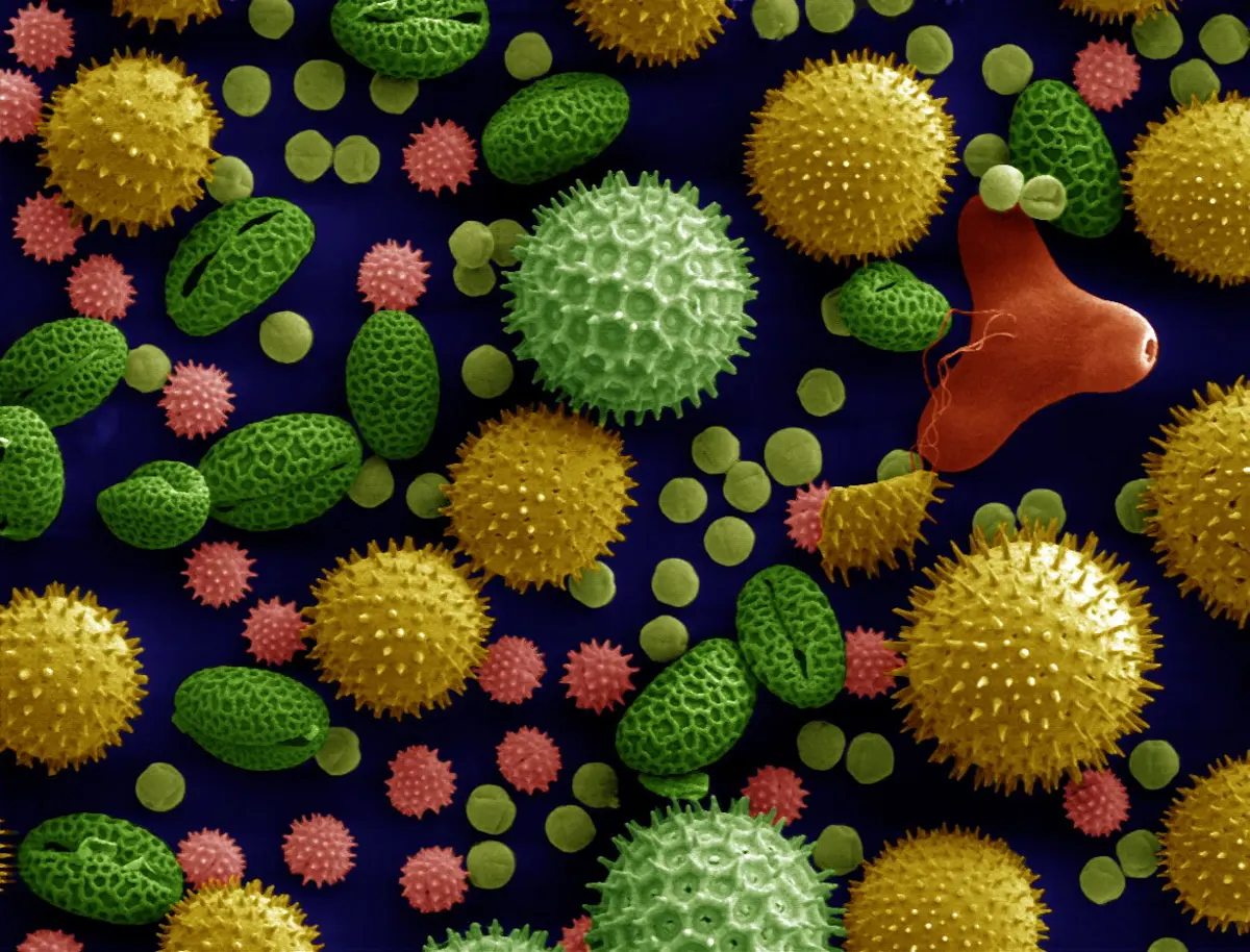
Inside the wall is the cytoplasm, which at shedding holds two or three cells (below).
Microgametogenesis: the male gametophyte
Microgametogenesis is the development of the haploid microspore into the mature male gametophyte, the pollen grain with its gametes. It begins inside the anther:
- The microspore enlarges, and a large vacuole pushes its nucleus to one side, against the wall.
- The nucleus divides by mitosis, and the division is unequal. It gives:
- a large vegetative cell (tube cell), with plenty of cytoplasm and food reserves and a large, irregular nucleus. It will form the pollen tube.
- a small generative cell, with a spindle-shaped nucleus and little cytoplasm, at first against the wall. It soon comes away and floats inside the cytoplasm of the vegetative cell: a cell inside a cell.
- In over 60% of flowering plants the pollen is shed at this two-celled stage. The generative cell divides later, by mitosis, inside the growing pollen tube, into two male gametes (sperm cells).
- In the rest, about 40% (grasses such as wheat and rice, and the mustard family among them), the generative cell divides before shedding, and the pollen is shed at the three-celled stage: the vegetative cell and two male gametes.
Either way, the mature male gametophyte is three cells: one vegetative (tube) cell and two male gametes, which have no flagella and can’t swim. They are carried to the egg by the pollen tube.
When a compatible pollen grain lands on a stigma, it absorbs water and sugars from the stigma’s surface and swells; the exine gives way at a germ pore, and the intine grows out as the pollen tube. The tube nucleus moves near its tip, and the generative cell or the two gametes follow it down the tube.
In a two-celled pollen grain, which cell divides to make the two male gametes?
Pick an answer to see why.
How many sets of chromosomes does a male gamete of a flowering plant have?
Pick an answer to see why.
2. Megasporogenesis
Megasporogenesis is the formation of haploid megaspores from a diploid megaspore mother cell by meiosis, inside the ovule. The ovule lies in the ovary. A carpel is one unit of the gynoecium: stigma, style and ovary; a pistil may be one carpel or several joined together. The ovules are attached inside the ovary along a cushion of tissue, the placenta.
Structure of the ovule
The ovule (the future seed) is a small oval body attached to the placenta by a stalk, the funiculus (funicle). The point where the body of the ovule meets the funiculus is the hilum. Its parts:
- Nucellus: the central mass of tissue, rich in food. It holds the embryo sac (the female gametophyte), usually one per ovule.
- Integuments: one or two protective coats (inner and outer integument) around the nucellus. They will become the seed coat.
- Micropyle: a small opening at the tip, where the integuments don’t meet.
- Chalaza: the base of the ovule, at the opposite end from the micropyle, where the integuments and nucellus join and the vascular strand from the funiculus ends.
How the body of the ovule is turned on its stalk gives the kinds of ovules. The commonest, found in most families of flowering plants, is the anatropous ovule: the body is turned right round, so the micropyle lies close to the hilum and both face the placenta; the funiculus runs along one side of the ovule as a ridge, the raphe. In an orthotropous ovule (Polygonum, black pepper) the ovule stands straight on its stalk, micropyle at the top.
Formation of the megaspore
- Early in the ovule’s growth, one cell of the nucellus just under its surface, at the micropylar end, enlarges, with dense cytoplasm and a big nucleus: the archesporial cell.
- It either becomes the megaspore mother cell (MMC, 2n) directly, or first divides, cutting off a wall cell, and its inner daughter becomes the MMC.
- The MMC divides by meiosis, forming four haploid megaspores in a row: a linear tetrad.
- Usually three degenerate and only one, the functional megaspore, survives. In most plants it is the one at the chalazal end, farthest from the micropyle.
Because one megaspore makes the embryo sac, this is called monosporic development. It is the commonest pattern, called the Polygonum type after the plant in which Eduard Strasburger first followed it in 1879.
Megagametogenesis: the female gametophyte
Megagametogenesis is the development of the functional megaspore into the mature female gametophyte, the embryo sac:
- The functional megaspore enlarges; it is now the young embryo sac.
- Its nucleus divides by mitosis, and the two nuclei move to opposite ends (poles) of the sac.
- Each divides twice more, by mitosis, so that there are four nuclei at each pole: eight nuclei in all. These three divisions are free nuclear: no walls form between the nuclei.
- One nucleus from each pole moves to the middle of the sac. These are the two polar nuclei.
- Now walls form, and the sac is organized into seven cells:
- At the micropylar end, three cells: the egg cell and two synergids beside it. Together they are the egg apparatus. Each synergid has, at its micropylar tip, a thickened, finger-like wall called the filiform apparatus, which guides the pollen tube in.
- At the chalazal end, three antipodal cells.
- In the middle, the large central cell, with the two polar nuclei. In many plants they fuse, before fertilization, into a diploid secondary nucleus.
So the typical mature embryo sac is 7-celled and 8-nucleate: egg 1, synergids 2, antipodals 3, and the central cell with 2 polar nuclei. It is the whole female gametophyte of a flowering plant, a tiny plant of seven cells, living inside the ovule.
How many meiotic and how many mitotic divisions does it take to go from the megaspore mother cell to a mature, eight-nucleate embryo sac?
Pick an answer to see why.
In a typical embryo sac, which cells lie at the micropylar end?
Pick an answer to see why.
4.2.3 Pollination
Pollination is the transfer of pollen grains from an anther to a stigma, of the same flower or of another flower. It brings the male gametophyte to the female part, and without it there is no fertilization in seed plants. (In the gymnosperms, such as Pinus, the pollen lands directly on the ovule, since there is no stigma: Grade 11 Biology.)
There are two kinds, by where the pollen comes from: self-pollination and cross-pollination.
1. Self-pollination
Self-pollination is the transfer of pollen from an anther to the stigma of the same flower or of another flower on the same plant. It has two forms:
- Autogamy: pollen falls on the stigma of the same flower. This needs a flower with both stamens and pistil (a bisexual flower), and it is helped by:
- Homogamy: the anthers and the stigma ripen at the same time, often with the anthers close to the stigma (wheat, rice, pea, tomato).
- Cleistogamy (Greek kleistos, closed): the flowers never open, so the pollen can only reach its own stigma. Some plants, such as Commelina, Viola (violets) and Oxalis (chari amilo), make both these closed flowers and ordinary open ones, which ensures some seed even when no pollinator comes.
- Geitonogamy (Greek geiton, neighbor): pollen goes to another flower of the same plant. It needs an agent, wind or an insect, just like cross-pollination; but genetically it is self-pollination, because the pollen comes from the same plant. It happens with bisexual flowers and also in plants whose male and female flowers are separate but on the same plant, such as maize and cucumber.
Advantages of self-pollination
- It keeps a variety pure (a pure line): the offspring are like the parent, so useful characters are kept.
- It is economical: little pollen is wasted, so the plant needs to make less.
- It doesn’t depend on pollinators or other outside agents, or on a second plant nearby.
- The flowers don’t need to make showy petals, scent or nectar.
- Seed production is almost certain.
- It doesn’t bring in new, untested combinations of genes.
Disadvantages of self-pollination
- It doesn’t bring in new characters, so there is little variation.
- The offspring are less able to adapt to a changing environment.
- Over many generations, harmful hidden (recessive) characters can come together, and the plants may become weaker (inbreeding depression): smaller, less fertile, less resistant.
- Unwanted characters are passed on to the offspring along with the good ones.
- It gives natural selection little variation to act on, so it contributes little to evolution.
A gardener puts paper bags over the flower heads of one maize plant, and shakes its tassel over the silks of the same plant. Which kind of pollination is this?
Pick an answer to see why.
2. Cross-pollination
Cross-pollination (xenogamy, Greek xenos, stranger) is the transfer of pollen from an anther of a flower on one plant to the stigma of a flower on another plant of the same species, which is genetically different. It is the only kind of pollination that brings new genes to the seed, so it is the main source of variation in flowering plants. It always needs an outside agent to carry the pollen.
Many plants have ways of avoiding self-pollination and making cross-pollination more likely:
- Unisexual flowers on separate plants (dioecy): papaya, date palm, spinach, Cannabis.
- Dichogamy: the anthers and the stigma of a flower ripen at different times, the anthers first (protandry: sunflower, coriander) or the stigma first (protogyny: custard apple).
- Self-incompatibility: pollen landing on a stigma of the same plant is recognized, and its tube is stopped; tea, cabbage, apple and pear can’t fertilize themselves.
- Herkogamy: a barrier of form between anthers and stigma, such as a stigma held well above the anthers.
Cross-pollination is grouped by the agent that carries the pollen. Abiotic agents are wind and water; biotic agents are animals.
a. Anemophily: pollination by wind
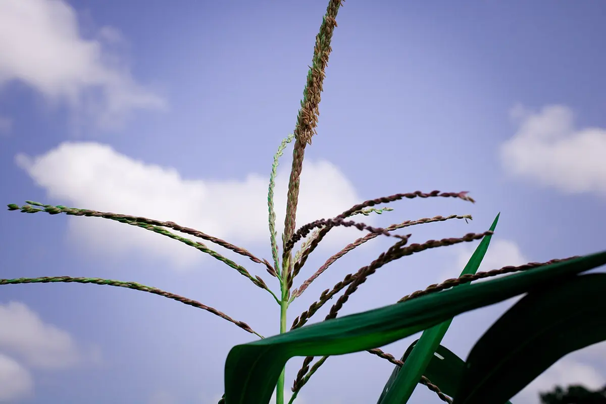
Anemophily (Greek anemos, wind) is pollination by wind. It is common in grasses (maize, wheat, rice, sugarcane, bamboo), in many trees (walnut, birch, alder, oak) and in all conifers such as pine. Wind-pollinated flowers:
- are small and inconspicuous, often without petals, with no color, scent or nectar;
- make huge amounts of pollen, which is light, dry, smooth and non-sticky, so it floats in air;
- have anthers on long filaments, hanging out of the flower, that the wind can shake;
- have large, feathery or sticky stigmas, hanging out to catch the pollen (maize’s silks);
- often have a single ovule in each ovary and many flowers packed into an inflorescence (the maize tassel, a grass spikelet).
b. Hydrophily: pollination by water
Hydrophily (Greek hydor, water) is pollination by water. It is rare: it is found in only about 30 genera, mostly monocots, such as Vallisneria (tape grass), Hydrilla and the sea grass Zostera. Not all aquatic plants are hydrophilous: water hyacinth and the lotus (kamal), for example, raise their flowers above the water and are pollinated by insects. The pollen of hydrophilous plants is usually protected from wetting by a mucilaginous covering.
In Vallisneria the female flowers rise to the surface of the water on long, coiled stalks; the male flowers break off under water and float up, open, and drift on the surface. When a floating male flower touches a female flower, its pollen is carried to the stigma. In Zostera, which flowers under the sea, the pollen grains are long and thread-like and drift inside the water until they wrap round a stigma.
c. Zoophily: pollination by animals
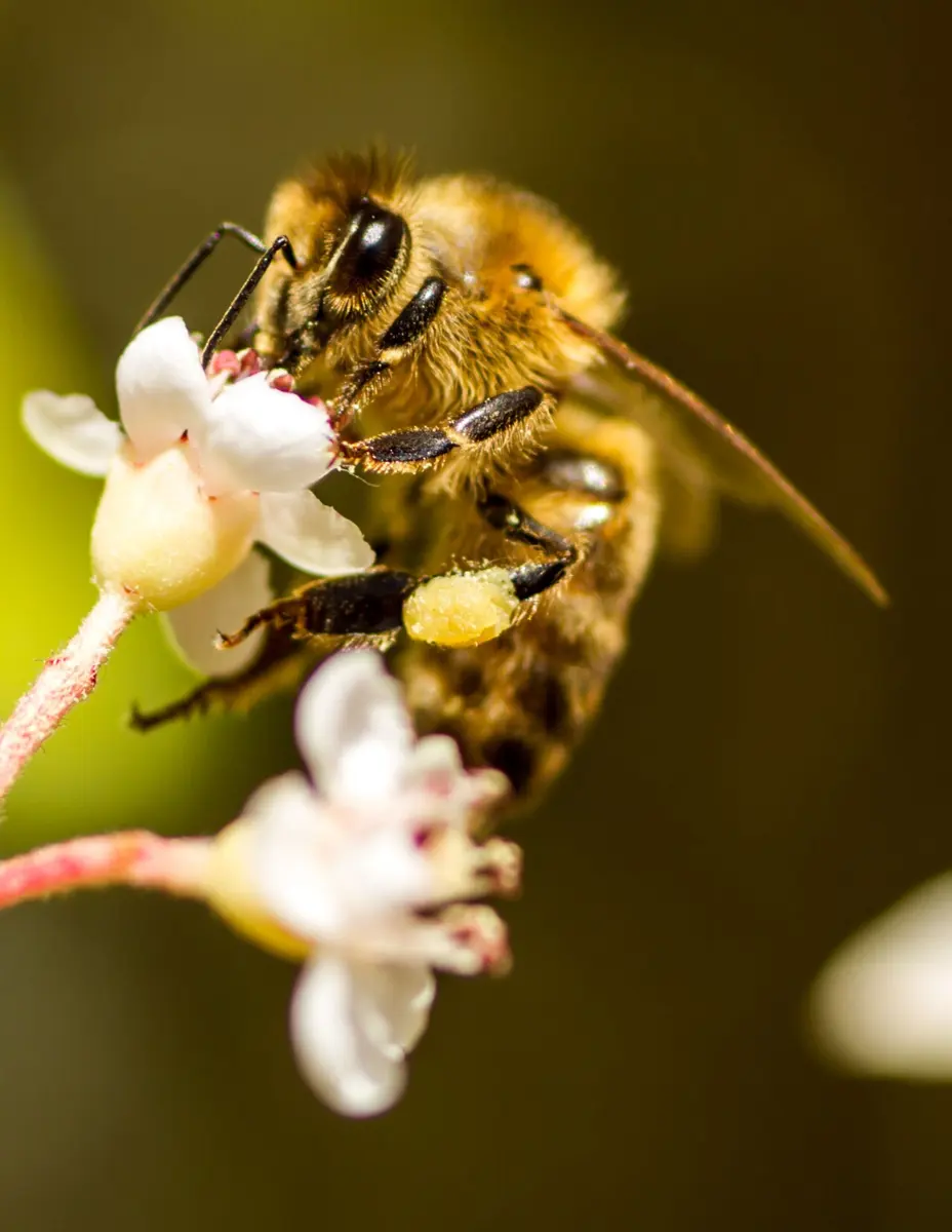
Zoophily (Greek zoon, animal) is pollination by animals. It is the commonest kind of cross-pollination: about 9 out of 10 flowering plants are pollinated by animals. Animal-pollinated flowers usually have large, colorful petals (or many small flowers packed into a showy cluster, as in a sunflower head), scent and nectar or edible pollen; their pollen is often sticky or spiny, so it clings to the animal. The animal gets food; the flower gets its pollen carried. Over millions of years many flowers and their pollinators have shaped each other, so different flowers suit different animals:
- Entomophily: by insects, the commonest kind: bees, butterflies, moths, flies, beetles, wasps. Bee flowers are often blue, yellow or purple, scented, with a landing platform and nectar guides (lines or patterns, many visible only in ultraviolet, which bees see). Mustard, sunflower, apple and citrus are pollinated mainly by bees.
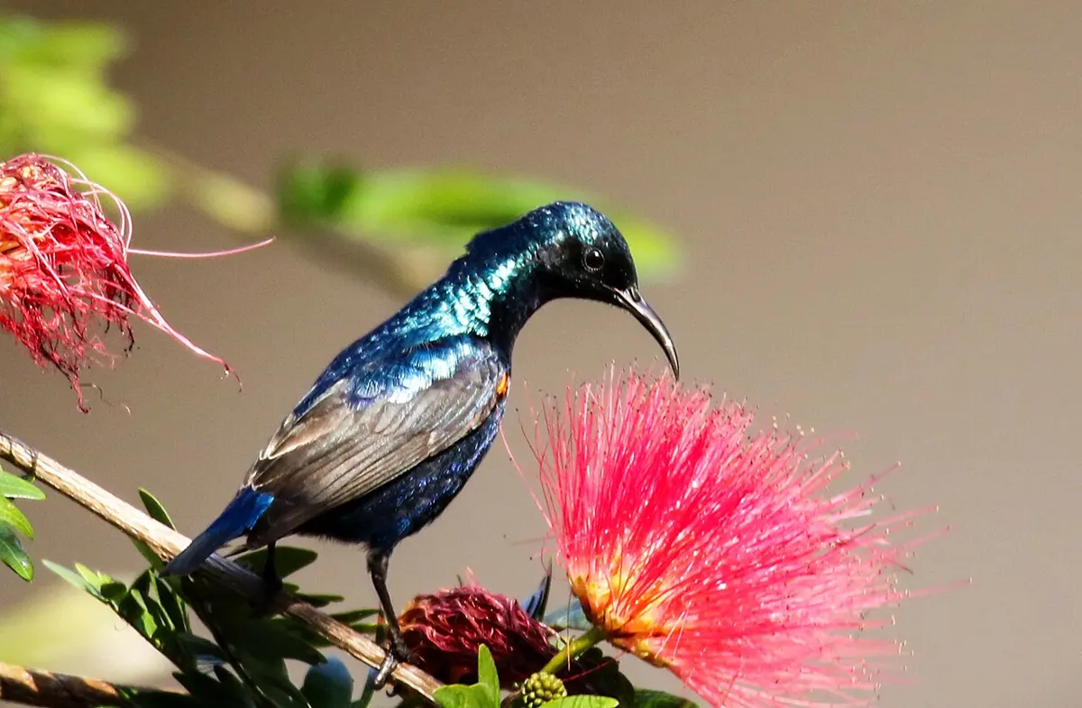
- Ornithophily: by birds, such as sunbirds in Asia and Africa and hummingbirds in the Americas. Bird flowers are usually red or orange, tubular, sturdy, with plenty of dilute nectar and little or no scent (birds have a poor sense of smell). In Nepal, the red flowers of the silk-cotton tree (simal, Bombax ceiba), the coral tree (Erythrina) and the rhododendron (laliguras) are visited by many nectar-feeding birds.
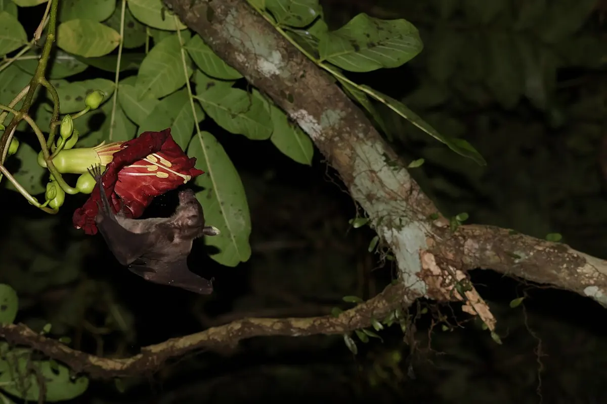
- Chiropterophily: by bats. Bat flowers open at night, are large and strong, pale or dull in color (white, cream, greenish, or dark maroon like the sausage tree’s), with a strong musty smell and much nectar and pollen, as in the sausage tree (Kigelia), the baobab and many wild bananas.
- Anthropophily: by people. Vanilla, an orchid, has its natural pollinators only in Central America, so in Madagascar and India every vanilla flower is pollinated by hand with a small stick; date palm growers have pollinated their female trees by hand since ancient times, as Assyrian carvings almost 3,000 years old show; and in some apple orchards in China, where bees have become scarce, people now pollinate each flower with a brush.
Other animals pollinate a few plants: snails and slugs (malacophily), lizards and geckos on some islands, and small mammals such as rodents.
Advantages of cross-pollination
- Plants that can’t fertilize themselves (self-sterile or dioecious plants) can still make seed and continue their race.
- It brings genes from two parents together, so it gives variation.
- Variation gives natural selection something to act on, so it helps evolution.
- The varied offspring are better able to adapt to changing conditions and new diseases.
- Plant breeders use it to make new, more useful varieties, such as hybrids.
- Cross-pollinated plants and their seeds are often more vigorous and productive (hybrid vigor).
- Harmful recessive characters are hidden or, over generations, weeded out.
Disadvantages of cross-pollination
- It is expensive for the plant: a lot of pollen is wasted, and nectar, scent and petals cost food to make.
- It depends on outside agents (wind, water, animals), which may fail: no bee, no seed.
- Undesirable characters of the other parent may be brought in.
- Good characters of a variety may be lost or mixed, so a pure variety can’t be kept pure.
- The plant has to make attractive parts (petals, scent, nectar), or large amounts of pollen.
- Seed production is less certain than with self-pollination.
A flower opens at night, is large, pale and strong-smelling, hangs on a long stalk outside the leaves, and has a lot of nectar. Which pollinator is it most likely adapted to?
Pick an answer to see why.
Why are wind-pollinated flowers usually small and green, without nectar or scent?
Pick an answer to see why.
4.2.4 Fertilization
Fertilization is the fusion of a male gamete with a female gamete. In flowering plants it is a double fertilization: of the two male gametes in a pollen tube, one fuses with the egg and the other with the central cell. Double fertilization that forms an endosperm is found in all flowering plants and in no other group, so it is one of the defining features of angiosperms.
Germination of the pollen and growth of the tube
A pollen grain that lands on a stigma is first “checked” by the stigma: pollen of another species, or of the same plant in self-incompatible species, is stopped. A compatible grain absorbs water and sugars from the stigma, swells, and puts out a pollen tube through a germ pore. The tube grows down through the style, between its cells or along a canal, feeding on the style’s secretions, towards an ovule. The vegetative (tube) nucleus is near its tip, followed by the generative cell, which divides into two male gametes if it hasn’t already. Because the male gametes can’t swim and are carried in a tube, this way of fertilization is called siphonogamy (Greek siphon, tube). In mosses and ferns the male gametes swim to the egg in water (zoidogamy).
The steps of double fertilization
- Entry of the pollen tube into the ovule. The tube reaches an ovule and enters it, usually through the micropyle: this is porogamy (Greek poros, an opening). Entry by any other way is aporogamy: through the chalaza (chalazogamy, as in Casuarina, where it was discovered in 1891) or through the integuments or the funiculus (mesogamy, as in pumpkin, Cucurbita).
- Entry into the embryo sac. Guided by the filiform apparatus, the tube enters one of the synergids, which degenerates. The tip of the tube bursts and releases the two male gametes into the synergid, and from there into the embryo sac.
- Syngamy. One male gamete fuses with the egg. This forms a diploid zygote (2n), which will grow into the embryo. This is the true fertilization, as in animals.
- Triple fusion. The second male gamete moves to the central cell and fuses with its two polar nuclei (or the diploid secondary nucleus, if they have already fused). Three haploid nuclei fuse, so this is called triple fusion, and it forms a triploid primary endosperm nucleus (PEN, 3n). The central cell becomes the primary endosperm cell, which grows into the endosperm, the food tissue for the embryo.
Syngamy and triple fusion together are double fertilization. Afterwards, the remaining synergid and the antipodals degenerate.
After fertilization, the flower changes into a fruit with seeds:
| Before fertilization | After fertilization |
|---|---|
| Zygote | Embryo |
| Primary endosperm nucleus | Endosperm |
| Integuments | Seed coat: testa (from the outer) and tegmen (from the inner) |
| Micropyle | A tiny pore in the seed coat, through which water enters at germination |
| Funiculus | Seed stalk (it breaks off, leaving the hilum as a scar on the seed) |
| Ovule | Seed |
| Ovary | Fruit |
| Ovary wall | Fruit wall (pericarp) |
| Synergids and antipodals | Degenerate |
Sepals, petals and stamens usually wither and fall, though in some plants the sepals stay on the fruit (tomato, brinjal).
Which nucleus is formed by triple fusion?
Pick an answer to see why.
A pollen tube enters an ovule through its chalaza. What is this called?
Pick an answer to see why.
4.2.5 Embryogenesis
The development of the embryo from the zygote is embryogenesis (embryogeny). It starts only after the endosperm has begun to form, since the embryo needs its food. In monocots and dicots the first steps are much alike, a zygote dividing into a terminal cell and a basal cell, but the later stages differ.
1. Development of the embryo in monocotyledonous plants
- The zygote divides transversely into a small terminal cell and a larger basal cell.
- The basal cell forms the suspensor, a short stalk that holds the young embryo and pushes it into the endosperm. In many monocots it doesn’t divide again, and stays a single large, swollen (vesicular) cell; it later degenerates.
- The terminal cell divides transversely into two cells. The cell at the tip, after many divisions, forms the single cotyledon and the plumule. The cotyledon grows fast and pushes the plumule, the shoot tip, to one side, so that it comes to lie in a notch (a depression) on the side of the embryo. The middle cell, after many divisions, forms the hypocotyl and the radicle, and adds a few cells to the suspensor.
- In grasses and cereals, the single cotyledon is a large shield-shaped organ, the scutellum, pressed against the endosperm. At germination it makes enzymes and absorbs the digested food from the endosperm for the growing seedling. The plumule is enclosed in a protective sheath, the coleoptile, and the radicle in another, the coleorhiza. At germination the coleoptile pushes up through the soil round the young leaves, so the tender shoot tip isn’t damaged; the radicle breaks out through the coleorhiza.
Maize grain: traced from Sarah Greenwood, CC BY-SA 4.0 (this drawing CC BY-SA 4.0 too)
2. Development of the embryo in dicotyledonous plants
The classic example is shepherd’s purse, Capsella bursa-pastoris, a small weed of the mustard family:
- The zygote divides transversely into two unequal cells: a large basal cell, towards the micropyle, and a small terminal cell (apical cell), towards the chalaza.
- The basal cell divides transversely again and again into a row of 6 to 10 cells, the suspensor, which pushes the young embryo deep into the endosperm. Its cell at the micropylar end grows large and acts as a haustorium (an absorbing cell). Its cell next to the embryo, the hypophysis, will form the tip of the radicle and the root cap.
- The terminal cell divides twice vertically, at right angles, into four cells (the quadrant stage), then once transversely, into eight cells (the octant stage), in two tiers of four.
- The tier next to the suspensor (the hypobasal tier) forms the hypocotyl and the radicle (except its tip, from the hypophysis).
- The tier away from the suspensor (the epibasal tier) forms the two cotyledons and the plumule.
- The eight cells divide in all directions, and the outermost layer of cells becomes the future epidermis. The embryo becomes a ball of cells: the globular stage.
- Two bulges, the future cotyledons, grow out on either side of the top, and the embryo becomes heart-shaped.
- The cotyledons and the hypocotyl lengthen (the torpedo stage), and in Capsella the embryo, growing inside a curved ovule, bends over like a horseshoe. The mature embryo has two cotyledons, the plumule (the shoot tip) between them, and the radicle (the root tip) at the other end, towards the suspensor and the micropyle.
What does the scutellum of a maize grain do when the grain germinates?
Pick an answer to see why.
In the embryo of Capsella, what does the hypophysis form?
Pick an answer to see why.
4.2.6 Development of the endosperm
The endosperm is the food tissue of the seed. It forms from the primary endosperm cell, whose nucleus, the primary endosperm nucleus (PEN), came from triple fusion: a haploid male gamete and the diploid secondary nucleus (or the two polar nuclei), so it is triploid (3n). The endosperm is rich in stored food (starch, oils, proteins), which feeds the growing embryo. Its development begins before the embryo’s: the PEN starts dividing soon after fertilization, often before the zygote divides at all, so there is food ready for the embryo.
There are three types of endosperm development:
1. Nuclear type
The PEN divides again and again by mitosis without walls forming, giving a large number of free nuclei in the central cell. A large central vacuole appears and pushes the nuclei to the edge. Later, walls form around the nuclei, from the edge inward, and the endosperm becomes cellular. This is the commonest type, found in most cereals (maize, wheat, rice) and in Capsella.
The coconut is the classic example. Its coconut water (the clear liquid in a young, green coconut; some books call it coconut milk) is endosperm still at the free-nuclear stage: a liquid with thousands of nuclei floating in it. The white flesh (the kernel, dried as copra) is the outer, cellular endosperm, formed as walls grow in from the edge. As the coconut ripens, more and more of the water becomes flesh.
2. Cellular type
A wall forms after every division of the PEN, right from the first, so the endosperm is cellular from the start, with no free-nuclear stage. Examples: Petunia, Datura (dhaturo) and the garden balsam (Impatiens).
3. Helobial type
This type is between the other two. The first division of the PEN is followed by a wall, which divides the central cell into two unequal cells: a large micropylar cell and a small chalazal cell. The micropylar cell then develops like a nuclear endosperm: its nucleus divides freely, and walls form later. The chalazal cell divides little or not at all. This type is found mostly in monocots (it is named after an old order of water plants, the Helobiales), for example Eremurus.
What happens to the endosperm. In many seeds the embryo uses up the endosperm before the seed is ripe, storing the food in its own cotyledons instead: these are non-endospermic (exalbuminous) seeds, such as pea, gram, bean and groundnut. In others a large endosperm remains in the ripe seed and is used at germination: endospermic (albuminous) seeds, such as maize, wheat, rice, castor and coconut. Rice, wheat flour and maize flour are mostly endosperm: it feeds much of the world.
A ripe pea seed has no endosperm. What happened to it?
Pick an answer to see why.
Which type of endosperm is cellular from its very first division?
Pick an answer to see why.
Exercise
A. Tick the correct alternatives
- Which of the following is the female reproductive part of a flower?
Pick an answer to see why.
- Which layer of the anther provides nutrition to the developing pollen grains?
Pick an answer to see why.
- How many microsporangia are typically found in a bilobed anther?
Pick an answer to see why.
- How many male gametes are carried by the pollen tube?
Pick an answer to see why.
- Which of the following develops into the embryo in a flowering plant?
Pick an answer to see why.
- If the micropyle of an ovule is blocked, which process will be directly affected?
Pick an answer to see why.
- A plant breeder wants to maintain a pure line of a particular plant variety. Which pollination strategy would be most effective for this purpose?
Pick an answer to see why.
- Which of the following correctly represents the nuclear condition after fertilization events in angiosperms?
Pick an answer to see why.
B. Answer the following questions
What is microsporogenesis? Where does it occur?
Answer
Describe the structure of a mature female gametophyte (embryo sac).
Answer
Why are cross-pollinated plants better adapted for evolution?
Answer
What type of pollination is practiced by flowers pollinated by bats? Mention some floral adaptations supporting it.
Answer
Mention the advantages and disadvantages of self-pollination.
Answer
Compare self-pollination and cross-pollination.
Answer
Explain the process of fertilization with a diagram.
Answer
Write short notes about the endosperm.
Answer
Explain microgametogenesis and the formation of the male gametophyte.
Answer
Explain the development of the female gametophyte in angiosperms with a necessary diagram.
Answer
Observe the diagram above carefully and answer the following questions.
- a.Name the figure.
- b.Label B, C, D and E.
- c.Which one of the labeled parts becomes triploid after fertilization?
- d.Which one of them forms the zygote after fertilization?
Answer
Describe the role of the suspensor in embryo development, highlighting the differences observed between monocot and dicot plants.
Answer
Printable PDFs
- Study notes 4 pages
- Worksheet 4 pages Answer key
Quiz yourself: Life Processes and the Human Body, Grades 5–12 →
Practice every question in the book, and keep track of the ones you miss →
A companion to your textbook, not a replacement for it: study from the book your school uses, and where the two differ, follow your teacher. Found a mistake? Please tell us.