
In 1953 two young scientists in Cambridge, James Watson (1928–2025) and Francis Crick (1916–2004), built a model out of metal plates and rods and announced that they had found “the secret of life”. Using X-ray photographs of DNA taken by Rosalind Franklin and Maurice Wilkins, they had worked out its shape: two chains twisted round each other into a double helix, held together by pairs of bases that fit like a lock and its key. The shape explained at once how a living thing copies its instructions and hands them on. Watson, Crick and Wilkins shared the 1962 Nobel Prize in Physiology or Medicine. Franklin had died in 1958, and a Nobel Prize is not given to someone who has died.
DNA is one molecule. Your body, a rhododendron, a yak and the bacteria in your curd are built from a few thousand kinds of molecules like it, and nearly all of them are made by joining a handful of small units into long chains. This unit is about those molecules of life: what they are made of, how their units are joined, and what each one does.
1.1.1 General introduction
Cells and organisms are made of many kinds of molecules, which together are called biomolecules: the molecules found in living things. They range in size from small ones like water (H₂O, 3 atoms) and carbon dioxide to giant ones like proteins (thousands of atoms) and DNA (billions of atoms in one human chromosome).
Small molecules dissolve easily and move about the cell, so they take part in the cell’s chemical reactions all the time. The sum of all the chemical reactions in a living thing is called metabolism: breaking food down to release energy (catabolism) and building the cell’s own molecules (anabolism). Large molecules are mostly used for structure (cellulose in a plant’s cell walls, collagen in your skin) or carry information: nucleic acids (DNA and RNA) store and pass on the genetic instructions, and proteins carry them out.
All the biomolecules present in a cell at a time, dissolved or suspended in its water, are together called the cellular pool. The cell takes many of them in from outside, across its plasma membrane and the membranes of its organelles.
The biomolecules of the cellular pool are of two kinds:
- Organic compounds: compounds of carbon, combined mainly with hydrogen and oxygen, and often nitrogen, phosphorus and sulphur. Examples: carbohydrates, proteins, lipids, nucleic acids, hormones and vitamins.
- Inorganic compounds: compounds also found in the non-living world, without carbon chains. Examples: water, minerals, salts and their ions (Na⁺, K⁺, Ca²⁺, Cl⁻), and gases such as O₂ and CO₂.
The biomolecules can also be grouped by size (molecular mass) and by how easily they dissolve:
- a.Micromolecules: small molecules of low molecular mass that dissolve in water, such as water itself, gases, minerals, sugars, amino acids and nucleotides.
- b.Macromolecules: large, complex molecules of high molecular mass. They are of four classes: carbohydrates (polysaccharides), lipids, proteins and nucleic acids. Except lipids, each is a polymer: a long chain of many small units called monomers (sugars, amino acids, nucleotides). Lipids are counted with them because they are large and gather into big structures such as membranes, though they are not long chains.
How much of a cell is each kind? The table gives the rough share of a cell’s total mass (bacteria and animal cells are much alike).
| Component | Share of the cell’s mass (%) |
|---|---|
| Water | 70–90 |
| Proteins | 10–15 |
| Nucleic acids | 5–7 |
| Carbohydrates | about 3 |
| Lipids | about 2 |
| Ions (minerals) | about 1 |
Take the water away and protein is more than half of what is left: proteins are the cell’s main working material.
Which of these is a macromolecule?
Pick an answer to see why.
Which group lists only inorganic biomolecules?
Pick an answer to see why.
1.1.2 Carbohydrates
What did you eat this morning? Roti, chiura, rice, a banana, a cup of sweet tea? Each of these is mostly carbohydrate, and it is what keeps you going until lunch.
Carbohydrates (Latin carbo = carbon, Greek hydor = water) are biomolecules made of carbon, hydrogen and oxygen, usually with hydrogen and oxygen in the ratio 2 : 1, as in water. That is why they were named “hydrates of carbon”, and why their general formula is written Cₓ(H₂O)ᵧ: glucose, C₆H₁₂O₆, is C₆(H₂O)₆. They are also called saccharides (Greek sakcharon = sugar).
Chemically, carbohydrates are polyhydroxy aldehydes or ketones (compounds with many –OH groups and one aldehyde or ketone group), or substances that give these when broken down by water.
- A sugar whose carbonyl group is an aldehyde group (–CHO), at the end of the chain (carbon 1), is an aldose. Glucose is an aldose.
- A sugar whose carbonyl group is a ketone group (>C=O), at the second carbon (not at the end), is a ketose. Fructose is a ketose.
Classification of carbohydrates
Carbohydrates are sorted into three groups by how many sugar units they have:
| Group | Number of sugar units | Kinds | Examples |
|---|---|---|---|
| Monosaccharides (monomers) | 1 | trioses, tetroses, pentoses, hexoses, heptoses | glyceraldehyde, erythrose, ribose, ribulose, glucose, fructose, galactose |
| Oligosaccharides (oligomers) | 2 to 10 | disaccharides, trisaccharides, tetrasaccharides, pentasaccharides… | sucrose, maltose, lactose, raffinose, stachyose |
| Polysaccharides (polymers) | more than 10, often thousands | homopolysaccharides, heteropolysaccharides | starch, glycogen, cellulose, chitin, glucans, fructans, arabinogalactans |
1. Monosaccharides
Monosaccharides (formula CₙH₂ₙOₙ, with n from 3 to 7) are the simplest carbohydrates: they can’t be broken down by water (hydrolyzed) into smaller sugars. Each is a chain of carbons; every carbon carries an –OH group except one, which is part of the aldehyde or ketone group. They are white, crystalline, dissolve easily in water and taste sweet.
They are named by their number of carbons, and as aldoses or ketoses:
| Monosaccharide | Aldose | Ketose |
|---|---|---|
| Trioses (C₃H₆O₃) | glyceraldehyde | dihydroxyacetone |
| Tetroses (C₄H₈O₄) | erythrose | erythrulose |
| Pentoses (C₅H₁₀O₅) | ribose | ribulose |
| Hexoses (C₆H₁₂O₆) | glucose, galactose | fructose |
| Heptoses (C₇H₁₄O₇) | glucoheptose | sedoheptulose |
Glucose, an aldohexose (an aldose with six carbons), is the most abundant monosaccharide in nature: it is the sugar plants make in photosynthesis and the sugar in your blood. Its open chain has five –OH groups and one aldehyde group on its six carbons. Fructose, the sugar of fruits and honey, is a ketohexose with the same formula, C₆H₁₂O₆, arranged differently: the two are isomers. Galactose (in milk sugar) is another aldohexose, differing from glucose only in which side one –OH points to, at carbon 4.
Rings, not chains
In water, a sugar of five or more carbons spends almost all its time as a ring. The –OH on one carbon reaches round and joins the carbonyl carbon: in glucose, the oxygen of carbon 5 bonds to carbon 1, giving a ring of five carbons and one oxygen. The new –OH on carbon 1 can point down (α, alpha) or up (β, beta) in the usual drawing. Fructose closes from carbon 5 onto carbon 2, giving a ring of four carbons and one oxygen; so does ribose, from carbon 4 onto carbon 1.
Biologists draw these rings as Haworth projections: the ring is seen tilted, lying almost flat, its near edge drawn thick, and each group on a ring carbon points straight up or straight down.
The α and β forms of glucose change into each other in water, through the open chain, all the time. But once a glucose is locked into a chain by its carbon 1, it stays α or β, and that one difference decides whether the chain is starch, which you can digest, or cellulose, which you can’t (Figure 1.2).
Reducing sugars
The free aldehyde (or ketone) group of a monosaccharide can give electrons to other substances: it can reduce copper(II) ions, Cu²⁺, to copper(I), Cu⁺. A sugar that can do this is a reducing sugar. In Benedict’s test, a blue solution of Cu²⁺ is boiled with the sugar; a reducing sugar turns it green, yellow, then brick-red, as red copper(I) oxide, Cu₂O, settles out. All monosaccharides are reducing sugars.
Which pair are both aldoses?
Pick an answer to see why.
A sugar has the formula C₅H₁₀O₅ and a ketone group. What is it?
Pick an answer to see why.
2. Oligosaccharides
Oligosaccharides (Greek oligos = a few) are short chains of 2 to 10 monosaccharides joined by glycosidic bonds. Most taste sweet and dissolve in water.
A glycosidic bond forms when the –OH on carbon 1 of one sugar (the carbonyl carbon, carbon 2 in fructose) reacts with an –OH of another sugar. A molecule of water is removed, and the two sugars are left joined through an oxygen atom (C–O–C). A reaction that joins two molecules by removing water is a condensation (dehydration synthesis); the opposite, splitting a bond by adding water, is hydrolysis. Digestion is hydrolysis.
The bond is named after the carbons it joins and the α or β form of the first sugar: in maltose, carbon 1 of an α-glucose is joined to carbon 4 of the next glucose, an α(1→4) glycosidic bond.
| Number of sugar units | Oligosaccharide | Examples |
|---|---|---|
| 2 | disaccharide | sucrose (glucose + fructose), maltose (glucose + glucose), lactose (galactose + glucose) |
| 3 | trisaccharide | raffinose (galactose + glucose + fructose), mannotriose |
| 4 | tetrasaccharide | stachyose (2 galactose + glucose + fructose) |
| 5 | pentasaccharide | verbascose (3 galactose + glucose + fructose) |
The most abundant oligosaccharides in plant and animal cells are the disaccharides:
- Sucrose (cane sugar, the sugar in your tea) is made in plants and carried round them in the phloem. Its bond joins carbon 1 of α-glucose to carbon 2 of β-fructose, an α1→β2 bond, using up the carbonyl group of both sugars. So sucrose is not a reducing sugar.
- Maltose (malt sugar) is made when starch is broken down, as in germinating grain and in your mouth: two glucoses joined α(1→4). It is a reducing sugar.
- Lactose (milk sugar) is made only in mammals’ milk: galactose joined β(1→4) to glucose. It is a reducing sugar.
Raffinose (C₁₈H₃₂O₁₆) is sucrose with a galactose joined to the glucose’s carbon 6 by an α(1→6) bond. Stachyose (C₂₄H₄₂O₂₁) is raffinose with one more galactose, again α(1→6): two galactoses, one glucose and one fructose. Both are stored in seeds, especially of legumes (beans, peas, chickpeas, soya), as food for the growing embryo.
Why is sucrose not a reducing sugar, while maltose is?
Pick an answer to see why.
3. Polysaccharides
Polysaccharides (also called glycans) are long chains of more than ten, usually hundreds to many thousands, of monosaccharides joined by glycosidic bonds. Because they are so big:
- they are not sweet;
- most are insoluble in water (some, like glycogen and amylose, form cloudy colloids instead of true solutions);
- they are practically non-reducing: a whole chain has only one free reducing end among thousands of units.
Examples are starch, glycogen, cellulose, chitin and dextrins (short pieces of starch made when it is partly broken down, as in toasted bread).
By their job, polysaccharides are of two kinds:
- a.Structural polysaccharides build walls and skeletons: cellulose in plant cell walls; chitin in fungal cell walls and in the outer skeletons of insects and crabs; mannan in yeast cell walls; peptidoglycan (sugar chains cross-linked by short peptides) in bacterial cell walls.
- b.Storage (nutrient) polysaccharides store food: starch in plants (and some bacteria), and glycogen in animals and fungi.
By their make-up, they are also of two kinds:
- a.Homopolysaccharides (homoglycans) are made of only one kind of monosaccharide: glucans, made of glucose (starch, glycogen, cellulose), and fructans, made of fructose (inulin, stored in garlic and onion).
- b.Heteropolysaccharides (heteroglycans) are made of two or more kinds of monosaccharide: arabinogalactans (arabinose and galactose, in plant gums), hyaluronic acid in the fluid of your joints, and peptidoglycan.
Starch, glycogen and cellulose: one sugar, three materials
All three are chains of glucose. The differences are in the bonds:
- Starch is a mixture of two glucans of α-glucose. Amylose is an unbranched chain joined α(1→4), which coils into a helix. Amylopectin has the same chain with branches joined α(1→6) about every 24 to 30 glucoses.
- Glycogen is built like amylopectin but branched much more, about every 8 to 12 glucoses. The many branch ends let enzymes add or remove glucose at many points at once, so the liver and muscles can store glucose and release it fast.
- Cellulose is an unbranched chain of β-glucose joined β(1→4). Each glucose is turned over relative to the last, so the chain is straight and flat. Neighboring chains lie side by side and hydrogen-bond to each other into strong microfibrils, the fibers of wood, cotton and paper.
Functions of carbohydrates
- Energy. Carbohydrates are the main fuel of most living things. About half of the energy in a typical diet comes from them, and more in a rice-based Nepali meal. In the cell, glucose is broken down in respiration, giving about 17 kJ (4 kcal) of energy per gram: + energy (about 2870 kJ per mole).
- Structure. Ribose and deoxyribose are parts of the nucleotides of RNA and DNA (and of ATP). Cellulose and hemicellulose build plant cell walls; chitin builds fungal walls and insect skeletons.
- Storage. Starch is the storage polysaccharide of plants; glycogen (“animal starch”) is the reserve food of animals, stored in the liver and muscles.
- Raw material. Carbohydrates are the starting material from which cells make fats and some amino acids.
- Burning fat properly. Fats are broken down completely only when some glucose is being broken down at the same time. With too little carbohydrate (in starvation, or uncontrolled diabetes), fats are oxidized only partly and acidic ketone bodies build up in the blood. Carbohydrates prevent this.
- Sugars joined to proteins and lipids on the outside of cells act as name tags: your blood group (A, B, AB or O) is set by a few sugars at the tips of such chains.
Which is a storage polysaccharide of animals?
Pick an answer to see why.
Starch and cellulose are both made of glucose. What makes cellulose so different?
Pick an answer to see why.
1.1.3 Amino acids
Amino acids are the units proteins are made of. Each is built round one carbon atom, the α-carbon (alpha carbon), which carries four different things:
- an amino group, –NH₂, which is basic;
- a carboxyl group, –COOH, which is acidic;
- a hydrogen atom;
- a side chain, written R, different in each amino acid.
The side chain is what makes each amino acid itself. In glycine, the simplest, R is just a hydrogen atom, –H; in alanine it is –CH₃; in others it is acidic, basic, sulphur-containing or a ring.
Because one molecule has both a basic group and an acidic group, an amino acid can act as an acid or as a base: it is amphoteric. In fact, in water and in your cells the carboxyl group gives its H⁺ to the amino group, so the molecule carries a + charge and a − charge at once (H₃N⁺–CHR–COO⁻), a zwitterion.
Some features of amino acids:
- They are the units that join to form proteins.
- Every amino acid has at least one acidic carboxyl group (–COOH) and one basic amino group (–NH₂).
- They are colorless, crystalline solids that melt (and break down) at high temperatures, because their charged molecules hold each other tightly.
- They dissolve in water but hardly at all in organic solvents such as ether.
- Twenty amino acids are coded by the genes and used to build proteins in all living things. (Nature has hundreds more, which are not used in proteins, and two rare extra ones, selenocysteine and pyrrolysine, are built into a few proteins.)
- Examples: glycine, alanine, serine, leucine, isoleucine, valine, tryptophan.
Formation of the peptide bond
Two amino acids join when the carboxyl group (–COOH) of one reacts with the amino group (–NH₂) of the next. A molecule of water is removed (a condensation, as in the glycosidic bond), and the bond left between them, –CO–NH–, is a peptide bond (or peptide linkage).
Two amino acids make a dipeptide, three a tripeptide, and many make a polypeptide chain. The chain still has a free amino group at one end (the N-terminus) and a free carboxyl group at the other (the C-terminus), and it is always written and built from the N-terminus to the C-terminus.
Categories of amino acids
Of the 20 amino acids, your body can make some for itself from other molecules, but not all.
i. Essential amino acids can’t be made by the human body (or not fast enough), so they must come from food. There are nine: histidine, isoleucine, leucine, lysine, methionine, phenylalanine, threonine, tryptophan and valine. Histidine was long counted as essential only for growing children; it is now known that adults need it in food too.
ii. Non-essential amino acids can be made by the body, so food doesn’t have to supply them. There are eleven: alanine, arginine, asparagine, aspartic acid, cysteine, glutamic acid, glutamine, glycine, proline, serine and tyrosine.
Functions of amino acids
- They are the building blocks of proteins, including enzymes.
- Tryptophan is used to make the vitamin niacin (vitamin B₃), the nerve messenger serotonin, and, in plants, the plant hormone indole-3-acetic acid (IAA), an auxin.
- Tyrosine is used to make the hormones thyroxine and adrenaline (epinephrine) and the skin pigment melanin.
- Glycine is used to make heme, the iron-holding part of hemoglobin, through the ring compound protoporphyrin.
- Several amino acids not used in proteins are raw materials for antibiotics: penicillin, for one, is built by the mold that makes it from three amino acids, one of them (α-aminoadipic acid) never found in proteins.
- In the cell walls of bacteria, short peptides cross-link sugar chains into peptidoglycan.
Which is an essential amino acid for humans?
Pick an answer to see why.
What is lost when a peptide bond forms between two amino acids?
Pick an answer to see why.
1.1.4 Proteins
Proteins (Greek proteios = of the first rank) are long chains of amino acids joined by peptide bonds, folded into particular shapes. They are the most varied macromolecules and make up more than half of a cell’s dry mass. Almost everything a cell does is done by a protein: enzymes, which are proteins (apart from a few RNAs), run nearly every reaction in the body.
A protein is made of the 20 amino acids in a particular order, and that order is set by its gene. The order decides how the chain folds, and the shape decides what the protein does.
Categories of proteins
A. By chemical make-up and solubility
a. Simple proteins give only amino acids when broken down by water. Examples: albumins (egg white, blood serum), globulins (in blood and seeds), histones (the proteins DNA is wound round in chromosomes) and protamines (which pack DNA in sperm).
b. Conjugated proteins are simple proteins joined to a non-protein part, called the prosthetic group:
- Glycoproteins: protein + carbohydrate (chains of sugars). Example: mucin of saliva, which makes it slippery.
- Phosphoproteins: protein + phosphate groups. Examples: casein of milk and ovovitellin (vitellin) of egg yolk.
- Lipoproteins: protein + lipids. They carry fats and cholesterol in the blood, and are found in cell membranes and the brain. Example: high-density lipoprotein (HDL).
- Nucleoproteins: protein + nucleic acid. Example: chromatin, the DNA of the chromosomes wound round histones, and ribosomes (RNA + proteins).
- Chromoproteins: protein + a colored pigment, often holding a metal such as Fe, Cu or Mg. Examples: hemoglobin (red, with iron), the visual pigment rhodopsin of the retina, and hemocyanin (blue, with copper) in the blood of snails and crabs.
c. Derived proteins are made by the partial breakdown (hydrolysis) of simple or conjugated proteins, by heat, acid or enzymes. Examples: proteoses and peptones, formed when pepsin digests protein in the stomach.
B. By shape
- Fibrous proteins are long chains (or bundles of chains) lying side by side as fibers or sheets. They are tough, often elastic, and insoluble in water. They do structural jobs in animals: keratin (hair, nails, horn, wool), collagen (skin, bone, tendons), elastin (artery walls) and myosin (muscle). Silk’s fibroin is fibrous too.
- Globular proteins are chains folded up into compact, roughly round shapes. Most dissolve in water. They do the cell’s chemical work and are found in all living things: enzymes, hormones such as insulin, antibodies, hemoglobin, egg albumin, the albumins and globulins of blood serum, and the glutelins of wheat.
Structure of proteins
A protein’s shape is described at four levels, each built on the one before.
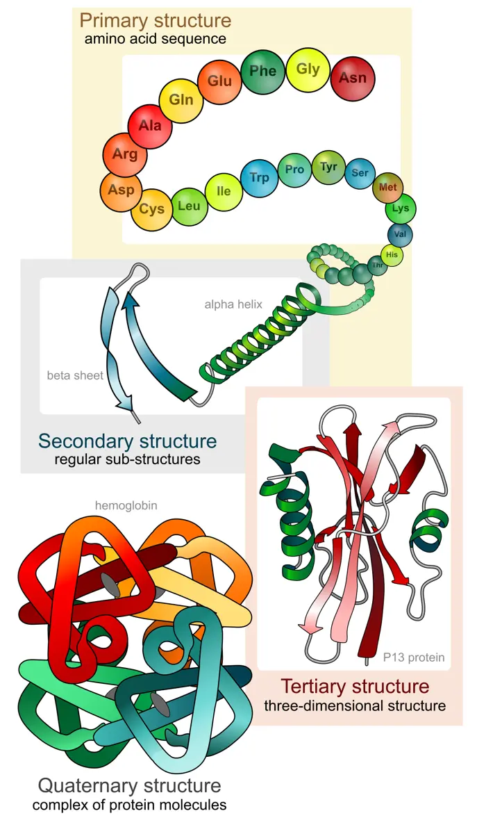
1. Primary structure is the sequence (order) of amino acids in a polypeptide chain, held by peptide bonds. It is set by the gene, and everything else follows from it. Insulin, a small hormone with 51 amino acids in two chains, was the first protein whose primary structure was found: Frederick Sanger worked out its sequence between 1949 and 1955, and won the 1958 Nobel Prize in Chemistry for it.
2. Secondary structure is a regular, repeating shape in parts of the chain, held by hydrogen bonds between the backbone’s C=O and N–H groups.
- In the α-helix, the chain coils into a right-handed spiral, and the C=O of each amino acid hydrogen-bonds to the N–H of the amino acid four places along. The α-keratin of hair and wool is mostly α-helix.
- In the β-pleated sheet, stretches of chain lie side by side, zigzagged like a folded fan, hydrogen-bonded across from one stretch to the next. Silk fibroin is mostly β-sheet.
3. Tertiary structure is the whole three-dimensional shape of one chain: its helices, sheets and the loops between them folded back on themselves into a compact shape. It is typical of globular proteins. It is held by bonds between the side chains: ionic bonds (between + and − side chains), hydrogen bonds, hydrophobic interactions (water-fearing side chains huddling together in the middle, away from water) and strong covalent disulphide bonds (–S–S–) between two cysteines.
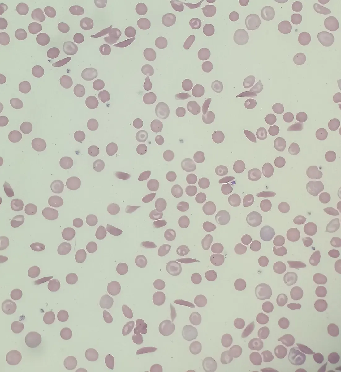
4. Quaternary structure is how two or more folded chains (called subunits) fit together into one working protein, and what holds them. Hemoglobin is four chains (two α and two β), each with a heme; antibodies (immunoglobulins) are four chains, two heavy and two light, joined by disulphide bonds. A protein of only one chain, such as the enzyme ribonuclease (124 amino acids) or myoglobin, has no quaternary structure.
Functions of proteins
- Structure, growth and repair. Structural proteins build tissues: collagen in skin, bone and tendons, keratin in hair and nails, actin and myosin in muscle.
- Energy, when needed. Protein can be burned for energy: burned completely in a calorimeter it gives about 5.65 kcal per gram, but in the body only about 4 kcal per gram (its physiological fuel value), because the body can’t burn the nitrogen and loses it in urine as urea.
- Enzymes. Several thousand kinds of enzymes are known; each speeds up one reaction of life.
- Hormones. Insulin, a protein hormone from the pancreas, lowers the glucose level of the blood.
- Defense. Antibodies (immunoglobulins) are proteins that recognize and bind germs and other foreign substances, so the body can destroy them.
- Blood clotting. At a cut, the enzyme thrombin cuts the soluble blood protein fibrinogen into fibrin, whose insoluble threads trap blood cells into a clot that stops the bleeding.
- Transport. Hemoglobin carries oxygen; proteins in the cell membrane carry substances in and out of the cell.
A protein made of a single polypeptide chain has which levels of structure?
Pick an answer to see why.
Which bonds hold the α-helix in shape?
Pick an answer to see why.
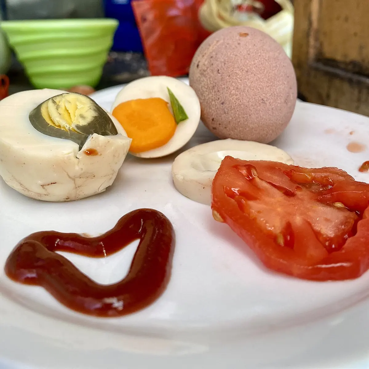
Boiling an egg white denatures its proteins. What does this change?
Pick an answer to see why.
1.1.5 Lipids or fats
Why does your body need fat? Which foods you eat are rich in it? Ghee, butter, mustard oil, groundnuts, sesame, eggs and meat come to mind.
Lipids are fats and fat-like substances. Like carbohydrates they are made of carbon, hydrogen and oxygen, but they have much less oxygen for their size: tristearin, a common fat, is C₅₇H₁₁₀O₆. They are insoluble in water but dissolve in organic solvents such as acetone, ether, chloroform and alcohol. Because their carbons are so little oxidized, burning them takes a lot of oxygen and gives a lot of energy.
Most true lipids are esters. An ester is the compound formed when an acid (here a fatty acid) joins an alcohol, giving off water:
Here R and R′ stand for the carbon chains: R may be –CH₃, –C₂H₅, … and in a real fatty acid it is a long chain such as C₁₇H₃₅–.
Fatty acids
Fatty acids are long chains of carbon and hydrogen with a carboxyl group, –COOH, at one end: general formula R–COOH. Natural ones mostly have an even number of carbons, 14 to 22, most often 16 or 18. They are of two kinds:
a. Saturated fatty acids have only single bonds between their carbons: every carbon holds as many hydrogens as it can (it is “saturated” with hydrogen). Their straight chains pack tightly, so they have higher melting points and are solid at room temperature. Animal fats (ghee, butter, lard, meat fat) are rich in them, and so are a few plant fats, coconut oil and palm oil, which go solid in a cold Kathmandu winter. Examples: palmitic acid (16 carbons) and stearic acid (18 carbons, melts at 69 °C).
b. Unsaturated fatty acids have one or more double bonds (C=C) in the chain. In natural fats each double bond is cis: the chain bends there, so the molecules can’t pack closely, and they have lower melting points and are liquid at room temperature, as clear oils. They are rich in plant oils (sunflower, mustard, soybean, olive) and in nuts, seeds and fish. Examples: oleic acid (one double bond; melts at about 13 °C), linoleic acid (two) and linolenic acid (three).
Classification of lipids
| Group | What it is | Examples |
|---|---|---|
| 1.Simple lipids | esters of fatty acids with an alcohol | neutral fats (glycerides), oils, waxes |
| 2.Complex (compound) lipids | fatty acids and an alcohol, plus another group | phospholipids, glycolipids, lipoproteins |
| 3.Derived lipids | made from the first two by hydrolysis or other changes | fatty acids, glycerol, mono- and diglycerides, steroids (cholesterol) |
1. Simple lipids
Simple lipids are esters of fatty acids with an alcohol. Digestion breaks them apart again by hydrolysis, using the enzyme lipase:
They are of three kinds: neutral fats, oils and waxes.
- i.Neutral fats (glycerides) are esters of fatty acids with the alcohol glycerol, which has three –OH groups. Each –OH can take one fatty acid:
- a.Monoglyceride: one glycerol + one fatty acid.
- b.Diglyceride: one glycerol + two fatty acids.
- c.Triglyceride (triacylglycerol, a triester): one glycerol + three fatty acids, with three molecules of water removed.
The three fatty acids of one triglyceride can be the same or different. Nearly all the fat in your food and in your body’s fat stores is triglyceride. Fats rich in saturated fatty acids are solid at room temperature.
ii. Oils are triglycerides rich in unsaturated fatty acids, so they are liquid at room temperature and usually clear: olive, sunflower, mustard and soybean oil. Hydrogen gas can be added across their double bonds, with nickel as a catalyst, turning them into solid, more saturated fat: this is hydrogenation, the way vanaspati ghee and margarine are made from vegetable oil. Partial hydrogenation also turns some cis double bonds into trans ones, and these trans fats both raise the harmful LDL cholesterol in the blood and lower the protective HDL, which makes them worse for the heart than any other fat; that is why the World Health Organization asks countries to remove them from food.
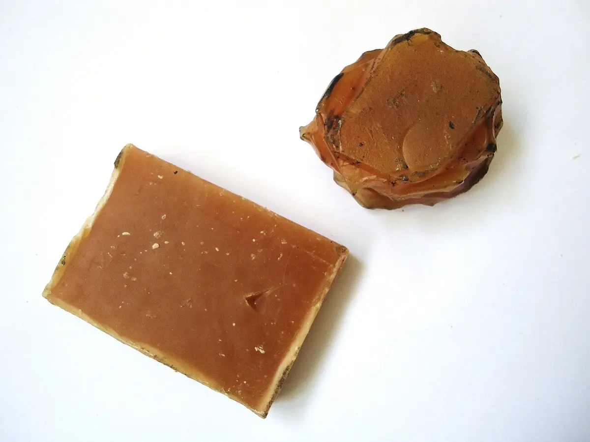
iii. Waxes are esters of long-chain fatty acids with long-chain alcohols other than glycerol. They are chemically inert and repel water, so they form protective, waterproof coatings on skin, fur and feathers, and on the stems, leaves and fruits of plants.
- Plant wax (the cuticle) coats leaves and fruits, cutting down the loss of water by transpiration and keeping out germs: it is the bloom you can rub off a plum.
- Beeswax is made by glands on the underside of the abdomen of young worker honeybees, who build the honeycomb with it.
- Lanolin (wool fat) is made by the sebaceous glands of sheep’s skin and coats the wool fibers.
- Paraffin wax, the wax of candles, comes from petroleum. It is a mixture of long-chain saturated hydrocarbons (alkanes), with no ester groups, so it is a wax only in its feel and use, not a lipid. Besides candles it is used in cosmetics and to coat some foods.
2. Complex (compound) lipids
Complex lipids have fatty acids and an alcohol, plus another group: a sugar, a phosphate or a protein.
a. Phospholipids (lipid + phosphate): in the commonest kind, glycerol carries two fatty acids and, in place of the third, a phosphate group joined to a small charged molecule (such as choline). The phosphate end is charged and hydrophilic (water-loving); the two fatty-acid tails are hydrophobic (water-fearing). In water such molecules line up tail to tail in a double layer, the lipid bilayer, which is the basic structure of the plasma membrane and of every membrane inside the cell. Examples: lecithin (phosphatidylcholine), cephalin, and plasmalogens.
b. Glycolipids (lipid + carbohydrate) have a sugar or chain of sugars in place of the phosphate head. They sit in the outer layer of cell membranes with their sugars facing out, and are plentiful in the myelin sheath of nerve fibers (the white matter of the brain). Examples: cerebrosides and gangliosides.
c. Lipoproteins (lipid + protein) are tiny balls with a fat core and a coat of protein, phospholipid and cholesterol, which carry fats and cholesterol in the blood (fat alone would not mix with the watery blood). Their kinds, from the largest and least dense: chylomicrons, very low density lipoprotein (VLDL), low density lipoprotein (LDL) and high density lipoprotein (HDL).
3. Derived lipids
Derived lipids are made from simple and complex lipids by hydrolysis or other changes: fatty acids, glycerol, mono- and diglycerides, and the steroids. They store energy, carry signals in the body (hormones) and build cell structures.
Steroids are lipids built on four joined rings of carbon atoms, not on fatty acids. The best known is cholesterol, a sterol (a steroid with an –OH group).
Cholesterol is part of every animal cell membrane, keeping it from becoming too fluid or too stiff. And it is the raw material for:
- the steroid hormones: the sex hormones progesterone, testosterone and estrogen, and the adrenal cortex’s hormones cortisol and aldosterone;
- vitamin D: in the skin, 7-dehydrocholesterol, made from cholesterol, is changed by the ultraviolet rays of sunlight into vitamin D₃, needed to absorb calcium for growing bones;
- bile acids (bile salts), made in the liver, which emulsify fats so they can be digested.
Functions of lipids
- They are the stored food of plants (oils in seeds such as mustard and groundnut) and animals.
- They give more than twice the energy of the same mass of carbohydrate: about 9 kcal (37 kJ) per gram against 4.
- Fat stored under the skin (subcutaneous fat) insulates the body against heat loss; a yak and a seal keep warm this way.
- Triglycerides stored in adipose tissue are the body’s main reserve of energy.
- Fats are the solvent that carries the fat-soluble vitamins A, D, E and K: a meal with no fat lets these pass unabsorbed.
- Pads of fat cushion organs such as the eyeballs and kidneys against knocks.
- Phospholipids form the membranes of every cell and organelle.
- Glycolipids form part of cell membranes and of the myelin round nerve fibers.
- Bile salts, made from cholesterol, emulsify fats so that they can be digested.
- Lipoproteins carry fats and cholesterol in the blood; they can also bind some bacterial poisons (endotoxins) and help clear them.
- Cholesterol is the raw material of vitamin D, needed for strong bones, and of the steroid hormones.
- Waxes waterproof leaves, fruits, fur and feathers.
A triglyceride is formed from:
Pick an answer to see why.
How is vanaspati ghee made from vegetable oil?
Pick an answer to see why.
Which part of a phospholipid faces the water in a membrane?
Pick an answer to see why.
1.1.6 Nucleic acids
How does your body store the instructions for making you, and pass them on to every new cell and to your children? The answer is the nucleic acids.
Nucleic acids are the molecules that store and carry genetic information. They are made of C, H, O, N and P; they are long chains, larger than most proteins (the DNA of the largest human chromosome has about 250 million base pairs). They are polymers of nucleotides: a nucleic acid is a long chain of nucleotides, a polynucleotide.
Structure of a nucleotide
A nucleotide has three parts:
- i.a pentose sugar (five carbons): ribose or deoxyribose;
- ii.a nitrogenous base (a ring compound containing nitrogen);
- iii.a phosphate group, from phosphoric acid.
The base is joined to carbon 1′ of the sugar, and the phosphate to carbon 5′. (The sugar’s carbons are numbered with a prime, 1′ to 5′, “one-prime” to “five-prime”, to tell them from the base’s atoms.)
A nucleoside is a base joined to a sugar, without phosphate. So:
- nucleoside = nitrogenous base + pentose sugar (for example adenosine = adenine + ribose);
- nucleotide = nucleoside + phosphate group (for example adenosine monophosphate, AMP).
The cell’s energy carrier, ATP (adenosine triphosphate), is a nucleotide too: adenosine with three phosphates in a row.
i. Pentose sugar
RNA (ribonucleic acid) contains the sugar ribose; DNA (deoxyribonucleic acid) contains deoxyribose, which has an H in place of the –OH on carbon 2′: “deoxy”, one oxygen less.
That one missing oxygen matters. The 2′ –OH of ribose can attack the chain’s own bonds, so RNA breaks down far more easily than DNA. DNA, without it, is stable enough to keep a library of instructions for a lifetime, while RNA makes good short-lived working copies.
ii. Nitrogenous bases
The bases are of two kinds:
- Purines have two rings, a six-membered ring joined to a five-membered one: adenine (A) and guanine (G).
- Pyrimidines have one six-membered ring: cytosine (C), thymine (T) and uracil (U). Thymine is found only in DNA and uracil only in RNA.
The bases of two strands pair in a fixed way, a purine always with a pyrimidine: adenine pairs with thymine (A=T, by two hydrogen bonds) and guanine pairs with cytosine (G≡C, by three hydrogen bonds). In RNA, where there is no thymine, adenine pairs with uracil. Bases that pair like this are complementary.
iii. Phosphoric acid
Phosphoric acid, H₃PO₄, gives the phosphate group. In a chain, each phosphate joins carbon 5′ of one sugar to carbon 3′ of the next, by two ester bonds: a phosphodiester bond. These bonds make the sugar–phosphate backbone of the chain.
Which is a purine?
Pick an answer to see why.
What makes a nucleoside into a nucleotide?
Pick an answer to see why.
Types of nucleic acids
The two kinds are deoxyribonucleic acid (DNA) and ribonucleic acid (RNA).
A. Deoxyribonucleic acid (DNA)
DNA is a molecule of two polynucleotide strands coiled round each other into a double helix. It is found in almost every cell: mostly in the nucleus, and a little in mitochondria and chloroplasts. (Mature red blood cells of mammals have none, and some viruses carry RNA instead.) It holds the genetic instructions for growth, development, working and reproduction. Each of its nucleotides has deoxyribose, a phosphate, and one of four bases: A, T, G or C.
Features of DNA (the Watson and Crick model):
- Two polynucleotide strands are twisted round a common axis into a right-handed double helix.
- The two strands are antiparallel: they run in opposite directions, one 5′ → 3′ and the other 3′ → 5′.
- The sugar–phosphate backbones are on the outside, and the bases are on the inside, stacked flat like the steps of a spiral staircase.
- The strands are held together by hydrogen bonds between complementary bases, a purine opposite a pyrimidine: A with T, G with C. Each hydrogen bond is weak, but millions together hold the strands firmly, yet they can still be opened a few at a time to copy or read the DNA.
- The helix is about 2 nm wide. Neighboring base pairs are 0.34 nm apart, and one full turn of the helix is about 3.4 nm long, with about 10 base pairs in a turn (DNA in water has about 10.5).
- Because A always pairs with T and G with C, the amount of A in any DNA equals the amount of T, and G equals C (Chargaff’s rule). Human DNA, for example, is about 30 % A, 30 % T, 20 % G and 20 % C.
- Within each strand, nucleotides are joined by phosphodiester bonds.
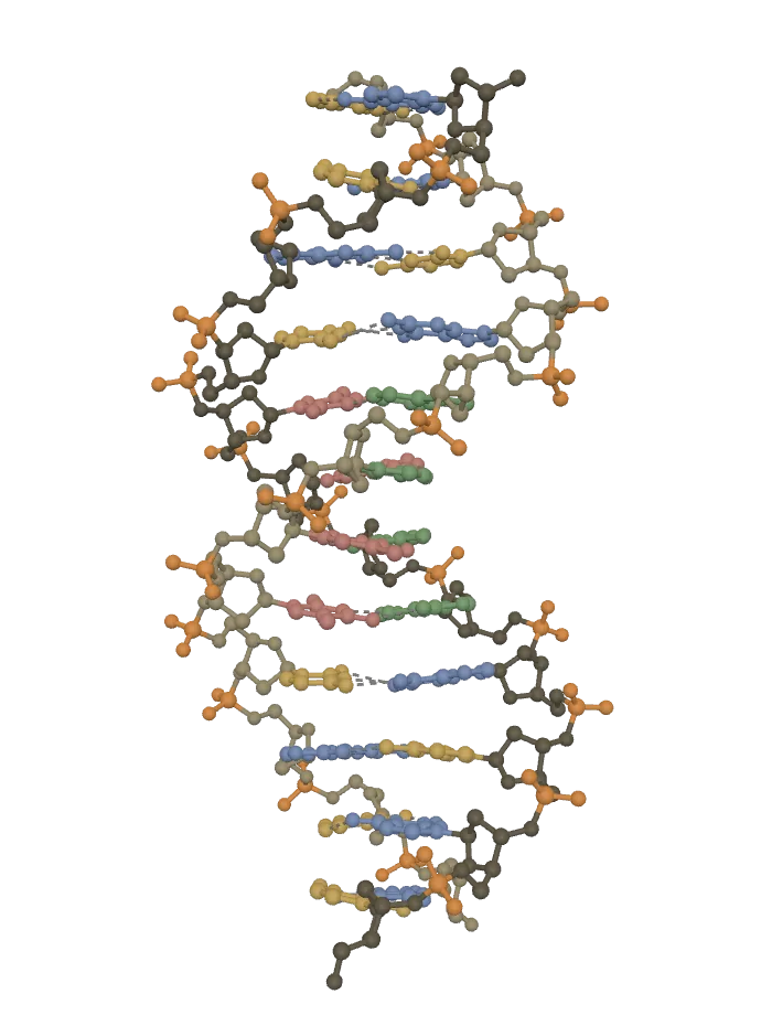
This browser can’t show the 3D model. The picture above is the same model.
Twelve base pairs of real DNA: a right-handed helix, the backbones outside, the bases stacked inside 0.34 nm apart, one turn in about 10 pairs. Turn it side-on for the wide major and narrow minor grooves. (X-rays don’t place hydrogen atoms, so none are shown.)
Sugar and phosphateThe sides of the ladder: each strand is a chain of deoxyribose sugars (brown; one strand darker) joined through phosphate groups (orange).
Adenine (A)Always paired with thymine (T) on the other strand, by two hydrogen bonds (dashed).
Thymine (T)Always paired with adenine (A) on the other strand, by two hydrogen bonds (dashed).
Guanine (G)Always paired with cytosine (C) on the other strand, by three hydrogen bonds (dashed).
Cytosine (C)Always paired with guanine (G) on the other strand, by three hydrogen bonds (dashed).
Drag to turn it. Tap a part, or a name below, to pick it out; tap it again to see the whole.
DNA: PDB 1BNA, Drew, Dickerson and others (1981), RCSB Protein Data Bank, CC0: www.rcsb.org/structure/1BNA
Functions of DNA
- It is the genetic material: it stores genetic information as the order (sequence) of its bases.
- It copies itself (replication) before a cell divides, so each new cell, and each new generation, gets a copy.
- Crossing over between chromosomes during meiosis swaps pieces of DNA, producing new combinations and so variation in the offspring.
- A change in its sequence is a mutation, a change that can be inherited.
- It is transcribed into the RNAs the cell needs.
- Through RNA and proteins it controls the cell’s metabolism: DNA → RNA → protein, the central dogma of molecular biology.
B. Ribonucleic acid (RNA)
RNA is usually a single strand of ribonucleotides. It is made in the nucleus (copied from DNA) and found in the nucleus, the nucleolus, the cytoplasm and the ribosomes. Its main work is making proteins; in some viruses (the influenza and polio viruses, the coronaviruses) it is the genetic material itself.
Structure of RNA:
- It is much smaller than DNA.
- It is a single polynucleotide strand, though the strand can fold back and pair with itself in places.
- Its nucleotides are joined by 3′–5′ phosphodiester bonds.
- Each ribonucleotide has ribose, a phosphate, and one of the bases adenine, guanine, cytosine or uracil.
Types of RNA:
- Messenger RNA (mRNA) carries the copy of a gene’s instructions from DNA in the nucleus to the ribosomes, where they are read to make a protein. It makes up only a few percent of a cell’s RNA, and is short-lived.
- Ribosomal RNA (rRNA) is part of the ribosomes, together with proteins. It is the most abundant RNA, about 80 % of a cell’s RNA, and it is stable. It is rRNA that actually joins the amino acids into a chain.
- Transfer RNA (tRNA) is the adapter: each tRNA carries one particular amino acid to the ribosome and pairs with the matching three bases (codon) of the mRNA.
Functions of RNA:
- mRNA carries the coded instructions for making polypeptides (proteins and enzymes).
- tRNA brings the right amino acids to the ribosome in the right order.
- rRNA, in the ribosome, joins the amino acids by peptide bonds.
- In eukaryotes, mRNA has a cap at its 5′ end, which the ribosome binds to when it starts reading.
- In genetic engineering, mRNA is copied back into DNA by the enzyme reverse transcriptase, giving a compact gene without its non-coding parts (complementary DNA, cDNA).
- RNA is the genetic material of some viruses.
| DNA | RNA |
|---|---|
| Two strands, a double helix | Usually one strand |
| Sugar: deoxyribose | Sugar: ribose |
| Bases A, G, C and T | Bases A, G, C and U |
| Mostly in the nucleus (some in mitochondria and chloroplasts) | Made in the nucleus; works mostly in the cytoplasm and ribosomes |
| Stable; lasts the cell’s lifetime | Less stable; mRNA lasts minutes to hours |
| Copies itself | Copied from DNA (except in some viruses) |
| Stores genetic information | Uses it to make proteins |
1.1.7 Minerals
Minerals are inorganic elements, taken in as ions or salts, that the body needs to work. Altogether they are only about 4 % of the body’s mass, most of it calcium and phosphorus in the bones, yet without them the body can’t grow, its nerves can’t signal and its enzymes stop. The body loses minerals all the time, in urine and sweat, so they must come regularly from food.
Your body depends most on calcium, phosphorus, potassium, sulphur, sodium, chlorine and magnesium, and needs trace elements such as iron, zinc, copper and iodine in much smaller amounts.
Categories of minerals
About 25 elements are needed by living things. By the amount needed, they are of two groups:
a. Macro-elements (major minerals) are needed in large amounts, more than about 100 mg a day for an adult: calcium (Ca), phosphorus (P), potassium (K), sulphur (S), sodium (Na), chlorine (Cl) and magnesium (Mg). Plants also need nitrogen (N) as a major nutrient (we take ours in as protein).
b. Micro-elements (trace elements) are needed in small amounts, less than about 100 mg a day, some only micrograms: iron (Fe), zinc (Zn), copper (Cu), manganese (Mn), iodine (I), selenium (Se), molybdenum (Mo), cobalt (Co), chromium (Cr), fluorine (F) and others.
| Macro-element | Symbol | Functions |
|---|---|---|
| Calcium | Ca | Gives strength and hardness to bones and teeth (as calcium phosphate). Forms the hard shells of snails and other mollusks and the skeletons of corals (as calcium carbonate, CaCO₃). Needed for blood clotting and muscle contraction. In plants, calcium pectate forms the middle lamella that cements neighboring cell walls. Bone meal is used as a fertilizer. |
| Magnesium | Mg | Part of the bones and teeth. At the center of every chlorophyll molecule. Needed by many enzymes, including all that use ATP (ATPases). |
| Potassium | K | Keeps the electrical potential across membranes (more K⁺ inside cells than outside); needed for nerve impulses and muscle contraction. Opens and closes the stomata of leaves. |
| Sulphur | S | Part of the amino acids cysteine and methionine, so of proteins; its disulphide bonds make keratin tough. Part of coenzyme A and some vitamins. |
| Sodium | Na | Works with potassium in nerve impulses. The main positive ion outside cells: keeps the balance of ions and water (osmotic balance) in body fluids. Helps the gut absorb glucose and amino acids. |
| Nitrogen | N | Part of every amino acid, so of all proteins, and of nucleic acids, ATP and chlorophyll. |
| Phosphorus | P | Part of nucleic acids and ATP, and of phospholipids in membranes. With calcium, forms bones and tooth enamel. |
| Chlorine | Cl | The main negative ion (Cl⁻) of blood plasma and body fluids. Makes the hydrochloric acid of the stomach. Helps carry CO₂ in the blood (the chloride shift). |
| Trace element | Symbol | Functions |
|---|---|---|
| Copper | Cu | Carries electrons in cytochrome oxidase in respiration, and in plastocyanin in photosynthesis. Needed to make the pigment melanin. |
| Iron | Fe | Holds oxygen in hemoglobin and myoglobin (respiratory pigments). Carries electrons in the cytochromes of respiration and photosynthesis. Needed by plants to make chlorophyll. Lack of iron causes anemia. |
| Iodine | I | Part of the thyroid hormone thyroxine. Lack of it causes goiter, and in babies cretinism (stunted growth and brain). |
| Manganese | Mn | Needed by enzymes of respiration and photosynthesis (it helps split water in photosynthesis); an enzyme of the mitochondria that destroys harmful oxygen radicals has manganese. Helps, with phosphatase enzymes, in forming bone. |
| Molybdenum | Mo | Part of nitrogenase, the enzyme of nitrogen fixation, and of nitrate reductase, so plants need it to make proteins. |
| Zinc | Zn | Needed by many enzymes (over 300), such as carbonic anhydrase, which helps carry CO₂ in the blood; needed for growth, healing and immunity. |
Which mineral is at the center of the chlorophyll molecule?
Pick an answer to see why.
1.1.8 Enzymes
How does your body break down the food you eat? The starch of a grain of rice, left in a glass of water, would take millions of years to break down into glucose by itself; in your gut it is done within a few hours. The difference is made by enzymes.
Enzymes are proteins that speed up the chemical reactions in living things, without being used up or changed themselves. They are biological catalysts, or biocatalysts. The substance an enzyme acts on is its substrate; when more than one substance takes part in the reaction, they are called its reactants.
By where they work, enzymes are of two kinds:
- a.Exoenzymes (extracellular enzymes) are made in a cell and work outside it, as the enzymes of the digestive juices do (amylase in saliva, pepsin in the stomach).
- b.Endoenzymes (intracellular enzymes) work inside the cell that made them, as the enzymes of the Krebs cycle inside mitochondria do.
How enzymes speed up reactions
Most reactions need a push to get started, an energy hill to climb before they can roll downhill: the activation energy. An enzyme gives the substrate an easier path, with a lower hill. The substrate fits into a pocket of the enzyme, the active site, shaped to hold it; there its bonds are strained and the reaction goes, then the products leave and the enzyme is free for the next molecule. An enzyme doesn’t change how much energy the reaction gives out, nor where its equilibrium lies: it only makes it get there much faster.
Characteristics of enzymes
- Enzymes are proteins, mostly globular. Many also need a non-protein helper to work: a metal ion (such as Zn²⁺ or Mg²⁺) or an organic molecule (a coenzyme, often made from a vitamin).
- They are hydrophilic and, free in the cell, are dissolved in its water as a colloid (a hydrosol).
- They are specific: each enzyme acts on one substrate, or one kind of bond, and catalyzes one reaction. Different enzymes can act on the same substrate to give different products.
- In principle, every enzyme reaction can go both ways; which way it runs depends on the amounts of substrate and product, the energy needed, and the pH.
- They are very efficient: a small amount of enzyme acts on a huge amount of substrate. One molecule of catalase breaks down millions of molecules of hydrogen peroxide a second.
- They don’t start reactions that couldn’t happen anyway; they speed them up by lowering the activation energy, and don’t change the reaction’s equilibrium.
- Being proteins, each is made by the cell from the instructions of its own gene.
- Heat, the wrong pH, heavy metals (mercury, lead, silver) and strong radiation denature them, so they stop working.
Biological importance of enzymes
- Enzymes carry out almost every chemical reaction in living things, from digestion to copying DNA.
- They work at body temperature: reactions that would need boiling or strong acid in a laboratory happen gently in the cell.
- Each works best at its own pH, so enzymes suited to each part of the body work there: pepsin in the acid stomach, trypsin in the alkaline small intestine.
- They let reactions go quickly without harsh conditions that would damage the cell.
- Because they work only in a narrow range of temperature and pH, living things must keep their insides near those conditions (homeostasis): a fever of 42 °C and a blood pH below 7.0 are both dangerous.
- The cell can control which enzymes it makes, by switching their genes on and off, and so control its own chemistry precisely.
Pepsin works best at pH 2. Why does it stop working when food passes into the small intestine?
Pick an answer to see why.
How does an enzyme make a reaction faster?
Pick an answer to see why.
1.1.9 Water
Water is the most plentiful substance in every cell: it is about 60–70 % of the mass of the human body and 70–90 % of protoplasm, and jellyfish are 95 % water. It gives no energy, but no life can go on without it.
A water molecule, H₂O, has an oxygen atom joined to two hydrogen atoms by covalent bonds, at an angle of 104.5°. Oxygen pulls the shared electrons towards itself, so the oxygen end is slightly negative (δ−) and the hydrogen ends slightly positive (δ+): water is a polar molecule, a dipole. The δ+ hydrogen of one molecule is attracted to the δ− oxygen of another: a hydrogen bond. Each molecule can hydrogen-bond to up to four others, so liquid water is a loose, ever-changing network of molecules, and in ice they lock into an open lattice, which is why ice floats.
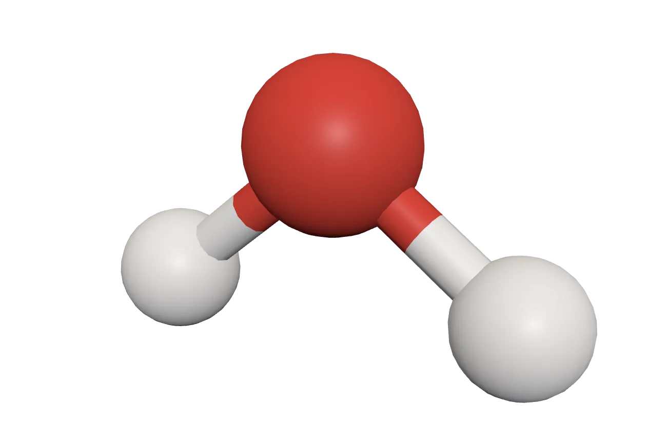
This browser can’t show the 3D model. The picture above is the same model.
Bent, not straight: the two O–H bonds are 104° apart in this computed model (about 104.5° in real water). Oxygen’s two lone pairs, not drawn, are on the side away from the hydrogens.
Oxygen atom: one single bond to each hydrogen.
Hydrogen atom: one single bond.
Drag to turn it. Tap an atom to see what it is.
Molecules: computed 3D conformers from PubChem, National Library of Medicine (public domain): pubchem.ncbi.nlm.nih.gov, CID 962
Biological functions of water
- Water is the best solvent known: salts, sugars, amino acids and gases dissolve in it, so the cell’s reactions happen in water.
- It takes part in reactions: it splits molecules in hydrolysis (digestion), and ions form and move in it.
- It takes a lot of heat to evaporate (about 2.4 kJ per gram at body temperature), so sweating cools the body well with little water lost. It also takes a lot of heat to warm, so it keeps body temperature (and lakes and seas) steady.
- Its molecules cling to each other (cohesion) and to the walls of narrow tubes, so a high surface tension and cohesion let unbroken columns of water be pulled up the xylem to the tops of tall trees.
- It is the medium in which substances are carried round the body: nutrients, hormones and wastes in blood plasma, sugar in phloem sap.
- It is the home of all the living things of ponds, rivers and seas; and because ice floats, lakes freeze from the top down and life survives below.
- It removes wastes in solution, mainly as urine and partly as sweat, and so helps keep the body’s inside steady (homeostasis).
Why can water dissolve salt and sugar so well?
Pick an answer to see why.
Exercise
A. Choose the best alternative.
- Which group is wrongly labeled?
Pick an answer to see why.
- What is the general formula of carbohydrates?
Pick an answer to see why.
- Which of the following is a disaccharide?
Pick an answer to see why.
- Polysaccharides are:
Pick an answer to see why.
- What is the importance of monosaccharides like ribose and deoxyribose in the cell?
Pick an answer to see why.
- Why is glycogen important in animals?
Pick an answer to see why.
- Which group of amino acids must come from the diet, because the human body can’t make them?
Pick an answer to see why.
- What does the body use tyrosine for, besides building proteins?
Pick an answer to see why.
- Which category of proteins is formed by the combination of amino acids and nucleic acids?
Pick an answer to see why.
- Which type of protein has long chains twisted together into fibers?
Pick an answer to see why.
- What is the main feature of fibrous proteins?
Pick an answer to see why.
- Which function of proteins involves controlling the glucose level of the blood?
Pick an answer to see why.
- Fats found in meat and milk, and in coconut and palm oil, belong to which category of lipids?
Pick an answer to see why.
- Which fatty acids generally have lower melting points and are liquid (oils) at room temperature?
Pick an answer to see why.
- Which complex lipid has sugars joined to it and is plentiful in the white matter of nerve tissue?
Pick an answer to see why.
- Which derived lipid is the raw material for sex hormones such as progesterone and testosterone, and for cortisol?
Pick an answer to see why.
- Which type of RNA carries the coded instructions for making polypeptide chains?
Pick an answer to see why.
- Which sugar is found in RNA nucleotides?
Pick an answer to see why.
- Which part is present in both DNA and RNA nucleotides?
Pick an answer to see why.
- Which mineral is part of the chlorophyll pigment?
Pick an answer to see why.
- Which trace element is part of the hormone thyroxine, and its lack causes goiter?
Pick an answer to see why.
- Which feature of enzymes lets each catalyze only one particular reaction?
Pick an answer to see why.
- What is the most important biological function of water?
Pick an answer to see why.
B. Very short questions
What are biomolecules?
Answer
Define metabolism.
Answer
Define aldoses and ketoses.
Answer
Give an example of a monosaccharide and a disaccharide.
Answer
What is the main role of amino acids in the body?
Answer
Which amino acid is the precursor of the plant hormone indole-3-acetic acid (IAA)?
Answer
What is the main function of enzymes in the body?
Answer
Name one fibrous protein and one globular protein.
Answer
What is the main role of antibodies in the body?
Answer
What is the main difference between unsaturated and saturated fatty acids?
Answer
What is the main function of the triglycerides stored in adipose tissue?
Answer
What are the two kinds of pentose sugar found in nucleic acids?
Answer
Name one macro-element that helps keep the electrical potential across membranes.
Answer
C. Short questions
What is the significance of the telomeres of chromosomes?
Hint
Answer
How do autosomes differ from sex chromosomes?
Answer
Explain the difference between oligosaccharides and polysaccharides in terms of their structure and properties.
Answer
Describe the role of carbohydrates in the cell’s metabolism, with a reaction.
Answer
Write the differences between oligosaccharides and polysaccharides.
Answer
Distinguish between essential and non-essential amino acids.
Answer
Describe the primary structure of a protein and its significance in the protein’s function.
Answer
Explain the difference between the primary and quaternary structures of proteins, with examples of proteins that show each.
Answer
Explain how thrombin and fibrinogen prevent bleeding by clotting the blood.
Answer
Describe the main function of messenger RNA (mRNA) in protein synthesis.
Answer
Distinguish between RNA and DNA.
Answer
Describe the function of phospholipids in the structure of cells.
Answer
Describe the functions of iron (Fe) in the human body.
Answer
Design a lunch for a child so that it gives all the essential nutrients. Use foods from different food groups and give reasons for your choices.
Answer
D. Diagrammatic questions
The diagram above shows a structure of protein. Identify the type of protein structure.
Answer
The diagram above shows the structure of an amino acid. Identify the labeled parts x, y and z.
Answer
Referring to the structure of a nucleotide above, explain the structure of a nucleic acid.
Answer
Project work
Printable PDFs
- Study notes 4 pages
- Worksheet 5 pages Answer key
Quiz yourself: Living Things and Their Structure, Grades 5–12 →
Practice every question in the book, and keep track of the ones you miss →
A companion to your textbook, not a replacement for it: study from the book your school uses, and where the two differ, follow your teacher. Found a mistake? Please tell us.