
Look at the tip of your finger. It is made of about a billion cells, each too small to see, and each one alive: taking in food and oxygen, building proteins, getting rid of wastes, and, when the time comes, dividing in two. A single bacterium, a yeast cell in a fermenting jar of jaand, an Amoeba in a pond, a leaf of rice and an elephant are all built from the same kind of unit. Whatever their size or shape, every living thing is made of cells, and everything a living thing does is done, in the end, by its cells.
Why are cells called the basic units of life, and what do all living things have in common? This unit answers by opening a cell up. You will meet the two great kinds of cell, the simple prokaryotic cell of bacteria and the far more complex eukaryotic cell of plants, animals, fungi and protists, and then go through the eukaryotic cell part by part: its wall and membrane, the organelles that make its energy, its food and its proteins, and the nucleus that holds the instructions for everything else.
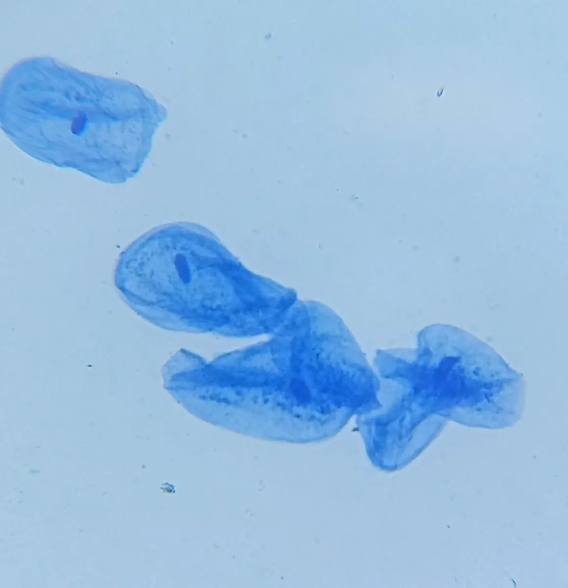
1.2.1 General introduction
All living things, past and present, are made of special compounds called biomolecules (Unit 1), packed into a structure called a cell. The cell is the basic building block of life: it is the smallest unit that can carry out all the activities of life on its own, and it is enclosed by a protective membrane. A cell can reproduce itself, and it carries out the vital functions of life: nutrition, respiration, growth, excretion and response.
- In a unicellular organism, such as a bacterium, Amoeba or yeast, a single cell does everything. The cell is the organism.
- In a multicellular organism, cells share the work. Muscle cells contract, nerve cells carry signals, red blood cells carry oxygen, and the life of the whole body comes from the combined actions of its cells.
That is why the cell is called the fundamental unit of life: nothing smaller than a cell is alive on its own, and everything alive is a cell or is made of cells.
The study of the structure and function of cells and their parts is cytology (Greek kytos, a hollow vessel, a cell, and logos, study), today usually called cell biology. The German biologist Oscar Hertwig, who first saw sperm and egg nuclei fuse, made it a subject of its own with his book Die Zelle und die Gewebe (The Cell and the Tissues, 1893–1898).
Why is the cell called the basic unit of life?
Pick an answer to see why.
1.2.2 Prokaryotic and eukaryotic cell
Cells are of two basic types, based on whether they have a true nucleus and membrane-bound organelles, and on how complex their genetic material is: prokaryotic and eukaryotic. The words (Greek pro, before, eu, true, and karyon, kernel or nucleus) were first used by the French biologist Édouard Chatton in 1925; Dougherty (1957) and other microbiologists made the division the basis of how cells are classified.
Cell drawings adapted from Servier Medical Art (smart.servier.com), CC BY 4.0
Mariana Ruiz Villarreal, LadyofHats, Public domain, via Wikimedia Commons
1. Prokaryotic cell
A prokaryotic cell is a simple cell with no organized nucleus and no membrane-bound organelles (Figure 2.3). It was the first kind of cell on Earth. Its features:
- It has no nuclear membrane, no membrane-bound organelles (no mitochondria, chloroplasts, ER or Golgi), no spindle (mitotic apparatus) and no nucleolus. Its flagella, where it has them, are simple threads of a protein called flagellin, without the 9 + 2 pattern of microtubules of a eukaryotic flagellum.
- Its DNA is a single naked circular molecule, not wrapped round histone proteins, folded into a region of the cytoplasm called the nucleoid. Many also carry small extra rings of DNA, plasmids.
- Its cell wall is made of peptidoglycan (murein), a mesh of sugar chains cross-linked by short chains of amino acids.
- It has no mitochondria: the enzymes of respiration are on its plasma membrane.
- In photosynthetic prokaryotes the chlorophyll lies on folded membranes, lamellae or thylakoids, in the cytoplasm, not inside chloroplasts.
- Its ribosomes are of the smaller 70S type (50S + 30S).
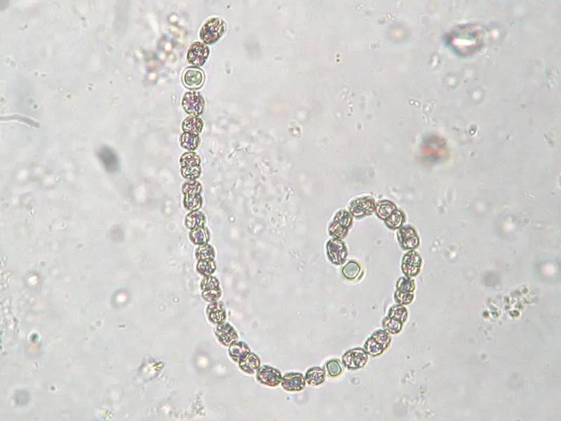
Examples: bacteria and cyanobacteria (blue-green algae), such as Nostoc, Anabaena and Lyngbya.
2. Eukaryotic cell
A eukaryotic cell has a true nucleus, enclosed by a nuclear envelope, and membrane-bound organelles such as mitochondria, endoplasmic reticulum, Golgi complex and vacuoles (Figure 2.2). Its features:
- Its DNA is enclosed in a nuclear envelope, forming a true nucleus.
- Its DNA is linear, in several chromosomes, wound round histone proteins.
- Its cells divide by mitosis and meiosis (and in a few cases by amitosis), with a spindle.
- Where it has a cell wall, the wall is of cellulose and hemicellulose with pectin, sometimes lignin, in plants, and of chitin in fungi.
- Its flagella and cilia are made of microtubules in a 9 + 2 pattern.
- Its cytoplasm streams (cyclosis), and in plants photosynthesis happens in membrane-bound chloroplasts.
- Its cytoplasm has the larger 80S ribosomes (60S + 40S), while its mitochondria and chloroplasts have 70S ribosomes, like bacteria.
Examples: all plants, animals, fungi and protists.
Which of these does a prokaryotic cell have?
Pick an answer to see why.
1.2.3 Structure of eukaryotic cell
Shapes and sizes
The shape of a cell depends on its function, its age, whether it has a cell wall, the pressure of the cells round it, its position, and the tension and viscosity of its contents. Cells can be irregular (Amoeba, white blood cells), round, oval, cylindrical, flat like plates (the cells lining the cheek), tubular, branched (nerve cells) or long and thin (muscle fibers).
Most eukaryotic cells are between about 10 and 100 µm across, though they range from about 1 µm to well over 1 mm. The smallest known cells are bacteria called mycoplasmas, about 0.2–0.3 µm across. The largest single cell is the egg of an ostrich, about 15 cm long; its yolk is one enormous cell, swollen with stored food. The longest cells are nerve cells, whose fibers can be a meter long in a person and several meters in a giraffe or a whale.
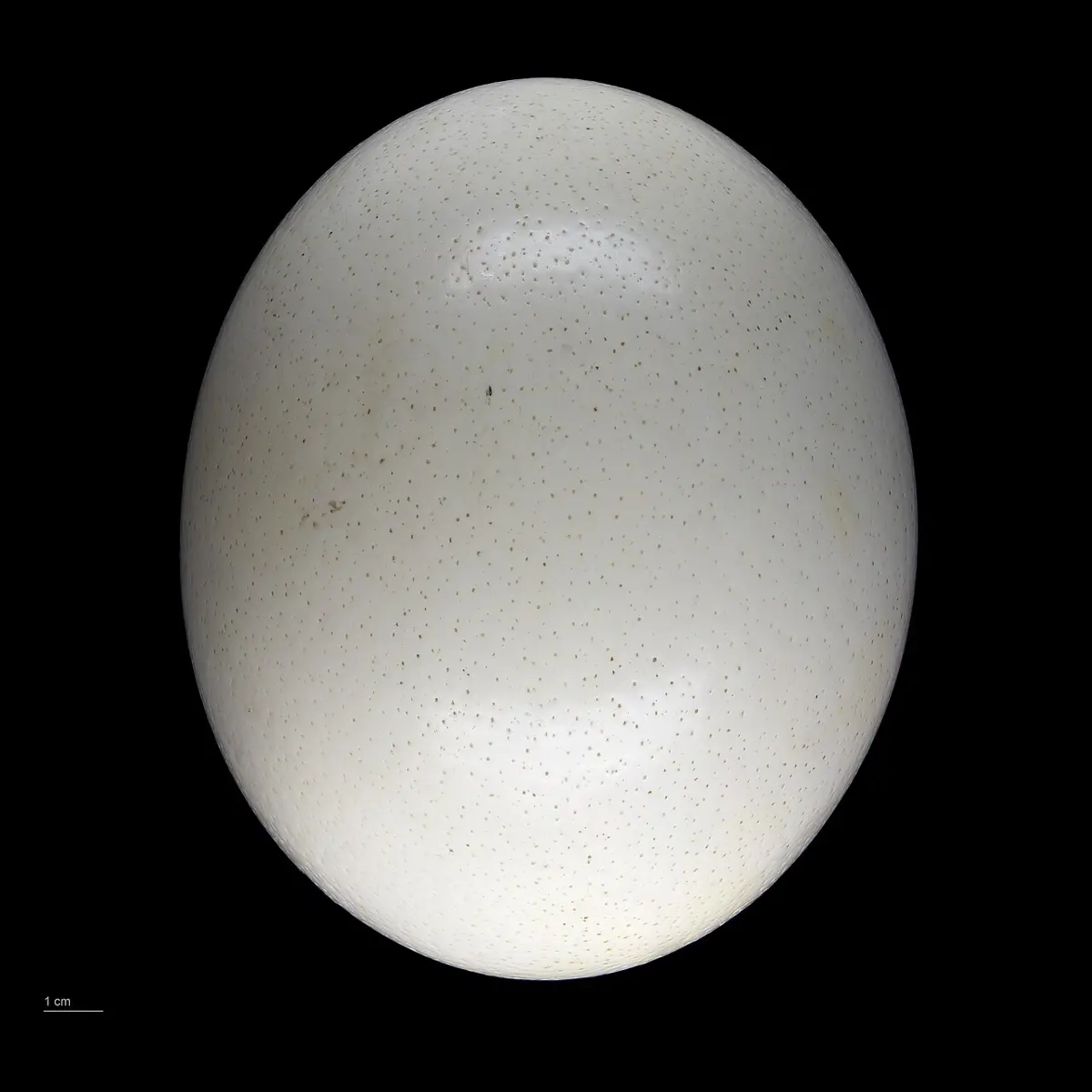
Why are most cells so small? Because everything a cell takes in and gets rid of must pass through its surface, while the amount it needs grows with its volume. As a cell grows bigger, its volume grows faster than its surface.
A typical eukaryotic animal cell is a mass of jelly-like, translucent protoplasm enclosed in a plasma membrane. In a plant cell, the plasma membrane is surrounded in addition by a rigid, porous cell wall, made mainly of cellulose. The fluid that fills the space between the plasma membrane and the nuclear envelope is the cytoplasm.
Parts of eukaryotic cells
The parts of a eukaryotic cell fit inside one another (Figure 2.6):
1. Cell wall
The cell wall is the outermost layer of a plant cell, a non-living, protective layer that gives the cell rigidity and mechanical support. Cell walls are found in plants, bacteria, cyanobacteria, fungi and some protists (many algae), but never in animal cells. Some plant cells have none: the naked gametes and zoospores of algae and lower plants. A wall is from about 0.1 µm to 10 µm thick. It was the cell wall that Robert Hooke saw in 1665, when he looked at thin slices of cork, the empty walls of dead cells, and called the little boxes “cells”.
The plant cell wall is built in layers (Figure 2.8), laid down from the outside in:
- Middle lamella. A thin layer between the walls of neighboring cells, the first layer formed, from the cell plate at cell division. It is made of pectin, mostly as calcium and magnesium pectates. Pectin is a hydrophilic, colloidal substance that absorbs water and acts as the cement that holds the cells together. (When a fruit ripens, enzymes dissolve its middle lamella, and it goes soft.)
- Primary cell wall. A thin, elastic and permeable layer, laid down by the protoplast on each side of the middle lamella while the cell is still growing. It is a network of cellulose microfibrils in a matrix of hemicellulose and pectin, with some protein. Because it can stretch, the cell can grow. Most cells of leaves, fruits, the cortex and the pith have only a primary wall and the middle lamella.
- Secondary cell wall. A thick, rigid and inelastic layer, laid down inside the primary wall once the cell has stopped growing. It is built of three layers (S₁, S₂ and S₃), in each of which the cellulose microfibrils run in a different direction, which makes the wall very strong, like plywood. It is of cellulose and hemicellulose, often hardened by lignin, and usually 1–10 µm thick. It is found in mature, specialized cells that give support or carry water: tracheids, vessels and sclerenchyma (fibers and stone cells). Other substances may be laid down in or on the walls: cutin and waxes on the outer walls of the epidermis, suberin in cork cells, mucilage, and silica (in grasses).
- Tertiary cell wall. In some cells, such as the tracheids of gymnosperms, a thin extra layer of cellulose and xylan (a hemicellulose) lines the inside of the secondary wall.
- Plasmodesmata (singular plasmodesma). Fine channels through the cell walls, lined by the plasma membrane, that join the cytoplasm of neighboring cells. Through the middle of each runs a narrow tube of endoplasmic reticulum, the desmotubule. They let water, solutes and signals pass directly from cell to cell, so that the living parts of a plant are joined into one continuous system. Where a cell has a secondary wall, they cross at the pits, thin places where the secondary wall was not laid down.
Functions of the cell wall:
- It gives the cell and the whole plant mechanical strength and support.
- It protects the contents from mechanical injury and keeps out many germs.
- It is permeable, so water and dissolved substances pass freely between the cell and its surroundings.
- It gives the cell its shape, preventing too much expansion.
- In root hairs, the thin wall takes part in the absorption of water and minerals from the soil.
- Waterproof substances laid down on the wall, suberin (in roots and cork) and cutin (in the cuticle), cut the loss of water by evaporation.
- It stops the cell from bursting: when water enters by osmosis, the wall pushes back (wall pressure), balancing the pressure inside the cell.
- It holds enzymes that act outside the membrane, for example in building and loosening the wall as the cell grows.
Which layer of the plant cell wall acts as a cement between neighboring cells?
Pick an answer to see why.
2. Protoplast
A protoplast is what is left of a plant cell when its cell wall is removed: the plasma membrane and everything inside it. Biologists make protoplasts by digesting the wall away with enzymes (cellulase and pectinase); a protoplast can grow a new wall, and two protoplasts can even be fused, a way of making new plant hybrids.
3. Plasma membrane
Every living cell is enclosed by a thin, transparent, selectively permeable membrane, the plasma membrane or cell membrane: it lets some substances through and holds others back. It is an elastic, living structure that can repair itself. The term cell membrane was used by C. Cramer and C. Nägeli (1855), and the name plasmalemma was given by J. Q. Plowe (1931).
The plasma membrane and the membranes round all the organelles inside the cell share the same basic structure, and together they are called biomembranes. Each separates the inside of the cell, or of an organelle, from what lies outside it.
The plasma membrane is made chiefly of lipids and proteins, so it is called lipoproteinous, with a little carbohydrate attached to them on the outer face. In a typical plasma membrane they are present in roughly equal amounts by mass (a red blood cell’s membrane is about 43 % lipid, 49 % protein and 8 % carbohydrate). The share varies with the membrane’s job: the myelin sheath round nerve fibers, mainly an insulator, is about 80 % lipid, while the inner membrane of the mitochondria, packed with enzymes, is only about 25 % lipid.
The proteins of the membrane are of three kinds by their work:
- Structural proteins, which give the membrane its form and strength.
- Enzymes, catalytic proteins that speed up reactions on the membrane.
- Transport proteins (carriers and channels), which carry substances across the membrane. Some let substances move down their concentration gradient faster (facilitated diffusion); others, the pumps, move substances against their gradient using energy from ATP (active transport).
Many models have been put forward to explain how the lipids and proteins are arranged. The two best known are the sandwich model and the fluid mosaic model.
Structural models of the plasma membrane
a. Sandwich model. In 1935 J. F. Danielli and H. Davson, reasoning from the membrane’s physical and chemical properties (it lets fat-soluble substances through easily, and its surface tension is lower than a pure layer of lipid would have), proposed that the membrane is a sandwich (Figure 2.9):
- In the middle, a bilayer of phospholipids. Each phospholipid is polar: it has a hydrophilic (water-loving) head, of glycerol and phosphate, and two hydrophobic (water-hating) tails, of fatty acids. The heads of the two layers face outward, and the tails of the two layers face each other in the middle, held together by weak van der Waals forces.
- On each face, a continuous layer of globular proteins, attached to the heads.
- Tiny pores, about 7–10 Å across, lined with protein, through which water and small ions could pass.
So the membrane is trilaminar: protein, lipid, protein. Under the electron microscope, membranes do look like three layers, dark–light–dark, which seemed to support the model. But it could not explain why different membranes do very different jobs, or how active transport happens, and later work showed that most membrane proteins are not spread as sheets over the surface but sit in the lipid layer itself.
b. Fluid mosaic model. In 1972 S. J. Singer and G. L. Nicolson proposed the model accepted today (Figure 2.10). The proteins do not form continuous layers: they are scattered through a fluid bilayer of phospholipids in a mosaic, like icebergs floating in a sea.
- The phospholipid bilayer is fluid: its molecules are not fixed but drift about. They change places with their neighbors in the same layer all the time (lateral movement), and much more rarely move from one layer to the other (flip-flop).
- The proteins are globular and are of two kinds. Extrinsic (peripheral) proteins lie on the inner or outer surface of the bilayer, loosely attached. Intrinsic (integral) proteins are embedded in the bilayer, some partly, others right across it (transmembrane proteins). Many of these form channels and carriers for water, ions and other water-soluble substances, which cannot cross the oily middle of the bilayer on their own.
- A phospholipid molecule is amphipathic: it has a hydrophilic head and two hydrophobic fatty-acid tails. In water the molecules arrange themselves heads out and tails in, so the bilayer forms a barrier that water-soluble substances can’t cross except through the proteins.
- Short chains of sugars are attached to some of the proteins and lipids on the outer face (glycoproteins and glycolipids), and cholesterol in animal membranes sits between the phospholipids, controlling the fluidity.
| Sandwich model | Fluid mosaic model |
|---|---|
| Proposed by Danielli and Davson (1935). | Proposed by Singer and Nicolson (1972). |
| Proteins form continuous layers on both faces of the lipid bilayer. | Proteins are scattered in the bilayer, like icebergs in a sea. |
| The membrane is a fixed, rigid structure. | The membrane is fluid; lipids and proteins move about in it. |
| Proteins only on the surfaces. | Peripheral proteins on the surfaces and integral proteins in and across the bilayer. |
| Can’t explain the different jobs of membranes, or active transport. | Explains selective transport, channels, carriers and receptors. |
Functions of the plasma membrane:
- It gives the cell its shape (in animal cells) and protects it, separating its contents from the surroundings.
- It controls what enters and leaves the cell, by diffusion, osmosis, facilitated diffusion and active transport, and takes in large particles or drops of liquid by endocytosis (phagocytosis, “cell eating”, of solids; pinocytosis, “cell drinking”, of liquids) and sends out large molecules by exocytosis.
- It helps some cells move: Amoeba and white blood cells crawl by pushing out pseudopodia, and some cells, such as fibroblasts, move by wave-like ruffles of the membrane.
- Its surface molecules (antigens) identify the cell: the ABO blood groups are decided by sugar chains on the membrane of red blood cells, and the immune system tells the body’s own cells from invaders by them.
- It is folded into microvilli in the cells lining the small intestine, which increases the surface for absorbing digested food many times over.
- Its receptor proteins detect signals, such as hormones and antigens, and pass them on into the cell.
According to the fluid mosaic model, which proteins are embedded within the phospholipid bilayer?
Pick an answer to see why.
4. Protoplasm
Protoplasm is the living matter of a cell, a colloidal mass in which all the organelles lie. Thomas Huxley called it “the physical basis of life” (1868). It is a thick, translucent fluid, about 70–90 % water, in which are proteins, lipids, carbohydrates, nucleic acids and inorganic salts. The term protoplasm was given to the living contents of cells by J. E. Purkinje in the late 1830s. It is made of the cytoplasm and the nucleus:
5. Cytoplasm
The cytoplasm is the jelly-like, semi-fluid part of the protoplasm that fills the space between the plasma membrane and the nuclear envelope. It is made of three things:
- the cytosol, the fluid itself, a watery solution of salts, sugars, amino acids and enzymes, in which many of the cell’s reactions (such as the first stage of respiration, glycolysis) take place;
- the cell organelles;
- the cell inclusions, non-living materials such as stored food and crystals (section 16).
The cytoplasm keeps the cell’s shape, holds the substances the organelles need, and is the place of many of the cell’s reactions; in plants it holds the chloroplasts, where photosynthesis happens.
In living plant cells, the cytoplasm is never still: it flows round the cell in a steady stream, carrying the chloroplasts and other organelles with it. This streaming is called cyclosis (cytoplasmic streaming). It is driven by the motor protein myosin moving along threads of actin, and it carries materials round large cells much faster than diffusion could. It is easy to see in the leaf cells of the water plant Hydrilla or in the long internodal cells of the alga Chara, where the chloroplasts move along the walls.
6. Cell organelles
Cell organelles are the small, specialized living parts of a cell, each with its own structure and its own job, as the organs of the body each have theirs. Most are bound by a membrane. Some (mitochondria, chloroplasts, the nucleus) have a double membrane; some (the ER, Golgi complex, lysosomes, vacuoles) a single membrane; and a few (ribosomes, centrioles) have no membrane at all.
7. Mitochondria
Mitochondria (singular mitochondrion; Greek mitos, thread, and chondrion, granule) are granular or thread-like organelles in the cytoplasm, called the powerhouses of the cell, because they release the energy of food as ATP in aerobic respiration, the energy that powers the cell’s activities. They were first described in the muscle cells of insects by Albert von Kölliker in 1857 and named by Carl Benda in 1898.
Occurrence. Mitochondria are absent in prokaryotes, whose respiratory enzymes lie on the plasma membrane. They are present in all eukaryotic cells with very few exceptions, such as mature mammalian red blood cells, which lose them as they mature (and so respire without oxygen, never using the oxygen they carry). Their shape varies from rods to round bodies and threads, and changes with the cell’s condition. They are usually 0.5–1 µm wide and 1–10 µm long. After the nucleus (and in plants the chloroplasts), they are among the largest structures in the cell.
Number. A cell may have one mitochondrion (some algae) or thousands: a liver cell has about 1,000–2,000. They are most numerous in cells that are growing, dividing or very active, such as muscle, liver and kidney cells and sperm, and they sit where the energy is needed: packed between the fibers of muscles, wound round the middle piece of a sperm’s tail.
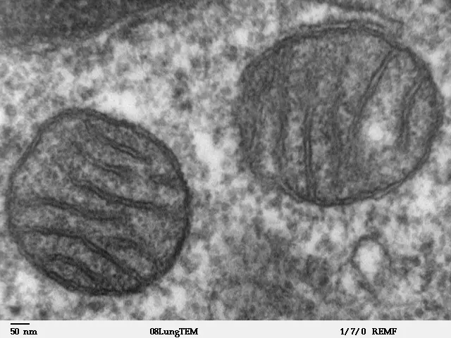
Structure (Figure 2.11). A mitochondrion has two membranes:
- a smooth outer membrane;
- an inner membrane, folded into many shelves or fingers, the cristae (singular crista), which greatly increase its surface for the reactions of respiration.
Between the two is the intermembrane space (the outer chamber), which runs on into the space inside each crista (the intercristal space). Inside the inner membrane is the matrix (the inner chamber), a dense fluid with the enzymes of the Krebs cycle, 70S ribosomes, granules, and the mitochondrion’s own circular DNA.
Cell drawings adapted from Servier Medical Art (smart.servier.com), CC BY 4.0
On the matrix side of the inner membrane stand thousands of tiny particles, each like a tennis racket: the elementary particles, F₁ particles or oxysomes (Figure 2.12). Each has a head (F₁), a stalk and a base (F₀) set in the membrane. Together the three parts make one enzyme, ATP synthase (also called ATP synthetase, and, because it can also run backwards and split ATP, ATPase). As hydrogen ions flow back through it into the matrix, it makes ATP from ADP and phosphate: the particles are where the cell’s ATP is made (oxidative phosphorylation), so they are the functional units of mitochondria.
Chemical composition. Mitochondria are about 65–70 % protein and 25–30 % lipid, with about 0.5 % RNA and a little DNA.
Semi-autonomous organelles. A mitochondrion has the things needed to make some of its own proteins: its own DNA, which can copy itself and be transcribed to RNA, its own 70S ribosomes, and ATP for energy. It also grows and divides on its own, by fission. But its DNA is small: in humans, a ring of 16,569 base pairs with 37 genes, only 13 of them for proteins (the rest for its rRNA and tRNA). That is far too few to build a mitochondrion: most of its proteins (more than 1,000) are coded by the DNA of the nucleus, made on ribosomes in the cytoplasm, and imported. So a mitochondrion is semi-autonomous: it can do some things on its own, but depends on the nucleus for most. (Chloroplasts are the same, below.)
Functions of mitochondria:
- They are the powerhouses (ATP factories) of the cell, the main site of aerobic respiration: the Krebs cycle in the matrix, the electron transport chain and ATP synthesis on the inner membrane.
- Intermediate compounds of respiration are used to make other substances, such as steroids, cytochromes and the porphyrin ring of chlorophyll and heme.
- They make some amino acids.
- They help control the concentration of calcium ions in the cell.
- They take part in the formation of yolk in developing egg cells.
- In a sperm, they gather round the base of the tail and form its middle piece, which powers its swimming.
In which part of the mitochondrion are the enzymes of the Krebs cycle found?
Pick an answer to see why.
8. Plastids
Plastids are double-membrane organelles of plants and of some protists, such as Euglena, which make or store food. The name was given by Ernst Haeckel in 1866. They are absent in fungi and animals, and in prokaryotes: photosynthetic bacteria and cyanobacteria have their pigments on membranes in the cytoplasm, called chromatophores, instead. Plastids are also missing from the sperm cells of many flowering plants, which is why a seedling usually gets its plastids from the egg alone. By their color, plastids are of three kinds: chloroplasts (green), chromoplasts (colored, but not green) and leucoplasts (colorless).
a. Chloroplasts
Chloroplasts (Greek chloros, green, and plastos, formed) are the commonest plastids, and the most important: in them, photosynthesis turns carbon dioxide and water into sugar with the energy of light, making the food on which nearly all life depends. They contain the green pigment chlorophyll, with orange and yellow carotenoids (carotene and xanthophyll). They were described in the 1830s by the German botanist Hugo von Mohl, and the name chloroplast was given by A. F. W. Schimper in 1883. They are found in the green, photosynthetic cells of plants and in algae and some protists like Euglena, and are absent in parts of plants not exposed to light, such as roots.
Shape, size and number. In flowering plants, chloroplasts are usually lens-shaped (biconvex or plano-convex), about 5–10 µm across and 2–3 µm thick. In algae their shapes vary greatly: cup-shaped in Chlamydomonas, girdle-shaped in Ulothrix, spiral ribbons in Spirogyra, star-shaped in Zygnema, a net in Oedogonium. A cell may have one (Chlamydomonas) or many: a leaf cell has 20 to 100, and the long internodal cells of Chara have many thousands.
Structure (Figure 2.14). A chloroplast has three main parts:
- The envelope: two membranes, outer and inner, with an intermembrane space between them.
- The stroma (matrix): the dense, colorless ground substance inside the inner membrane. It is rich in proteins (more than half the chloroplast’s protein is here, most of it the enzyme Rubisco), and contains 70S ribosomes, circular naked DNA, mRNA and tRNA, water, minerals, and the enzymes of the dark reaction (the Calvin cycle), where carbon dioxide is made into sugar. Starch grains and oil droplets are often stored in it.
- The thylakoids: flattened, disc-shaped membrane sacs. Many are piled one on another like coins in stacks called grana (singular granum), and the grana are joined to one another by flat membrane tubes running through the stroma, the stroma lamellae (intergrana lamellae). The thylakoid membranes hold the chlorophyll and all the other parts needed for the light reaction.
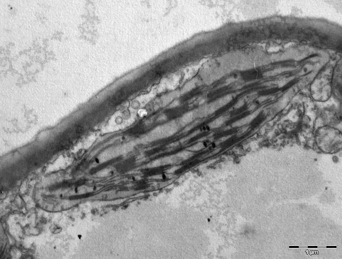
Like mitochondria, chloroplasts are semi-autonomous: they have their own DNA and ribosomes and make some of their proteins, and divide on their own, but most of their proteins are coded in the nucleus and made on the cell’s cytoplasmic ribosomes.
Functions of chloroplasts:
- They carry out photosynthesis, making the food of the plant: the cell’s kitchen.
- They give out oxygen, which aerobic organisms need for respiration.
- They help keep the balance of oxygen and carbon dioxide in the air.
- By taking in carbon dioxide, they help to slow global warming.
- In many algae, starch is stored round protein bodies in the chloroplast called pyrenoids, which are packed with the enzyme Rubisco and concentrate carbon dioxide round it.
- They give plants their green color.
b. Chromoplasts
Chromoplasts (Greek chroma, color) are plastids colored yellow, orange or red by carotenoids, found in the non-green parts of plants: petals, ripe fruits and some roots. They may develop from chloroplasts, as the chlorophyll breaks down and carotenoids build up (a green tomato or chilli turning red), or from leucoplasts that gain pigments (a carrot root). They are angular, needle-shaped or irregular, have no grana, and are bound by a double membrane. (The red and blue anthocyanins of many flowers and fruits are not in plastids at all: they are dissolved in the cell sap of the vacuole.)
Function: they give flowers and fruits their bright colors, which attract insects for pollination and animals that eat the fruits and disperse the seeds.
Many books list, with the chromoplasts, the plastids of algae and the pigment membranes of prokaryotes that are colored by pigments other than chlorophyll alone:
- Phaeoplasts: brown plastids, with fucoxanthin (a xanthophyll) masking their chlorophyll, in diatoms, dinoflagellates and brown algae.
- Rhodoplasts: red plastids, with the red R-phycoerythrin and blue R-phycocyanin, in red algae.
- Chromatophores of blue-green algae: membranes with C-phycocyanin, C-phycoerythrin and chlorophyll a.
- Chromatophores of photosynthetic bacteria: membranes with bacteriochlorophyll and purple-red carotenoids, in purple bacteria.
c. Leucoplasts
Leucoplasts (Greek leukos, white) are colorless plastids found in storage organs and other parts out of the light: roots, tubers, seeds. They may be rod-shaped, round or oval, are bound by a double membrane, and have a granular matrix with few or no thylakoids. If exposed to light, many turn into chloroplasts, which is why a potato left in the light turns green. By the food they store, they are of three kinds:
- Amyloplasts store starch: in potato tubers and grains of wheat, rice and maize.
- Elaioplasts (lipoplasts, oleoplasts) store oils and fats: in the seeds of mustard, castor, groundnut, sunflower and cotton.
- Proteinoplasts (aleuroplasts) store proteins: mainly in seeds, such as the aleurone layer of cereal grains.
Function: to store reserve food, carbohydrates, lipids and proteins.
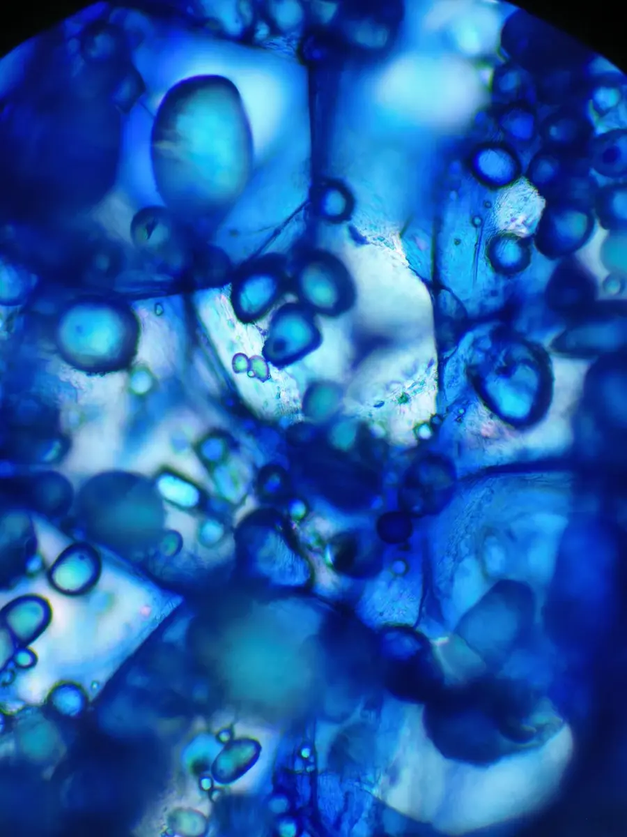
Which plastid stores starch in a potato tuber?
Pick an answer to see why.
9. Endoplasmic reticulum (ER)
The endoplasmic reticulum (ER) is a network of membrane-bound cisternae, tubules and vesicles spread through the cytoplasm, especially its inner part, the endoplasm (hence the name: a network in the endoplasm). It is like a factory with winding passages and chambers, where proteins and lipids are made, processed, packed and sent on. It is found only in eukaryotic cells; it is absent in mature mammalian red blood cells and in prokaryotes. How much ER a cell has depends on its activity: it is poorly developed in eggs, embryonic cells and the cells that form sperm, and well developed in active, specialized cells such as those of the pancreas and liver.
The ER was first seen as a stained material in gland cells by C. Garnier (1897), who called it ergastoplasm. Its structure was first shown with the electron microscope by K. R. Porter, A. Claude and E. F. Fullam (1945), and it was named endoplasmic reticulum by Porter in 1953.
Types of ER. By whether ribosomes are attached to its surface, ER is of two kinds:
- Smooth (agranular) ER (SER) has no ribosomes on its surface and is made mostly of tubules. It usually lies towards the cell membrane, and grows out from the rough ER, which loses its ribosomes. It is abundant in cells that make lipids and steroids: fat cells (adipocytes), the interstitial cells of the testes, the corpus luteum of the ovaries, the sebaceous (oil) glands, the cortex of the adrenal glands; in liver cells, where it breaks down drugs and poisons; and in muscle cells, where it stores calcium (as the sarcoplasmic reticulum). It makes and stores lipids.
- Rough (granular) ER (RER) has ribosomes on its outer surface, which make it look rough, and is made mostly of cisternae. It usually lies near the nucleus, and its membrane is continuous with the outer membrane of the nuclear envelope. It is abundant in cells that make proteins for export: the acinar cells of the pancreas (digestive enzymes), goblet cells (mucus), plasma cells (antibodies), and the Nissl granules of nerve cells, which are stacks of RER. It makes proteins and sends them on.
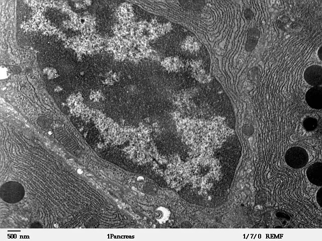
Structure. The ER is built of three kinds of part (Figure 2.18):
- Cisternae: long, flattened, unbranched sacs, usually near the nucleus, joined to one another and often lying in parallel stacks, in cells with a big job of making things, such as the pancreas and the notochord. Each is a closed sac of membrane, with a space, the lumen, inside.
- Vesicles: small, round or oval sacs scattered in the cytoplasm, abundant in pancreatic cells, which carry materials onward.
- Tubules: wider, branched, irregular tubes, which with the cisternae and vesicles make up the network. They constantly change, joining and dividing as membranes move round the cell.
Functions of the ER:
- The SER makes lipids (phospholipids, cholesterol and steroid hormones); the RER makes proteins for membranes, for lysosomes and for secretion.
- It forms an intracellular transport system: substances move through its lumen from one part of the cell to another, and to the Golgi complex.
- It divides the cytoplasm into compartments and gives the cytoplasm some mechanical support.
- Its membrane gives rise to the nuclear envelope after cell division, and to the membranes of other organelles; the enzymes of the first lysosomes are made on the RER.
- In plant cells, ER takes part with the Golgi vesicles in forming the cell plate at cytokinesis, and runs through the plasmodesmata.
- Its enzymes carry out many reactions: the breakdown of glycogen to glucose and the making of sterols in the liver.
- It helps carry messages and molecules from the nucleus to the rest of the cell.
- It stores some of the cell’s products, such as glycogen (near the SER of liver cells) and calcium.
- The SER of liver cells detoxifies drugs, alcohol and other foreign chemicals (xenobiotics), making them easier to excrete.
Which type of endoplasmic reticulum is mainly involved in making lipids?
Pick an answer to see why.
10. Golgi complex
The Golgi complex (Golgi apparatus, Golgi body) is a membrane-bound, cup-shaped organelle, usually near the nucleus, that processes, packs and sends on proteins and lipids in eukaryotic cells. It was discovered by the Italian scientist Camillo Golgi in 1898, in the nerve cells of the cat and the barn owl, with a silver stain he had invented. James Darnell and his colleagues (1986) called it the traffic police of the cell, because it sorts the cell’s proteins and membrane materials and directs each to its destination. It has also been called lipochondria, idiosome and the Dalton complex.
It is found in all eukaryotic cells except a few, such as mature mammalian red blood cells, the sperms (antherozoids) of bryophytes and pteridophytes, and mature sieve tube cells. Its shape and size vary with the kind of cell and its activity, so it is called pleomorphic. It is large in active, secreting cells such as nerve cells, pancreatic cells, liver cells and the cells that form eggs and sperm, and small in cells that secrete little, such as muscle cells. In animal cells it is usually one compact complex near the nucleus; in plant cells it is many separate small stacks scattered through the cytoplasm, called dictyosomes.
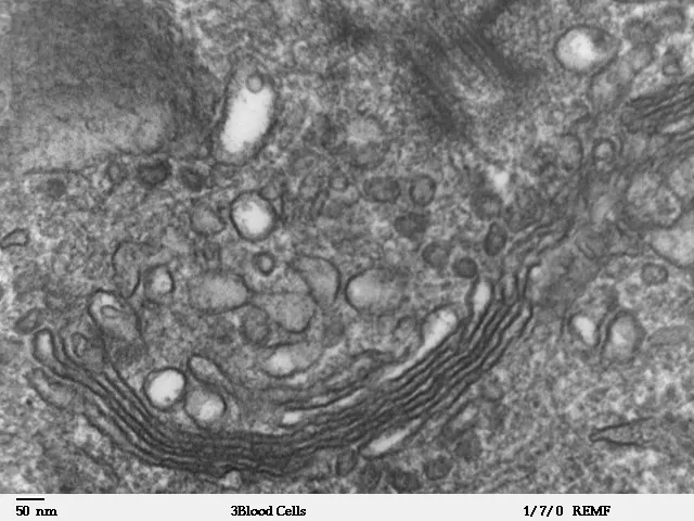
Structure. The Golgi complex is made of four kinds of smooth membrane parts (Figure 2.19):
- Cisternae (flattened sacs, saccules): flat, curved sacs filled with fluid, each bounded by a membrane, lying in parallel stacks with narrow spaces (sometimes holding fibrils) between them. A stack usually has 4 to 8 cisternae in animal cells, and in some plant and algal cells 20 or more. The cisternae are curved and often perforated (fenestrated) at their rims. Each stack has two faces:
- the cis face or forming face: convex, and facing the nucleus and ER, where materials arrive;
- the trans face or maturing face: concave, and facing the cell membrane, where finished products leave.
- Tubules: a network of fine tubes growing from the perforated rims of the cisternae, towards the edge of the stack, joined to the vesicles.
- Vesicles: small sacs at the tips of the tubules and round the stack, some arriving from the ER at the cis face, others leaving from the trans face. They are of two kinds, smooth vesicles and coated vesicles (covered in a coat of protein such as clathrin).
- Golgian vacuoles: large, round sacs budded from the trans face, formed by the swelling of the cisternae there. They contain secretions, and some hold hydrolyzing enzymes and become lysosomes.
Functions of the Golgi complex:
- It receives proteins, lipids and other materials from the ER, chemically modifies them (for example by adding sugar chains to proteins, making glycoproteins), and sends each to its destination.
- It stores, concentrates (condenses) and packs materials into vesicles.
- It takes part in secretion: vesicles full of secretions pinch off from the trans face, move to the cell membrane, fuse with it and empty their contents outside the cell (exocytosis). Digestive enzymes, mucus, milk proteins and hormones such as insulin leave cells this way.
- In plant cells, its vesicles carry pectin and other wall materials to the equator of a dividing cell and form the cell plate.
- It forms the primary lysosomes: it packs the digestive enzymes made on the RER into vesicles, which bud off.
- It takes part in making hormones in endocrine cells: in the thyroid gland it adds sugars to thyroglobulin, the protein from which thyroxine is made.
- Its vesicles add new membrane to the cell surface and take part in recycling the plasma membrane.
- It makes glycoproteins and glycolipids, and in plants the polysaccharides of the wall.
Which part of the Golgi complex modifies, packs and sends on proteins and lipids?
Pick an answer to see why.
11. Lysosomes
Lysosomes (Greek lysis, loosening or dissolving, and soma, body) are tiny sacs bounded by a single membrane, found in the cytoplasm, that carry out digestion inside the cell. Each holds dozens of kinds of hydrolytic (digestive) enzymes, proteases, lipases, nucleases and others, which break down almost any biological material. The enzymes work best in acid, at about pH 5, the pH inside the lysosome. If its membrane breaks, the enzymes can digest the cell itself (autolysis), so lysosomes are called suicidal bags. They were discovered by the Belgian biochemist Christian de Duve in 1955, in rat liver cells. They are of many shapes (pleomorphic), usually round.
Lysosomes are found in most animal cells (not in mammalian red blood cells), in some fungi (yeasts), in some protists (Euglena), and in some plant cells, such as the cells of the root tip of maize; in most plant cells the large vacuole does their work. They are most numerous in cells that destroy invaders and debris: phagocytes (white blood cells that eat bacteria) and the cells of the reticuloendothelial system, in the bone marrow, liver and spleen.
Kinds of lysosome. By what is inside them, lysosomes are of the following kinds (Figure 2.21):
- Primary lysosomes: newly formed lysosomes, budded off from the Golgi complex, holding enzymes made on the RER that have not yet digested anything. They are also called virgin lysosomes.
- Secondary lysosomes: formed when a primary lysosome fuses with a vacuole containing material to be digested, from outside or inside the cell. Because it is often hard to tell which kind a secondary lysosome is, they are together called digestive vacuoles.
- a. Heterophagosomes (heterophagic vacuoles): formed when primary lysosomes fuse with a food vacuole (phagosome or pinosome) holding material taken in from outside the cell, such as bacteria.
- b. Autophagosomes (autophagic vacuoles): formed when primary lysosomes digest the cell’s own parts. During growth, repair, the change of tissues and starvation, worn-out or unneeded mitochondria, ribosomes, peroxisomes and glycogen are wrapped in membrane and digested. The digestion of a cell’s own organelles is autophagy (“self-eating”).
- c. Residual bodies: when the contents of a digestive vacuole can’t all be digested (perhaps for want of some enzyme), the undigested remains collect as grains and whorls inside it. Such secondary lysosomes are called residual bodies, tertiary lysosomes or telolysosomes. They are usually emptied out of the cell by exocytosis; in long-lived cells that can’t do this, such as nerve and heart cells, they stay as a brown pigment, lipofuscin, which builds up with age.
Functions of lysosomes:
- They digest material taken in from outside: food particles, and bacteria and viruses engulfed by white blood cells (in heterophagosomes).
- They digest the cell’s stored food, glycogen, proteins and lipids, when it is starving.
- In autophagy they digest worn-out organelles, such as ER and mitochondria, so that their materials can be used again.
- At fertilization, the acrosome at the tip of a sperm, a special lysosome, releases its enzymes to digest a path through the coverings of the egg.
- They destroy whole cells and tissues when they are no longer needed: a tadpole’s tail is broken down as it turns into a frog.
- In bone, they help bone cells dissolve old bone so that it can be remodeled.
Which of these is not a function of lysosomes?
Pick an answer to see why.
12. Ribosomes
Ribosomes are tiny, compact, round granules, the organelles on which the cell makes its proteins. They are not bound by any membrane. Small particles rich in RNA were first separated from cells by spinning them in a centrifuge by Albert Claude (1943), and ribosomes were first clearly seen with the electron microscope by George Palade (1955).
Ribosomes are found in all cells, prokaryotic (70S) and eukaryotic (80S); the only cells without them are those, such as mature mammalian red blood cells, that have stopped making proteins. In prokaryotic cells they lie free in the cytoplasm. In eukaryotic cells they are of two kinds in the cytoplasm, free in the cytosol and bound to the rough ER and the outer membrane of the nuclear envelope, and there are smaller ribosomes inside mitochondria and chloroplasts. Their number depends on how much protein the cell makes: they are most numerous in cells that make a lot, such as plasma cells, liver cells, meristem cells, cancer cells and gland cells. A single bacterium (E. coli) has about 20,000 or more; a mammalian cell has millions.
Structure (Figure 2.22). A ribosome is a slightly flattened sphere made of two subunits, a larger one (50S or 60S) and a smaller one (30S or 40S), which come together only when they are making protein. The larger subunit is dome-shaped, and is the one attached to the ER; the smaller subunit sits on it like a cap. Between them is a groove for the mRNA. A ribosome is made of rRNA and protein: about two-thirds rRNA by mass in bacteria, a little over half in eukaryotes. When several ribosomes work along one strand of mRNA together, like beads on a string, the whole is called a polyribosome or polysome.
By their size, measured by how fast they sink in a centrifuge (their sedimentation coefficient, measured in svedberg units, S), ribosomes are of two types:
- 70S type: a large 50S and a small 30S subunit; about 20 nm across. Found in bacteria, cyanobacteria, and in mitochondria and chloroplasts.
- 80S type: a large 60S and a small 40S subunit; about 25 nm across. Found in the cytoplasm of eukaryotic cells, in fungi, protists, plants and animals.
The RNA of the cell
Ribosomes work with the cell’s three kinds of RNA. Of all the RNA in a cell, rRNA is by far the largest share, tRNA comes next, and mRNA is the least. The shares vary from cell to cell; typical figures are:
- a. ribosomal RNA (rRNA): about 80 % (books give 50–80 %)
- b. messenger RNA (mRNA): about 5 % (5–10 %)
- c. transfer RNA (tRNA): about 15 % (10–20 %)
Ribosomal RNA (rRNA) is the RNA of the ribosomes themselves: the factory where proteins are made. In eukaryotic cells the 60S subunit holds three rRNAs (28S, 5.8S and 5S) and about 47 different proteins; the 40S subunit, one rRNA (18S) and about 33 proteins. rRNA is needed for the mRNA to bind to the ribosome, and it is the rRNA, not the proteins, that joins the amino acids together: rRNA is an enzyme, a ribozyme.
Messenger RNA (mRNA) is a single, linear chain made in the nucleus by transcription of the DNA; its sequence of bases is complementary to the template strand of the DNA. It carries the genetic message, the code for a protein, from the chromosomes in the nucleus to the ribosomes in the cytoplasm. There it is joined by ribosomes to form polysomes, several to several dozen ribosomes on one mRNA, each making a copy of the same protein.
Transfer RNA (tRNA), also called soluble RNA, is a small molecule of about 75–90 nucleotides, folded into the shape of a clover leaf (Figure 2.23). Each tRNA carries one particular amino acid to the ribosome and, with its anticodon, matches it to the right codon of the mRNA. There are more than 20 kinds of tRNA, at least one for each of the 20 amino acids found in proteins. It is made in the nucleus from a DNA template.
Functions of ribosomes:
- They are the protein factories of the cell, the sites of protein synthesis.
- The proteins they make build the cell’s structures, and act as enzymes that run and regulate its work.
- Free ribosomes make proteins used inside the cell, in the cytosol (such as the enzymes of glycolysis); ribosomes bound to the RER make proteins that are sent out of the cell (such as digestive enzymes and hormones), or go into membranes or lysosomes.
- They hold the mRNA, tRNA and the growing protein together while the protein is made, and their subunits are stored in the cell ready to be used again.
Which type of ribosome is found in the cytoplasm of eukaryotic cells?
Pick an answer to see why.
13. Cilia and flagella
Cilia (singular cilium; Latin cilium, eyelash) and flagella (singular flagellum; Latin, a little whip) are fine, hair-like or whip-like threads that beat and move. They arise inside the cell from basal bodies and stick out from its surface, covered by the plasma membrane. They move cells and organisms, drive currents of water for feeding, and move fluids over surfaces. The German biologist T. W. Engelmann described their beating in the 1860s.
Occurrence.
- Cilia are found on all protists of the group Ciliata (such as Paramecium), the flame cells of flatworms, the swimming larvae of many animals (such as the bipinnaria larva of the starfish), and the ciliated epithelium of many animals, for example lining the windpipe (trachea) and the fallopian tubes.
- Flagella are found on the flagellate protists (such as Euglena), the collar cells (choanocytes) of sponges, some gastrodermal cells of cnidarians (coelenterates), the sperms of animals and of lower plants, the zoospores of algae, and many gametes.
Structure (Figure 2.24). Cilia and flagella have the same structure: a basal body inside the cell, with rootlets anchoring it, a basal plate where it passes out of the cell, and the shaft outside. A cilium is short (about 5–20 µm) and there are many on a cell; a flagellum is long (up to 100–200 µm), and there are only one or a few.
A cross-section of the shaft shows nine pairs of microtubules in a ring round two single ones in the middle: the 9 + 2 pattern. This bundle of microtubules is the axoneme (axial filament). It is covered by an extension of the plasma membrane, about 90 Å thick.
- Each of the nine outer (peripheral) microtubules is a doublet: an A tubule, a complete ring of 13 protofilaments, and a B tubule, an incomplete one sharing part of A’s wall.
- The A tubule of each doublet carries two dynein arms, an inner and an outer, reaching towards the B tubule of the next doublet. Dynein is a motor protein and an ATPase: it splits ATP and uses the energy to “walk” along the next doublet.
- The two central microtubules are single, and lie inside a central sheath.
- Radial spokes run from each A tubule inward to the central sheath.
The basal body has the same structure as a centriole: nine triplets of microtubules (A, B and C) set like the blades of a pinwheel round a cartwheel-shaped hub, with no central pair (9 × 3 + 0). Chemically, all these microtubules, the central ones and the peripheral ones alike, are made of the protein tubulin; the dynein arms are the only part made of dynein.
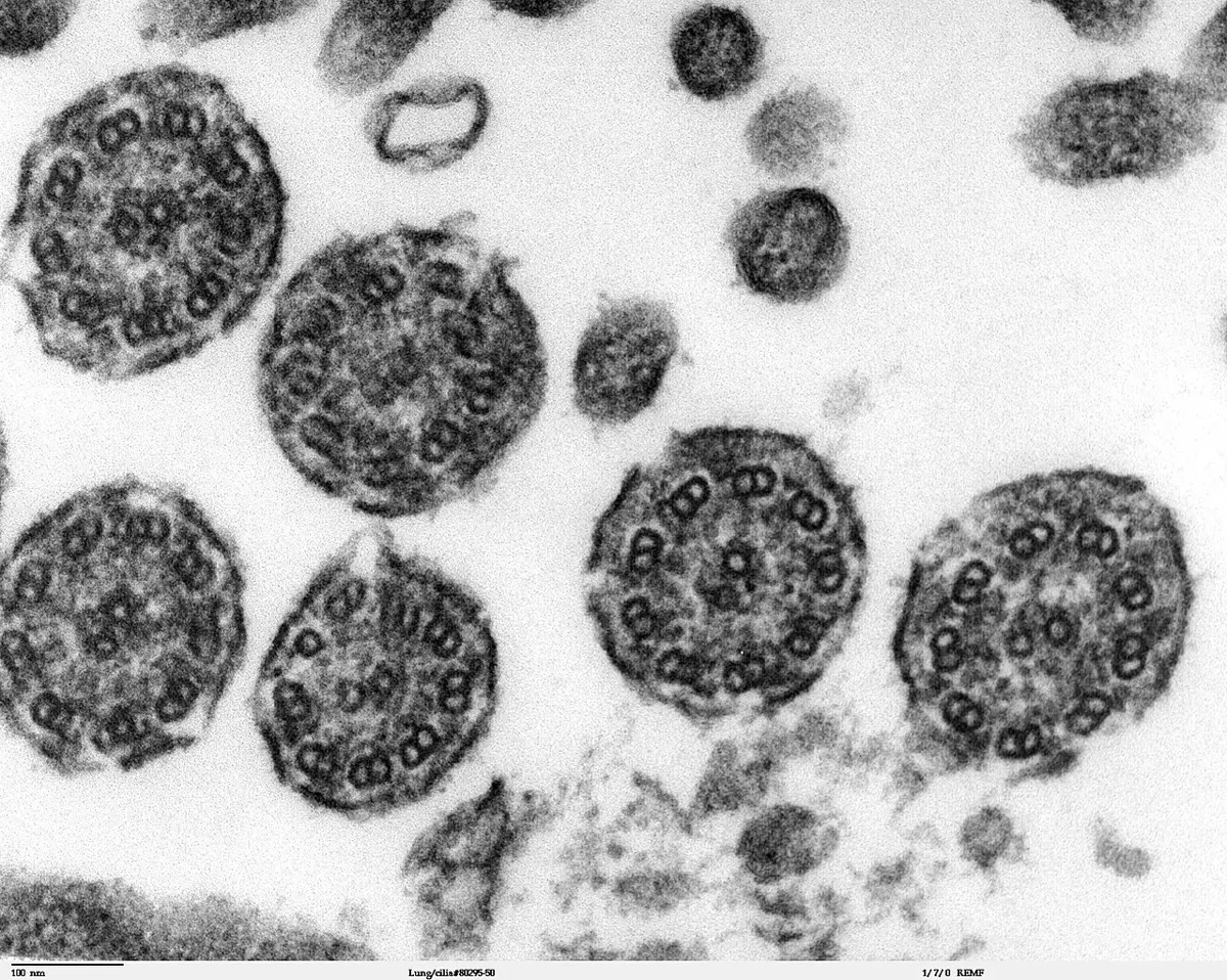
How they beat. When the dynein arms of one doublet walk along the next, the doublets would slide past each other; but they are held together by the radial spokes and by links between them, so instead of sliding they bend, and the whole cilium bends. The energy comes from ATP, made by mitochondria clustered at the base and reaching the axoneme from the cytoplasm. A cilium beats like an oar, with a fast, stiff power stroke and a slow, bent recovery stroke; a flagellum beats in waves that run from its base to its tip.
Functions of cilia and flagella:
- They move organisms and cells: Paramecium swims with its cilia, a sperm with its flagellum, by power strokes and recovery strokes.
- They set up currents of water that bring food and oxygen: the cilia round the mouth of Paramecium, the flagella of the collar cells of sponges.
- In the respiratory tract, cilia sweep mucus, with the dust and germs trapped in it, up and out of the airways.
- In the kidneys of some animals, cilia move fluid along the tubules.
- In the oviducts of amphibians and mammals, cilia move the egg towards the uterus.
- They help the larvae of many sea animals swim and spread.
Cells: Servier Medical Art (smart.servier.com), CC BY 4.0
14. Nucleus
The nucleus (Latin nucleus, kernel) is a large, round body in the protoplasm, bounded by a double membrane, that holds the cell’s genetic information. It controls all the activities of the cell and carries its hereditary information from generation to generation. It is the largest organelle of most cells. It was first named and described in plant cells by the Scottish botanist Robert Brown (1831, published 1833).
A true nucleus (eukaryon) is found in all eukaryotic cells, except a few that lose it as they mature: mammalian red blood cells, the sieve tube cells of phloem, and the tracheids and vessels of xylem (which are dead when they work). Prokaryotes have no true nucleus, only a nucleoid (prokaryon, genophore), the region where the DNA lies, without a nuclear membrane or nucleolus.
The shape of the nucleus usually follows that of the cell: round in a round cell, oval in a long cell, sometimes flat, lobed or irregular. Its size depends on the number of chromosomes it holds, and on the size of the cell (the ratio of nucleus to cytoplasm, the nucleo-cytoplasmic index): in a young cell it may fill about a quarter of the cell, in a mature cell about a tenth.
Structure (Figure 2.27). The nucleus of a cell in interphase (not dividing) has four parts:
- Nuclear membrane (nuclear envelope, karyotheca)
- Nuclear sap (nucleoplasm)
- Chromatin fibers
- Nucleolus
a. Nuclear envelope. The outer boundary of the nucleus is made of two membranes, with a fluid-filled space about 10–50 nm wide between them, the perinuclear space. The outer membrane is continuous with the rough ER, and like it carries ribosomes. The envelope is crossed by thousands of nuclear pores, each filled with a large ring of proteins (the pore complex), through which substances pass between the nucleus and the cytoplasm. Its functions: it separates the nucleus from the cytoplasm and controls what passes between them; small molecules and ions pass freely through the pores; large molecules pass only through the pores, by an active, controlled process: mRNA, tRNA and ribosomal subunits go out, and proteins made in the cytoplasm (histones, enzymes, ribosomal proteins) come in; and it keeps the nucleus in shape.
b. Nucleoplasm (nuclear sap, karyolymph). The space inside the envelope is filled with a clear, semi-fluid, gel-like, colloidal ground substance, slightly acidophilic (taking up acid stains). It contains water, nucleotides, sugars, minerals (such as manganese and magnesium), enzymes, lipids, proteins and RNA (mRNA, tRNA, and ribosomal subunits on their way out), all needed to make and run DNA and RNA.
c. Chromatin fibers (nuclear reticulum). In the nucleoplasm lies a network of very long, thin, coiled threads, the chromatin (Greek chroma, color, because they take up stains), made of DNA wound round histone proteins. They are seen as a network only in interphase; when the cell divides, the chromatin coils up into short, thick chromosomes. Chromatin is of two kinds:
- Heterochromatin: the tightly packed, darkly staining parts, usually lying against the nuclear envelope and round the nucleolus. Its DNA is so tightly packed that its genes are mostly inactive; much of it is DNA of repeated sequences.
- Euchromatin: the loosely packed, lightly staining parts. Its DNA is open and its genes are active, being transcribed into RNA; most of the cell’s working genes lie in it.
d. Nucleolus. Inside the nucleus are one or more large, round, dense bodies, the nucleoli (singular nucleolus), also called plasmosomes. A nucleolus has no membrane. It forms round the nucleolar organizer region (NOR) of particular chromosomes, which carries the genes for rRNA. It is made of RNA and proteins (mostly non-histone proteins), with the DNA of those genes. It is the site where ribosomes are made: the rRNA genes are transcribed there, and the rRNA is joined with ribosomal proteins brought in from the cytoplasm to form ribosomal subunits, which then leave through the nuclear pores. The nucleolus disappears when a cell divides and forms again afterwards.
Functions of the nucleus:
- It controls all the activities of the cell, through the proteins its genes code for.
- It controls the making of structural proteins.
- It controls the cell’s work by controlling the making of enzymes.
- It holds the genetic information for growth, development and reproduction, and passes it on at each division.
- It makes the ribosomes (in the nucleolus).
- Changes in its genes (mutations), and their recombination in sexual reproduction, give the variation on which evolution works.
- By switching different genes on in different cells, it controls cell differentiation.
15. Chromosomes
Chromosomes (Greek chroma, color, and soma, body) are thread-like bodies in the nucleus, each with its own structure, individuality and function, that carry the genes. The German botanist Eduard Strasburger (1875) saw thread-like structures appear in dividing cells, and Wilhelm Waldeyer (1888) named them chromosomes, because they stained so deeply. Their shape changes from phase to phase as the cell grows and divides: in interphase they are long, thin chromatin threads, and in division they coil into short, thick, visible chromosomes.
Chromosomes are made of DNA and proteins, mostly histones, with a little RNA. They carry the genes, the units of heredity, which code for proteins and so decide the organism’s characters. The number of chromosomes is constant for each species: 46 in humans, 16 in onion, 24 in rice, 42 in bread wheat, 8 in the fruit fly Drosophila.
By the number of chromosome sets they have, cells are of two kinds:
- Haploid (n) cells have one set of chromosomes, one of each kind. Gametes are haploid, as sexual reproduction requires.
- Diploid (2n) cells have two sets, so their chromosomes are in homologous pairs. In each pair, one chromosome came from the mother (maternal chromosome) and one from the father (paternal chromosome).
The diploid cells of humans have two kinds of chromosome:
- a. Autosomes, 22 pairs, which carry the genes for the body’s characters (and for most of its cells’ work);
- b. Sex chromosomes, one pair, X and Y, which decide the sex: XX in a female, XY in a male.
Structure (Figure 2.28). Under the light and electron microscope, a chromosome at metaphase shows the following parts: the chromatids and chromonemata, chromomeres, the centromere, the nucleolar organizer, secondary constrictions, the satellite and the telomeres.
a. Chromatid. At metaphase each chromosome is made of two identical halves, the sister chromatids, each containing one DNA molecule, a copy made in the S phase. They are joined at the centromere, and they separate at anaphase, moving to opposite poles (Unit 3).
b. Chromonema (plural chromonemata). In prophase, chromosomes first appear as thin coiled threads, the chromonemata (named by Vejdovsky, 1912). A chromonema is a chromatid in its early, thread-like stage: both words mean the same structure, a single long DNA molecule with its proteins, which carries the genes.
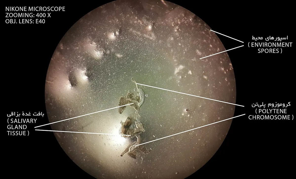
c. Chromomeres. Along the chromonema lie dark, bead-like swellings, the chromomeres: regions where the DNA is folded more tightly. They can be seen in interphase in some cells and early in prophase, and are especially clear in polytene chromosomes, the giant chromosomes of the salivary glands of fly larvae, which have copied their DNA many times without dividing, so that hundreds of copies lie side by side and their chromomeres line up as bands. At metaphase the chromosomes are so tightly coiled that the chromomeres can no longer be seen.
d. Centromere (primary constriction). Each chromosome has a narrow region, the centromere or primary constriction, where the two sister chromatids are joined. Its position is fixed for each chromosome. It differs in structure and function from the rest of the chromosome, and it works during cell division: on each side of it is a disc-shaped plate of protein in three layers, the kinetochore, to which the spindle fibers attach.
By the position of the centromere, chromosomes are of four types (Figure 2.30):
- Telocentric: the centromere at one end; one arm only.
- Acrocentric: the centromere very near one end; one very short and one long arm.
- Submetacentric: the centromere a little off the middle; arms of unequal length.
- Metacentric: the centromere in the middle; two equal arms.
e. Nucleolar organizer (secondary constriction I). In one or more chromosomes of a set, a narrow region near one end, a secondary constriction, carries the genes for rRNA. In interphase the nucleolus forms round it, so it is called the nucleolar organizer.
f. Secondary constriction II. Besides the centromere and the nucleolar organizer, the arms of chromosomes may show one or more other narrow places, called secondary constrictions II to tell them apart from the nucleolar organizer. They are places where the chromosome often breaks and rejoins, and they help to identify a chromosome.
g. Satellite. The part of a chromosome beyond its nucleolar organizer, a round or oblong knob joined to the rest by a thin thread, is the satellite. It keeps its shape and size. Chromosomes with satellites are called SAT chromosomes. The name was first read as an abbreviation of sine acido thymonucleinico, “without thymonucleic acid” (DNA), because the thread joining the satellite stains so weakly; in fact both the satellite and its thread contain DNA, like the rest of the chromosome.
h. Telomeres (Greek telos, end, and meros, part). The two ends of each chromosome are its telomeres. They are made of a short sequence of DNA repeated thousands of times (TTAGGG in humans), with special proteins, and they protect the ends: they stop the chromosomes from sticking to one another and from being treated as broken DNA. They get a little shorter each time a cell divides, which limits how often most body cells can divide.
Functions of chromosomes:
- Their DNA is the genetic material, carrying the genes from cell to cell and from parents to offspring.
- Through their genes they control the making of structural and enzyme proteins, and so the cell’s work and the organism’s characters.
- Changes in their DNA or structure are mutations, a source of new variation.
- Their exact sharing at mitosis keeps all body cells alike, and their halving at meiosis keeps the number constant from generation to generation (Unit 3).
A chromosome whose centromere lies near its middle, giving two arms of nearly equal length, is:
Pick an answer to see why.
16. Cell inclusions
As a result of the cell’s metabolism, several non-living substances collect in its cytoplasm. These metabolically inactive substances, such as stored proteins, amino compounds, starch grains, oils and fats, essential oils, latex, mineral crystals, inulin, sugars and pigments, are called cell inclusions, deutoplasmic bodies or ergastic substances. Unlike organelles, they are not alive and have no membrane of their own (though many lie in vacuoles or plastids).
Cell inclusions are of three kinds:
- Reserve materials
- Secretory materials
- Excretory materials
1. Reserve materials. The products of metabolism stored by a cell as reserve food, solid or dissolved:
- a. Nitrogenous reserve food, which contains nitrogen as well as carbon, hydrogen and oxygen: proteins, stored as solid grains (aleurone grains in seeds), and amino acids, in solution.
- b. Non-nitrogenous reserve food, of carbon, hydrogen and oxygen only, usually with hydrogen and oxygen in the ratio 2 : 1, as in water: the carbohydrates, such as starch, sugars (in the cell sap of sugarcane and beet), inulin (in the roots of dahlia), and glycogen (in fungi and animals), some soluble in water and some not, all ultimately products of photosynthesis. Oils and fats (in seeds) are non-nitrogenous reserves too, though with much less oxygen.
2. Secretory materials. Substances the cell makes for a use other than food:
- a. Pigments: colored substances such as the chlorophylls (a, b and c), carotene, lutein and flavoxanthin, fucoxanthin, phycocyanin and phycoerythrin.
- b. Enzymes: water-soluble biological catalysts, proteins and so nitrogenous.
- c. Nectar: a sugary liquid secreted by glands called nectaries, which attracts pollinators.
3. Excretory materials. By-products of metabolism of no further use to the plant, which it stores out of the way, since plants have no excretory organs:
- a. Alkaloids: bitter compounds of carbon, hydrogen, oxygen and nitrogen, often combined with organic acids, many of them poisonous or used as medicines: quinine (from the bark of Cinchona), nicotine (tobacco), morphine (opium poppy), atropine (belladonna) and daturine (Datura, धतुरो).
- b. Organic acids: citric (lemon), tartaric (tamarind, grape), oxalic and malic (apple) acids.
- c. Gums: substances formed by the breakdown of cell walls, which swell in water into a sticky mass (gum arabic from Acacia).
- d. Resins: solid, brittle, non-volatile complex substances in many plants, especially conifers such as pine (from whose resin turpentine and rosin are made).
- e. Tannins: complex compounds without nitrogen, bitter and astringent, common in unripe fruits, bark and tea leaves, dissolved in the cell sap of parenchyma cells.
- f. Essential oils: volatile, scented oils in fruits, flowers and glandular leaves, such as those of lemon, Eucalyptus, mint and tulsi.
- g. Latex: a milky emulsion of fine particles in a watery fluid, in which starch grains, sugars, proteins, enzymes and alkaloids may be dissolved; found in the latex cells of plants such as fig, papaya and the rubber tree, from whose latex rubber is made.
- h. Mineral crystals: crystals of calcium oxalate (as needles, raphides, in the leaves and corms of Colocasia, pindalu, which is why they itch in the throat if not cooked properly), calcium carbonate (cystoliths, in the leaves of fig) and silica (in grasses).
Which of these is a cell inclusion, not a cell organelle?
Pick an answer to see why.
Exercise
A. Tick the correct alternatives
- Which of the following is NOT a characteristic of prokaryotic cells?
Pick an answer to see why.
- Which of the following organelles is not membrane-bound?
Pick an answer to see why.
- How would you describe the property of the cell membrane?
Pick an answer to see why.
- What term describes the protoplasmic connection between two cells?
Pick an answer to see why.
- According to the fluid mosaic model, which type of proteins are embedded within the phospholipid bilayer?
Pick an answer to see why.
- Which process involves the expulsion of large-sized materials from the cell through the cell membrane?
Pick an answer to see why.
- What enzyme is responsible for the synthesis of ATP in mitochondria?
Pick an answer to see why.
- Chromosomes with the centromere in the middle, giving two arms of equal length, are classified as:
Pick an answer to see why.
- What was taken to be the primary function of mesosomes in prokaryotic cells?
Pick an answer to see why.
- Which type of ribosome is found in the cytoplasm of eukaryotic cells?
Pick an answer to see why.
- What type of endoplasmic reticulum (ER) is primarily involved in lipid synthesis?
Pick an answer to see why.
- Which structure in the Golgi complex is responsible for modifying, packaging and transporting proteins and lipids?
Pick an answer to see why.
- Which of the following is NOT a function of lysosomes?
Pick an answer to see why.
- Which pigment gives chloroplasts their green color?
Pick an answer to see why.
- Which type of plastid is involved in photosynthesis?
Pick an answer to see why.
B. Answer the following questions
Why is the cell considered as a basic unit of life?
Answer
What type of cell lacks a well-organized nucleus?
Answer
What type of ribosomes do prokaryotic cells have?
Answer
What is the primary composition of a plant cell wall?
Answer
What is the powerhouse of the eukaryotic cell?
Answer
Which type of protein is involved in transporting substances across the cell membrane?
Answer
Name the type of plastid that is colorless.
Answer
What pigment do phaeoplasts contain?
Answer
Which plastid is responsible for storing lipids?
Answer
Where are chromoplasts typically found?
Answer
What are the two main types of endoplasmic reticulum?
Answer
Name the primary function of Rough Endoplasmic Reticulum (RER).
Answer
What is the primary role of the Golgi complex in the cell?
Answer
What are the two types of ribosomes based on their sedimentation coefficient?
Answer
What do you mean by cell inclusions?
Answer
Write a brief note on the fluid mosaic model.
Answer
What is cyclosis?
Answer
Why is the mitochondrion called the powerhouse of the cell?
Answer
Name two semi-autonomous organelles.
Answer
Which organelle is called the “suicidal bag”? Why is it called so?
Answer
What is the basic difference between a ribosome and a mitochondrion?
Answer
Draw well-labeled sketches of the prokaryotic and eukaryotic cells and point out their differences.
Answer
Give an account of the structure and function of the mitochondrion.
Answer
Differentiate between leucoplasts and chromoplasts.
Answer
List two characteristics of prokaryotic cells.
Answer
What are mesosomes in prokaryotic cells, and their function?
Answer
Describe the composition of the primary cell wall in plants.
Answer
What is the fluid between the plasma membrane and the nuclear envelope in eukaryotic cells called?
Answer
Name the model that describes the plasma membrane as a mosaic of proteins in a fluid phospholipid bilayer.
Answer
List the three types of plastids.
Answer
What are the main components of a chloroplast’s structure?
Answer
Explain the function of pyrenoids in algal chloroplasts.
Answer
What types of pigments are found in chromoplasts?
Answer
Describe the structure of the endoplasmic reticulum.
Answer
What distinguishes the Smooth Endoplasmic Reticulum (SER) from the Rough Endoplasmic Reticulum (RER)?
Answer
Explain the process of autophagy in lysosomes.
Answer
What is the significance of the nucleolus within the nucleus?
Answer
How does the Golgi complex contribute to the formation of primary lysosomes?
Answer
Write briefly about the structure of the nucleus.
Answer
a. Name the different types of plastids and describe their structure and function.
b. Describe the structures and functions of any two major cytoplasmic organelles of the eukaryotic cell.
Answer
Describe the structure of a nucleotide based on the diagram of a part of a DNA given below.
Answer
Below is an image of the nucleus with labeled peripheral structures. Refer to the image and answer the following questions.
a. Identify the labeled parts A, B, C, D and E.
b. Write their functions.
Answer
Printable PDFs
- Study notes 6 pages
- Worksheet 5 pages Answer key
Quiz yourself: Living Things and Their Structure, Grades 5–12 →
Practice every question in the book, and keep track of the ones you miss →
A companion to your textbook, not a replacement for it: study from the book your school uses, and where the two differ, follow your teacher. Found a mistake? Please tell us.