
You began as one cell, a fertilized egg (a zygote) smaller than the full stop at the end of this sentence. Today you are made of roughly 30 to 40 trillion cells. They are not all alike: some are long and thin and carry messages, some are flat like floor tiles and cover you, some contract, some are full of fat. How does one cell become a body that can run, think, digest dal bhat and blush?
The answer is teamwork at every level. Cells of one kind work together as a tissue. Several tissues make an organ, like the heart. Organs that work together make an organ system, like the circulatory system. All the systems together make an organism: you.
Cell and tissue: Servier Medical Art (CC BY 4.0); heart and body: BodyParts3D (CC BY 4.0)
A one-celled (unicellular) living thing, such as an amoeba, does everything inside its single cell: it feeds, breathes, gets rid of wastes and divides. In a multicellular organism the work is shared out. All the activities that keep a living thing alive and let its kind continue, such as nutrition, respiration, photosynthesis (in plants), circulation, excretion and reproduction, are called life processes. The bigger and more complex the animal, the more its organs and systems are specialized to carry out these processes.
In this unit you will meet the tissues of plants and animals, the nervous system that carries messages in a flash, the glands that send slower chemical messages called hormones, and the hormones of plants, which make a seedling bend towards your window.
5.1 Tissue
Every living body is built of small units called cells. A cell is where the work of life actually happens: food is broken down for energy, new materials are made, wastes are formed. That is why the cell is called the structural and functional unit of life.
In a unicellular organism, one cell does every life process: respiration, nutrition, excretion, movement and reproduction. In a multicellular organism, groups of cells specialize. A group of similar cells (sometimes with a few different ones among them) that come from the same origin and work together to do one particular job is called a tissue. The branch of biology that studies tissues is histology.
There are two groups: plant tissues and animal tissues.
Which is the correct order, from the smallest to the largest?
Pick an answer to see why.
Plant tissue
Peel the thin skin from the inside of an onion scale and look at it under a microscope: you see neat rows of brick-like cells. Every part of a plant is built like this, of cells of the same or different kinds. These groups of cells are plant tissues. Depending on whether their cells can still divide, plant tissues are of two kinds: meristematic tissue and permanent tissue.
1. Meristematic tissue
Most cells in a plant stop dividing once they are mature. But some, in the growing parts, keep dividing all their lives. Making new cells by division is called cell division, and a tissue made of actively dividing cells is called a meristematic tissue or meristem (from the Greek meristos, divided).
Meristem cells are built for dividing:
- thin cell walls,
- a large nucleus and dense cytoplasm (few or no vacuoles),
- packed tightly together, with no spaces between the cells (no intercellular spaces).
Meristems are found in the growing regions of a plant, and their job is the growth and development of the plant. According to where they are, there are three kinds: apical, lateral and intercalary.
Mint: H. A. Köhler, Medizinal-Pflanzen (1887), public domain
Shoot tip: L. H. Bailey, Beginners' Botany (1909), public domain
a) Apical meristem
The apical meristem sits at the very tip of the stem and of the root (apex means tip). Its new cells make the stem and root longer, so the plant grows taller and its roots go deeper. If the tip of a stem is broken off, the stem stops growing in height.
b) Lateral meristem
The lateral meristem lies along the sides of the stem and root, in a ring parallel to their length, just inside the bark. The main one is called the cambium. Its new cells make the stem and root thicker (they increase the girth, or diameter). The rings you see on a cut tree trunk are the wood made by the cambium, one ring each year.
c) Intercalary meristem
The intercalary meristem is found at the base of the internodes, just above the nodes (a node is where a leaf grows from the stem; the part of the stem between two nodes is the internode). It makes the internodes longer. It is important in grasses, bamboo, maize and wheat.
Seedling: V. Rattan, A Popular California Flora (1888), public domain
Sugar cane: P. J. F. Turpin (c. 1820), public domain
A tree’s trunk becomes thicker year by year. Which meristem does this?
Pick an answer to see why.
2. Permanent tissue
When meristem cells stop dividing, they take on a fixed shape, size and job: they become permanent tissue. In general, cells of permanent tissue do not divide. Their walls may be thin or thick, and the cells may be living or dead.
There are three kinds of permanent tissue: simple permanent tissue, complex permanent tissue and special tissue.
(A) Simple permanent tissue
A simple permanent tissue is made of cells of one kind, and its structure is simple. Simple tissues protect and support the plant, and make and store food. By the structure of their cells, there are three kinds.
a. Parenchyma
- Living cells with thin walls, oval or rounded (sometimes elongated), with spaces between the cells.
- Found in the soft parts of the plant: the cortex and pith of stem and root, the flesh of leaves, flowers and fruits; also in xylem and phloem.
- Stores food (a potato is mostly parenchyma packed with starch).
- Chlorenchyma is parenchyma with chloroplasts, in leaves and green stems; it makes food by photosynthesis.
- Aerenchyma is parenchyma with large air spaces between the cells, in water plants such as lotus and water hyacinth; the trapped air helps them float.
b. Collenchyma
- Living cells whose walls are thickened at the corners with extra cellulose and pectin.
- Found just under the epidermis of young stems and in the leaf stalks (petioles).
- Gives the plant mechanical strength with flexibility: a young stem can bend in the wind without breaking.
- Some collenchyma cells have chloroplasts and make food too.
c. Sclerenchyma
- Dead cells with very thick walls, hardened by a substance called lignin; no protoplasm, only a narrow empty space (the lumen) in the middle.
- Found in stems, roots, the veins of leaves, the hard shells of nuts and seeds, and the husks of fruits.
- Two kinds: long, narrow, pointed fibers, and short, hard sclereids (stone cells).
- Makes the plant hard, strong and rigid, and lets it stand upright.
| Parenchyma | Collenchyma | Sclerenchyma | |
|---|---|---|---|
| Cells | living | living | dead |
| Wall | thin, cellulose | thickened at the corners | very thick, lignin |
| Spaces between cells | present | absent or few | absent |
| Main job | stores and makes food; helps water plants float | strength with flexibility | hardness and strength |
Which simple tissue is made of dead cells?
Pick an answer to see why.
(B) Complex tissue
A complex permanent tissue is made of more than one kind of cell, living and dead, working together as one unit. There are two: xylem and phloem. Because they conduct (carry) materials through the plant like pipes, they are called vascular (conducting) tissues.
a. Xylem
Xylem carries water and minerals from the roots up to every part of the plant. It has four kinds of cells:
- vessels: long tubes made of cells joined end to end, whose end walls have disappeared;
- tracheids: long, narrow cells with tapering ends and small thin areas (pits) in their walls;
- xylem fibers: thick-walled supporting cells;
- xylem parenchyma: living cells that store food.
Except for the xylem parenchyma, these cells are dead when they work: they are empty pipes and supports with thick, lignified walls. Xylem also makes the plant hard and strong; the wood of a tree is xylem. It runs through the inside of the root, stem and leaves (the veins).
b. Phloem
Phloem carries food (mainly sugar) made in the leaves to all the other parts of the plant, upwards and downwards: to the roots, the growing tips, fruits and seeds. It also has four kinds of cells:
- sieve tubes: tubes made of living cells joined end to end; the end walls between them, the sieve plates, are full of holes;
- companion cells: narrow cells beside the sieve tubes, with a nucleus, that keep the sieve tubes working;
- phloem fibers: thick-walled supporting cells (dead);
- phloem parenchyma: living cells that store food.
Except for the fibers, phloem is made of living cells. Phloem is also called bast, and its fibers give the plant some support (jute fibers are phloem fibers).
Plant cells: Encyclopaedia Britannica (1911), "Plants", public domain
A ring of bark is stripped from around a tree trunk. After some weeks the bark just above the ring swells. Why?
Pick an answer to see why.
(C) Special tissue
Some simple tissues are modified to make and give out (secrete) or store away (excrete) particular substances. These are special tissues, and there are two kinds.
a. Glandular tissue
Glandular tissue is made of groups of similar gland cells. Depending on the plant, it secretes enzymes, hormones, digestive juices, oils, resin, gum or nectar. Examples: the oil glands of tulsi (holy basil), whose oil gives it its smell; the glandular hairs of dhaturo; the resin that oozes from a pine tree’s bark.
b. Laticiferous tissue
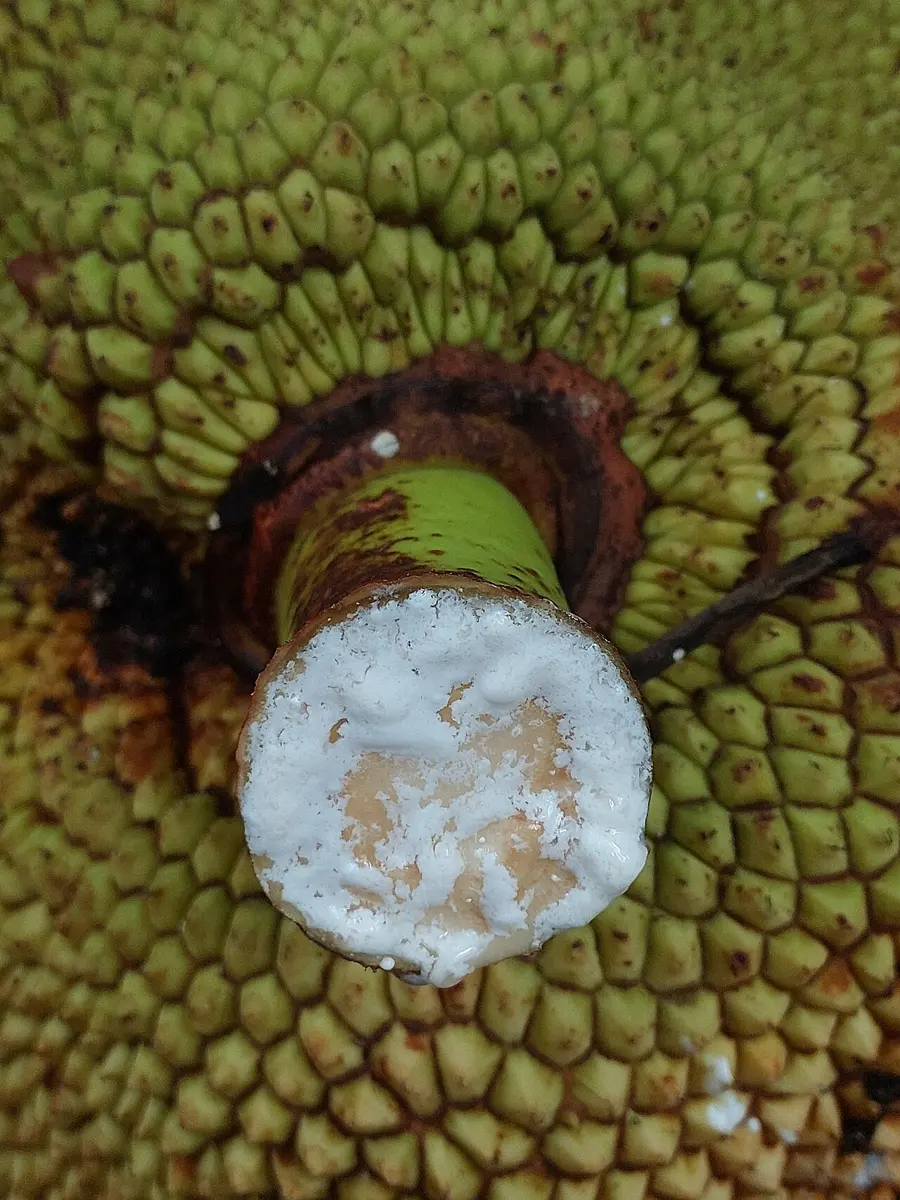
Laticiferous tissue (also spelled lactiferous) is made of special cells or tubes that contain a milky liquid called latex. Break a leaf or stem of these plants and white latex oozes out: pipal and other Ficus trees, lalupate (poinsettia), katahar (jackfruit), the rubber tree, and aank (Calotropis). Latex protects the plant: it seals wounds and many insects find it sticky or bitter.
Animal tissue
The tissues of animals are made of animal cells, and each kind does one main job. There are four basic kinds:
- epithelial tissue: covers and lines;
- muscular tissue: moves;
- connective tissue: joins and supports;
- nervous tissue: carries messages.
(A) Epithelial tissue
Epithelial tissue (epithelium) covers the outside of the body and lines the inside of organs, tubes and cavities. Its cells are packed closely together in sheets, and the sheet sits on a thin layer underneath called the basement membrane. Epithelium covers and protects, and in different places it also secretes, excretes and absorbs.
Epithelium is named by the shape of its cells. The main kinds are:
- Squamous (pavement) epithelium: thin, flat cells with many sides, fitted closely together like the paving stones of a courtyard, in a single layer. Its thinness lets substances pass through easily, so it lines the blood vessels and the air sacs (alveoli) of the lungs, and it covers the surfaces of organs such as the heart, liver and kidneys. Jobs: covering, protection, filtering and exchange. (The outer skin is many layers of squamous cells stacked up, which makes it tough.)
- Cuboidal (cubical) epithelium: a single layer of cube-shaped cells with round nuclei. It lines the tubules of the kidney, the follicles of the thyroid gland, and the ducts of glands such as the salivary and sweat glands. Jobs: secretion and absorption.
- Columnar epithelium: a single layer of tall, pillar-like cells, with the nuclei near the base. It lines the stomach, the intestine and the gall bladder. Jobs: secretion and absorption (in the intestine its cells absorb digested food). In the windpipe, the columnar cells carry tiny moving hairs called cilia, which sweep mucus, dust and germs up and out.
- Glandular epithelium: epithelial cells that make and pour out secretions: hormones, enzymes, mucus, saliva, sweat, digestive juices. Glands are made of it, often folded into pits or flasks that open through a duct.
Servier Medical Art (smart.servier.com), CC BY 4.0
Why are the walls of the lungs’ air sacs made of squamous epithelium?
Pick an answer to see why.
(B) Muscular tissue
Muscular tissue is made of long, thread-like cells called muscle fibers, which can contract (shorten) and relax. Its job is movement: of the whole body, and of the organs inside it. There are three kinds.
Servier Medical Art (smart.servier.com), CC BY 4.0
a. Skeletal muscle
- Long, cylindrical fibers, each with many nuclei.
- Light and dark stripes across them, so it is also called striped or striated muscle.
- Attached to the bones (by tendons, a connective tissue), so it moves the skeleton: arms, legs, jaw, eyes.
- Works when we want it to: it is voluntary muscle.
- Tires after hard work.
b. Cardiac muscle
- Found only in the wall of the heart.
- Striped like skeletal muscle, but its cells are branched, each with one nucleus, joined end to end by dark bands.
- Contracts and relaxes rhythmically all through life without tiring; each contraction is a heartbeat that pumps blood.
- Involuntary: you can’t stop or start your heart by wishing.
c. Smooth muscle
- Spindle-shaped cells (thick in the middle, pointed at both ends), each with one nucleus.
- No stripes, so it is called smooth or unstriped muscle.
- In the walls of the stomach and intestine (pushing food along), the ureters and bladder, the blood vessels, the uterus, and the iris of the eye.
- Involuntary: it can’t be controlled at will. Slow, steady and tireless.
| Skeletal | Cardiac | Smooth | |
|---|---|---|---|
| Shape of cells | long cylinders | branched | spindles |
| Nuclei | many per fiber | one per cell | one per cell |
| Stripes | yes | yes | no |
| Control | voluntary | involuntary | involuntary |
| Where | attached to bones | wall of the heart | stomach, intestine, blood vessels, ureter |
When you swallow, food moves down the food pipe and through the intestine without your thinking about it. Which muscle does this?
Pick an answer to see why.
(C) Connective tissue
Connective tissue connects and supports the other tissues and organs. Unlike epithelium, its cells are spread out, with a lot of material between them, the matrix. The matrix may be jelly-like, solid or liquid, and contains water, protein fibers and minerals. It is the matrix that makes bone hard and blood liquid. By their structure, connective tissues are of three kinds: soft, hard and fluid.
a. Soft connective tissue
The cells lie loosely in a matrix with plenty of fibers; some kinds store a lot of fat.
i. Fibrous connective tissue
Made of fibers, loosely or tightly packed.
- Loose (areolar) connective tissue is the simplest and most widespread. It lies in the dermis, joining the skin to the muscles below, and around blood vessels, nerves and muscles, filling the spaces between organs and holding them in place.
- Dense connective tissue has tightly packed fibers and is very strong. It forms the tough coverings around the kidneys, liver, glands and muscles, and two important cords:
- a tendon joins a muscle to a bone;
- a ligament joins a bone to a bone (at a joint); ligaments are a little elastic.
ii. Adipose connective tissue
A loose connective tissue with few fibers, whose cells are filled with fat. It is found under the skin and around organs such as the kidneys and heart. It stores energy, acts as a cushion that protects the organs inside, and keeps the body warm (fat is a poor conductor of heat).
b. Hard connective tissue
Bone and cartilage are the hard connective tissues: they make up the skeleton and support the body.
- Bone is very hard because calcium (as calcium phosphate) is laid down in its matrix, along with protein fibers that stop it from being brittle. Its living cells are called osteocytes, and the bone is wrapped in a thin membrane, the periosteum. The bones of the skull, limbs and backbone are made of strong, hard bone.
- Cartilage is firm but bendable; its cells are called chondrocytes. It is found in the tip of the nose, the outer ear, the larynx and windpipe in the neck, the discs between the vertebrae, and at the ends of long bones, where it reduces friction at the joints and absorbs shocks.
In an unborn baby, much of the skeleton is first made of cartilage; it slowly hardens into bone as calcium salts are laid down. That is why a baby is born with about 300 bones and soft spots in the skull, and an adult has 206: some bones fuse together as they harden.
c. Fluid connective tissue
Some connective tissues are liquid. They flow around the body, carry materials and link the systems together. There are two: blood and lymph.
i. Blood
Blood is a red, liquid connective tissue that carries materials from one part of the body to another. About 55 % of it is a pale yellow liquid, plasma, and 45 % is blood cells. Plasma is about 90 % water and 10 % dissolved substances; it carries digested food, enzymes, hormones and wastes. The blood cells are of three kinds:
- Red blood cells contain hemoglobin, which takes up oxygen in the lungs and carries it to all parts of the body; they also help carry carbon dioxide back to the lungs.
- White blood cells fight and destroy harmful germs (pathogens).
- Platelets help the blood to clot at a wound, so we don’t keep bleeding.
ii. Lymph
Lymph is a colorless liquid connective tissue that flows in thin tubes called lymph vessels. It forms from the liquid part of the blood: some plasma leaks out of the thin capillaries into the spaces between the cells, bathing them. Most of it goes back into the blood; the rest drains into lymph vessels and is called lymph. Lymph has no red blood cells and no hemoglobin; it has plasma and many white cells called lymphocytes. It protects the body against infection (the lymph nodes, which swell when you have a sore throat, are where germs are trapped), brings nutrients to the tissues, and carries away wastes.
(D) Nervous tissue
Nervous tissue receives messages, carries them and directs the organs of the body, so that they work together. It is made of nerve cells (neurons), the basic units of the nervous system, held in place, protected and fed by supporting cells called neuroglia (glial cells).
Neuron or nerve cell
A neuron has three main parts:
- the cell body (cyton), which holds the nucleus; cell bodies look gray (ash-colored);
- dendrites: short, branched threads that grow out from the cell body; they receive messages (from other neurons or from sense cells) and carry them towards the cell body;
- the axon: one long thread from the cell body that carries messages away from the cell body to the next neuron, a muscle or a gland; it ends in branched axon terminals.
Many axons are wrapped in a fatty, whitish myelin sheath (in the nerves of the body it is made by Schwann cells), with small gaps along it called nodes of Ranvier. The myelin insulates the axon and makes messages travel much faster. Because of the myelin, bundles of axons look white.
So a message always travels one way: dendrites → cell body → axon → axon terminals → next cell. When you stub your toe, a chain of neurons carries the message to your brain, and you feel the pain.
Pearson Scott Foresman, Public domain, via Wikimedia Commons
Which part of a neuron carries the message away from the cell body?
Pick an answer to see why.
Draw it: Meristem cells at a growing tip, Parenchyma cut across, Collenchyma cut across, Sclerenchyma cut across, A neuron
Exercise (5.1 Tissue)
1. Choose the correct answer
(i) Which kind of tissue has cells that can divide?
Pick an answer to see why.
(ii) Which tissue lines the tubules of the kidney and the ducts of the salivary glands?
Pick an answer to see why.
(iii) Where is meristematic tissue found in a plant?
Pick an answer to see why.
(iv) In which organ is cardiac muscle found?
Pick an answer to see why.
(v) What does a plant use its vascular tissue for?
Pick an answer to see why.
2. Give reasons
Blood is called a liquid (fluid) connective tissue.
Answer
A plant stops growing taller when its tip is cut off.
Answer
A white liquid comes out when a leaf of lalupate (poinsettia) is broken off.
Answer
Xylem is called a complex permanent tissue.
Answer
3. Write the differences
Muscular tissue and connective tissue
Answer
Xylem and phloem
Answer
Meristematic tissue and permanent tissue
Answer
Sclerenchyma and collenchyma
Answer
4. Answer the following questions
What is a life process? Describe it with examples.
Answer
Draw a concept map of the classification of plant tissues and animal tissues, with examples.
Answer
Explain meristematic tissue with a diagram.
Answer
What is permanent tissue? Describe its kinds briefly, with examples.
Answer
Draw neat, labeled diagrams of sclerenchyma, collenchyma and parenchyma, and list their functions.
Answer
Write the functions of epithelial tissue.
Answer
Prepare a chart of animal tissues and write where each is found.
Answer
Observe the figure and answer the questions.
Plant cells: Encyclopaedia Britannica (1911), "Plants", public domain
- i.Identify the figure.
- ii.Name the parts marked A, B, C and D.
- iii.Write the function of this tissue.
- iv.Write the importance of this tissue to plants.
Answer
Observe the figure and answer the questions.
Servier Medical Art (smart.servier.com), CC BY 4.0
- i.Name the muscular tissues shown.
- ii.Name one organ where each is found.
- iii.Write one function of each.
Answer
Study the figure and answer the questions.
Pearson Scott Foresman, Public domain, via Wikimedia Commons
- i.Identify the figure.
- ii.Name the parts a, b and c.
- iii.Write the function of a, b and c.
Answer
5. Project work
5.2 Human nervous system
How do you know that the dal is too hot, that a bus is coming, or that someone has called your name? And how does your stomach “know” to start digesting while your feet walk you home? Something must collect information, decide, and send orders, all the time.
We learn about the things around us through our sense organs (eyes, ears, nose, tongue, skin). The messages they pick up are carried by nervous tissue to the brain, which makes sense of them and decides what to do; then orders go out to the muscles and glands. A change around us that we react to is a stimulus (plural stimuli), and what we do is the response. The system of organs that does all this, the brain, the spinal cord and the nerves, is the nervous system. The sense organs are its outposts.
By its parts and their work, the human nervous system is divided into three parts:
- the central nervous system (CNS)
- the peripheral nervous system (PNS)
- the autonomic nervous system (ANS)
1. Central nervous system
The central nervous system is the part that runs down the middle of the body, from the head to the lower back. It is made of the brain and the spinal cord, and it is built of a huge number of neurons and their supporting cells. It is the control center: messages come in to it, and orders go out from it.
(a) Brain
The brain is the largest and topmost part of the central nervous system. It lies inside the skull (the cranium), which protects it. It controls and coordinates every part of the body. An adult brain weighs about 1.3 to 1.4 kg.
The brain is wrapped in three layers of membrane, the meninges. From the outside in:
- the dura mater, a tough layer stuck to the inside of the skull;
- the arachnoid, a thin layer with web-like threads (arachne is Greek for spider);
- the pia mater, a very thin layer that clings to the surface of the brain and follows all its folds.
The space between the arachnoid and the pia mater is filled with a clear, watery liquid, the cerebrospinal fluid (CSF). It contains water, minerals, glucose and a little protein, and is slightly alkaline. CSF protects the brain like a water cushion (the brain floats in it, so a bump on the head is softened), and brings it nutrients. The same meninges and CSF surround the spinal cord.
Servier Medical Art (smart.servier.com), CC BY 4.0
The main parts of the brain are the cerebrum, the cerebellum and the medulla oblongata.
Brain: Patrick J. Lynch (CC BY 2.5), recolored
(i) Cerebrum
The cerebrum (the large brain) is the largest part of the brain: about four-fifths of it. A deep groove from front to back divides it into a left and a right cerebral hemisphere (half-spheres). Its surface is folded into ridges and grooves (convolutions), which pack a much bigger surface into the skull. The outer layer is gray matter (cell bodies of neurons); inside it is white matter (axons with myelin).
The cerebrum is the seat of thinking and awareness. It:
- recognizes smells, and makes sense of what we see, hear, taste and touch;
- controls speech, memory and hearing;
- creates consciousness, thought, feelings and imagination;
- lets us analyze, put ideas together, predict and decide;
- controls our voluntary movements, and the other parts of the brain.
If the cerebrum is badly injured, a person may lose consciousness and fall into a coma, lose memory or imagination, or lose the power to coordinate and control movements.
(ii) Cerebellum
The cerebellum (the small brain) is the second largest part, about a tenth of the brain’s volume. It lies at the back, below the cerebrum and above the medulla oblongata, in two rounded halves. Like the cerebrum, it has gray matter outside and white matter inside, and fine parallel folds. It:
- coordinates muscular movements, getting their timing right;
- keeps the body balanced (in equilibrium) while we walk, run or ride a bicycle;
- helps control voluntary movements, making them smooth and accurate;
- keeps the muscle tone and posture of the body.
If the cerebellum is injured, a person’s movements become shaky and they lose their balance. Alcohol affects the cerebellum, which is why a drunk person staggers and can’t walk in a straight line.
(iii) Medulla oblongata
The brainstem joins the brain to the spinal cord, and has three parts: the midbrain, the pons and the medulla oblongata. The midbrain and pons act as bridges, passing messages between the spinal cord and the higher parts of the brain.
The medulla oblongata is the lowest part of the brain, between the pons and the spinal cord. It is shaped like a short stalk or tube. It passes out of the skull through a large opening in its base, the foramen magnum, and continues as the spinal cord. It controls the involuntary actions that keep us alive:
- it regulates breathing and the heartbeat;
- it controls vomiting, coughing, sneezing and swallowing;
- it controls the narrowing and widening of blood vessels, and so the blood pressure;
- it controls the flow of saliva and digestive juices.
Because breathing and the heartbeat depend on it, a serious injury to the medulla oblongata can kill a person almost at once.
A man has had too much alcohol. He can still talk and think, but he sways and can’t walk along a straight line. Which part of his brain is affected most?
Pick an answer to see why.
(b) Spinal cord
The spinal cord is a long, soft, jelly-like cord that runs down through the canal inside the backbone (vertebral column), which protects it. It starts at the bottom of the medulla oblongata and ends at about the first or second lumbar vertebra in the lower back. It is about 45 cm long and roughly 1–2 cm thick, and is covered by the same meninges as the brain, with CSF around it.
Unlike the brain, the spinal cord has white matter outside and gray matter inside (the gray matter is shaped like a butterfly, or the letter H). A tiny central canal full of CSF runs down its middle.
The spinal cord does two jobs:
- it carries messages between the brain and the body, up and down;
- it is the center for most reflex actions (see below).
Bones: BodyParts3D (CC BY 4.0); cord cut across: Polarlys (CC BY 2.5)
Nerve fibers
A nerve is a bundle of nerve fibers (axons), wrapped like the wires in a cable. Nerves carry messages to and from the brain and spinal cord. By their job, nerve fibers are of three kinds:
- Sensory (afferent) nerves carry messages from the receptors (the sense organs) to the brain or spinal cord.
- Motor (efferent) nerves carry orders from the brain or spinal cord to the muscles and glands, the effectors.
- Connecting neurons (interneurons, relay neurons) inside the brain and spinal cord link sensory neurons to motor neurons, passing the message from one to the other.
Which nerve carries an order from the spinal cord to the muscle of your arm?
Pick an answer to see why.
2. Peripheral nervous system
The peripheral nervous system (PNS) is made of all the nerves that branch out from the brain and the spinal cord to every part of the body (periphery means the outer edge). It links the central nervous system with the sense organs, muscles and glands. By where the nerves start, there are two kinds:
- Cranial nerves arise from the brain. There are 12 pairs. They connect the brain with the organs of the head and face, such as the eyes, ears, nose and tongue (one pair, the vagus nerve, goes further, down to the heart, lungs and stomach).
- Spinal nerves arise from the spinal cord. There are 31 pairs. Each carries messages both ways between the spinal cord and a part of the body: sensory fibers come in through the back (dorsal) root, and motor fibers go out through the front (ventral) root.
Reflex action
You slip on a wet path and your hands shoot out to catch you before you even think “I’m falling”. You touch a hot pan and your hand jerks away. Dust flies towards your eye and you blink. These are reflex actions: sudden, automatic, involuntary responses to a stimulus, made without thinking about them. They protect us from harm, and because they don’t wait for the brain to decide, they are very fast. Most reflex actions of the body are controlled by the spinal cord.
The path a message takes in a reflex action is called a reflex arc. For touching a hot object:
- Receptor: nerve endings in the skin of the finger sense the heat (the stimulus) and start a nerve impulse.
- Sensory nerve: carries the impulse from the receptor to the spinal cord.
- Connecting neuron (interneuron): inside the gray matter of the spinal cord, links the sensory nerve to a motor nerve.
- Motor nerve: carries the impulse from the spinal cord to the muscle.
- Effector: the muscle (here, the biceps of the upper arm) contracts and pulls the hand away.
Receptor → sensory nerve → spinal cord (connecting neuron) → motor nerve → effector. At the same time, a message goes up the spinal cord to the brain, and you feel the pain and know what happened, but only after your hand has already moved.
3. Autonomic nervous system
Some organs work day and night without our control: the heart, the lungs, the kidneys, the stomach and intestine, the glands. The part of the nervous system that controls these involuntary actions, whether we are asleep or awake, is the autonomic nervous system (ANS) (autonomic means self-governing). It controls smooth muscle, cardiac muscle and many glands. It has two parts, which work against each other, like an accelerator and a brake:
- Sympathetic nervous system: gets the body ready to face an emergency: to fight or to run (“fight or flight”). It speeds up the heartbeat and breathing, raises the blood pressure, widens the pupils and sends more blood and sugar to the muscles, so the body has more energy to protect itself. It slows digestion, which can wait.
- Parasympathetic nervous system: calms the body down once the danger has passed and brings the heartbeat, blood pressure and breathing back to normal. It runs the body’s quiet, everyday work, such as digestion (“rest and digest”).
| Sympathetic nervous system | Parasympathetic nervous system |
|---|---|
| Prepares the body for an emergency. | Calms the body after an emergency; keeps up normal work. |
| Speeds up the heartbeat; raises blood pressure. | Slows the heartbeat; lowers blood pressure. |
| Faster breathing. | Slower breathing. |
| Widens (dilates) the pupils. | Narrows the pupils. |
| Slows digestion; less saliva (dry mouth). | Speeds up digestion; more saliva. |
Draw it: The human brain from the side
Exercise (5.2 Nervous system)
1. Choose the correct answer
a. Which is the right order of the meninges, from outside to inside?
Pick an answer to see why.
b. Which part of the nervous system is ash-colored (gray)?
Pick an answer to see why.
c. Injury to which part of the brain can leave a person in a coma?
Pick an answer to see why.
d. Which nerve carries messages from the parts of the body to the brain?
Pick an answer to see why.
e. Which part of the nervous system controls reflex actions?
Pick an answer to see why.
2. Give reasons
A person may go into a coma when the large brain (cerebrum) is injured.
Answer
The hand is pulled away at once when a finger touches a hot object.
Answer
The body loses its balance when the cerebellum is injured.
Answer
The spinal cord is called the center of reflex action.
Answer
3. Write the differences
Cranial nerves and spinal nerves
Answer
Sensory nerves and motor nerves
Answer
Cerebrum and cerebellum
Answer
Sympathetic and parasympathetic nervous system
Answer
4. Answer the following questions
Draw a neat diagram of the brain and label its parts.
Answer
What are the functions of the medulla oblongata? Explain.
Answer
What is the work of the autonomic nervous system? Describe it with a diagram.
Answer
Describe the reflex action that happens when we touch a hot object.
Answer
What is the nervous system? Describe its kinds briefly.
Answer
Write the functions of the small brain, the large brain and the spinal cord.
Answer
What is a neuron? Draw a neat diagram, label its parts and write the function of each part.
Answer
Describe reflex action, with examples.
Answer
Study the diagram and answer the questions.
Brain: Patrick J. Lynch (CC BY 2.5), recolored
- i.Name the parts marked A, B and C.
- ii.Write the functions of A, B and C.
- iii.What happens if part C is injured?
Answer
5.3 Human glandular system
You hear a loud bang at night. In a moment your heart is pounding, and it keeps pounding for minutes after you know it was only a firecracker. A nerve message is over in a flash; what keeps the heart racing is a chemical carried in your blood. Chemicals like this are made by glands.
A gland is a cell or group of cells (an organ) that makes a useful substance and pours it out (secretes it): hormones, enzymes, and juices such as sweat, saliva and tears. These secretions control many activities and keep the body healthy, helping in digestion, reproduction, excretion and growth. All the glands of the body together make up the glandular system. By how they deliver their secretion, glands are of two main kinds: exocrine and endocrine.
A) Exocrine glands
An exocrine gland pours its secretion through a duct (a small tube) to the place where it is needed. Examples: sweat glands (onto the skin), salivary glands (into the mouth), tear glands (onto the eyes), the liver (bile into the intestine). They are also called duct glands. Their secretions do important work in digestion and excretion; many contain enzymes, which act as biological catalysts: they speed up chemical reactions in the body, such as breaking down food, without being used up.
B) Endocrine glands
An endocrine gland has no duct (it is a ductless gland). It releases its secretion straight into the blood, which carries it all over the body. This secretion is called a hormone. A hormone is made in one place but works in another, often far away: it stimulates and regulates particular body cells, organs and other glands. That is why hormones are called chemical messengers. The pituitary, thyroid, parathyroid and adrenal glands are endocrine glands, and together the endocrine glands make up the endocrine system.
Too much or too little of a hormone causes problems: it can upset growth and development, weaken the power to reproduce, or cause disease.
Mixed glands
Some glands do both: they release enzymes through a duct and hormones into the blood. They are called mixed glands (both exocrine and endocrine). The pancreas is the main example: it sends digestive juice down a duct to the intestine, and the hormones insulin and glucagon into the blood. The gonads (testes and ovaries) are also often counted as mixed glands: they release sex cells (sperm, ova) through tubes and hormones into the blood.
Body and glands: BodyParts3D (CC BY 4.0)
Functions of hormones
- They bring about physical, mental and emotional growth and development.
- They stimulate and control various organs.
- They control the production of sperm and ova (egg cells).
- They develop the sexual characters (the changes of puberty).
- They control the power to reproduce.
- They keep the calcium and phosphorus in the body balanced.
- They keep the level of sugar in the blood steady.
| Gland | Where it is | Hormone(s) | Main job |
|---|---|---|---|
| 1.Pituitary | at the base of the brain | growth hormone, stimulating hormones | growth; controls other glands |
| 2.Thyroid | in the neck, on the windpipe | thyroxine (also T₃ and calcitonin) | rate of body chemistry (metabolism), growth |
| 3.Parathyroid | behind the thyroid, in the neck | parathormone | raises calcium in the blood |
| 4.Adrenal | on top of each kidney | adrenaline, cortisol | emergencies, stress |
| 5.Pancreas | behind the stomach, in the loop of the duodenum | insulin, glucagon | blood sugar |
| 6.Gonads: testes | in the scrotum (men) | testosterone | sperm; male characters |
| Gonads: ovaries | in the lower abdomen (women) | estrogen, progesterone | ova; female characters |
Pituitary gland
The pituitary gland is about the size of a pea. It hangs from the base of the brain, inside the skull. It makes growth hormone and several stimulating hormones, which control the thyroid, adrenal glands and gonads. Because it coordinates and controls the other endocrine glands, it is called the master gland. (The pituitary itself takes orders from the part of the brain just above it, the hypothalamus.)
Growth hormone controls the growth of the body. If a child makes too little of it, the child grows up very short: dwarfism. If a child makes too much, the child grows unusually tall: gigantism.
Thyroid gland
The thyroid gland is shaped like a butterfly: two lobes, one on each side of the windpipe in the neck, joined by a thin band of tissue. It makes three hormones: thyroxine (T₄), triiodothyronine (T₃) and calcitonin.
Thyroxine controls how fast the body’s cells use food and oxygen (the metabolism), and it is needed for normal physical and mental growth. About two-thirds (65 %) of a thyroxine molecule by mass is iodine, so our food must contain iodine; that is why the salt sold in Nepal has iodine added (iodized salt). If there is too little iodine, the thyroid swells up: a goiter.
- Too little thyroxine: tiredness, weakness of body and mind, dry skin, little sweating, slow and slurred speech, a hoarse voice. In babies and children it slows growth and mental development.
- Too much thyroxine: the metabolism speeds up: heavy sweating, great hunger but loss of weight, nervousness and restlessness, and bulging eyes.
Calcitonin lowers the calcium in the blood by laying it down in the bones.
Parathyroid glands
There are four small parathyroid glands, at the back of the thyroid. They make parathormone, which moves calcium between the bones and the blood and raises the calcium level in the blood.
- Too much parathormone (usually because of a small tumor in one of the glands): too much calcium is taken from the bones into the blood; the bones become weak, and kidney stones may form.
- Too little: the calcium in the blood falls and the muscles go into painful cramps and spasms: tetany.
Adrenal glands
An adrenal gland sits on top of each kidney, like a cap. It makes adrenaline and cortisol (called cortisone in many books), along with a few other hormones.
- Adrenaline prepares the body to face a sudden emergency or danger: it makes the heart beat faster, raises the blood pressure and releases sugar into the blood. It works hand in hand with the sympathetic nervous system. Too much adrenaline raises the blood pressure.
- Cortisol helps the body cope with stress and controls the use of sugar; the adrenals also make small amounts of sex hormones.
When the adrenal glands make too little of their hormones, a person feels weak, the blood pressure and blood sugar fall, and they may feel sick and vomit.
Pancreas
The pancreas is one of the largest glands of the body, about 12–15 cm long. It lies behind the stomach, in the loop of the duodenum (the first part of the small intestine). It is a mixed gland:
- as an exocrine gland, it makes pancreatic juice, full of digestive enzymes, which flows through a duct into the duodenum;
- as an endocrine gland, small clusters of cells in it (the islets of Langerhans) release two hormones into the blood: insulin and glucagon.
Insulin lowers the blood sugar: it lets the body’s cells take in glucose, and the liver store it. Glucagon raises the blood sugar: it makes the liver release its stored sugar. Because they do opposite jobs, they are called antagonistic hormones, and together they keep the blood sugar steady.
- Too little insulin: the glucose in the blood stays high. This is diabetes (diabetes mellitus). A person with diabetes feels very hungry and thirsty and passes urine often.
- Too much insulin: the blood glucose falls too low. The person feels anxious, shaky and sweaty, may become confused and faint, and in severe cases may fall into a coma, with damage to the brain.
Gonads
The testes in males and the ovaries in females are called the gonads.
- Testes: two oval glands in a pouch of skin, the scrotum. They make sperm and the hormone testosterone. Testosterone helps make sperm and brings the secondary sexual characters of boys at puberty: hair on the face, a deeper voice, growth of the sex organs and muscles.
- Ovaries: two oval glands in the lower abdomen of a woman, beside the uterus and near the fallopian tubes. They make ova (egg cells) and the hormones estrogen and progesterone, which help the ova to develop and bring the female sexual characters at puberty, and prepare the uterus for a baby.
Which gland is called the master gland?
Pick an answer to see why.
A boy’s mother forgot to buy iodized salt for years. Which gland is most likely to be affected?
Pick an answer to see why.
Nerves and hormones work together
The nervous system and the glands are the body’s two message systems. Nerves send fast electrical messages along fixed wires to particular muscles and glands; hormones send slower chemical messages through the blood to every cell that can read them. They are linked: the hypothalamus in the brain controls the pituitary, and so most of the other glands; and in a fright, sympathetic nerves tell the adrenal glands to pour out adrenaline, which keeps the body ready long after the nerve messages have stopped.
| Nervous system | Endocrine (glandular) system |
|---|---|
| Messages are electrical impulses along nerves. | Messages are chemicals (hormones) in the blood. |
| Very fast. | Slower. |
| Effects are short-lived. | Effects can last a long time (growth lasts years). |
| Go to particular muscles and glands. | Reach the whole body; only the target cells respond. |
Draw it: The endocrine glands, The pancreas
5.4 Plant hormone
Put a pot of seedlings on a windowsill, and in a few days they all lean towards the window. Plants have no nerves and no glands. So how does a plant “know” where the light is, and how does it bend towards it?
Plants, too, respond to changes in their surroundings: to light, to gravity, to chemicals and to touch. A shoot grows towards the light (phototropism); a root grows down, with gravity (geotropism); a climbing bean’s tendril coils around a stick it touches. They need special substances at special times and places to grow, to flower, to set fruit, to ripen it and to drop their leaves and seeds. These substances are plant hormones.
Plant hormones are not nutrients. They are made in tiny amounts by ordinary cells in particular parts of the plant (plants have no glands like ours), and they carry messages from one cell to another: they are chemical messengers, like animal hormones.
The growth of a plant depends on external (environmental) factors, such as light, water and temperature, and on internal factors. The most important internal factor is its hormones. The hormones that support and regulate the growth and development of a plant are called plant growth hormones (plant growth regulators). Examples: auxin, gibberellin, cytokinin, ethylene and brassinosteroids.
Functions of plant growth hormones
The main job of growth hormones is to regulate the growth and development of the plant. Most are made in the growing regions, such as the tips of the shoot and root.
- Auxin is made in the tip of the shoot. It moves down the stem and makes the cells grow longer. When light falls on a shoot from one side, auxin moves to the shaded side; those cells grow longer than the ones on the lit side, so the shoot bends towards the light. This is phototropism. Auxin from the tip also keeps the side buds from growing, so the plant grows tall rather than bushy: this is apical dominance (Activity 5.1).
- Cytokinin is found in large amounts in the root tips, embryos (young plants in seeds) and fruits. It makes cells divide, helps side buds to grow, and keeps leaves green and fresh for longer.
- Gibberellin makes stems grow long and helps seeds sprout; farmers use it to make grapes and other fruits bigger.
- Ethylene is a gas. It makes fruits ripen, and makes old leaves and ripe fruits fall off (abscission).
- Brassinosteroids help cells grow and divide and help the plant cope with stress.
Uses of plant growth hormones
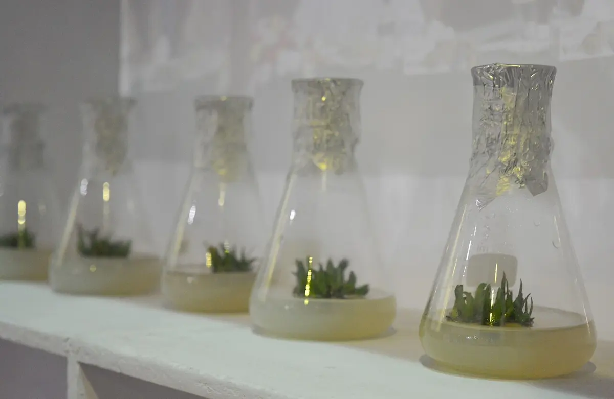
- Tissue culture. A few cells or a tiny piece of a plant are grown on a jelly of nutrients in a clean glass container, with auxin and cytokinin added to it. More auxin makes roots start; more cytokinin makes shoots grow; the right balance of the two gives fast, healthy growth. In this way, thousands of identical, disease-free plantlets (of bananas, potatoes, orchids, flowers) can be grown from one plant, in labs in Nepal too.
- Better and off-season crops. Hormones help farmers grow good-quality fruit and vegetables, and vegetables out of season: auxin helps cuttings take root and fruits set; gibberellin makes fruits bigger; ethylene ripens fruits evenly.
But hormones sprayed on vegetables and fruits in large amounts can harm the health of the people who eat them. They must be used only in the right amounts and at the right time, as experts advise.
Exercise (5.3 Glands and 5.4 Plant hormone)
1. Choose the correct answer
a. Which hormone controls the sugar level in the blood?
Pick an answer to see why.
b. Which hormone ripens fruit?
Pick an answer to see why.
c. Which hormone causes apical dominance?
Pick an answer to see why.
d. Which gland is called the master gland?
Pick an answer to see why.
e. Which hormone develops and controls the male sexual characters?
Pick an answer to see why.
f. Which hormone is involved in the ripening of fruit and the falling of leaves?
Pick an answer to see why.
2. Give reasons
The pancreas is called a mixed gland.
Answer
The pituitary gland is called the master gland.
Answer
A hormone is called a chemical messenger.
Answer
Glucagon and insulin work against each other.
Answer
An enzyme is called a biological catalyst.
Answer
3. Write the differences
Exocrine and endocrine glands
Answer
Cytokinin and auxin
Answer
Parathyroid and thyroid glands
Answer
4. Answer the following questions
What is a gland? Make a chart of its kinds, with examples.
Answer
What are plant hormones? Write about them.
Answer
Draw a diagram of the endocrine glands in the human body.
Answer
Describe the pituitary gland briefly.
Answer
List the plant hormones.
Answer
Write the uses of plant hormones.
Answer
Describe the pancreas, with a diagram.
Answer
What happens in the human body if too much of a hormone is made? Describe with examples.
Answer
Describe how the glandular system and the nervous system are related, with examples.
Answer
Observe the diagram and answer the questions.
Body and glands: BodyParts3D (CC BY 4.0)
- a.Name the glands marked A to F.
- b.Name the hormone made by each gland, with one function.
- c.Which hormone is responsible for tetany?
Answer
Worksheet: Body Structure and Life Process: tissues (PDF, 4 pages)
Worksheet: Body Structure and Life Process: nervous system, glands and plant hormones (PDF, 5 pages)
Quiz yourself: Life Processes and the Human Body, Grades 5–10 →
Practice every question in the book, and keep track of the ones you miss →
A companion to your textbook, not a replacement for it: study from the book your school uses, and where the two differ, follow your teacher. Found a mistake? Please tell us.