
Roshani’s grandfather has a painful knee. The doctor gives him a tablet, and he swallows it. A few hours later, the pain in his knee is less. But the tablet went into his stomach. How did the medicine find its way down to his knee?
The answer is a river that flows inside you, all the time, without ever stopping: your blood. It is pumped by a fist-sized muscle that has been beating since before you were born, about a hundred thousand times every day. In this unit you’ll follow that river: what blood is made of, how the heart pushes it, why blood groups matter when someone needs blood, what goes wrong in a heart attack, and what the two numbers of a blood pressure reading mean.
Blood circulation in the human body
Your body has several organ systems, each with its own job. The digestive system breaks food down into small, soluble pieces such as glucose. The respiratory system takes in oxygen. But the cells that need glucose and oxygen are everywhere: in your toes, your brain, the tips of your fingers. Something has to carry these things to every cell, and carry the wastes away. That is the job of the circulatory system.
It carries food, oxygen, water, hormones and medicines from one part of the body to another. Blood is made of blood cells (corpuscles) floating in a liquid called plasma, and it flows in one direction only, pushed by the pumping of the heart. The push of the blood on the walls of the arteries is the blood pressure, and you can feel it as your pulse.
Sometimes the blood pressure stays higher than normal, or fat and cholesterol build up on the walls of the arteries; both can damage the heart. Doctors today can open blocked arteries with angioplasty, or build a new route for the blood with bypass surgery. And not all blood is the same: there are four main blood groups, A, B, AB and O, and each can be Rh positive or Rh negative, so eight in all.
5.1 Human blood circulatory system
The blood circulatory system is one of the body’s organ systems, and it works closely with the others (digestive, respiratory, excretory…). It has three main parts:
- The heart: the pump. It collects blood from all over the body and pumps it out again.
- The blood vessels: the pipes (arteries, veins and capillaries) that carry blood to every cell.
- The blood: the liquid that does the carrying.
Blood carries digested food and the oxygen you breathe in to every cell. On the way back, it carries the carbon dioxide made by the cells to the lungs, and other wastes to the excretory organs (the kidneys, the skin).
5.2 Blood
A healthy adult has about 5 to 5.5 liters of blood, roughly 7 to 8 % of the body’s mass. Blood carries nutrients, oxygen and hormones to all the cells, and takes harmful wastes to the excretory organs.
Blood is slightly alkaline: its pH is 7.35 to 7.45. The body keeps it in this narrow range very carefully; if it moves outside it, a person becomes seriously ill.
Structure of blood
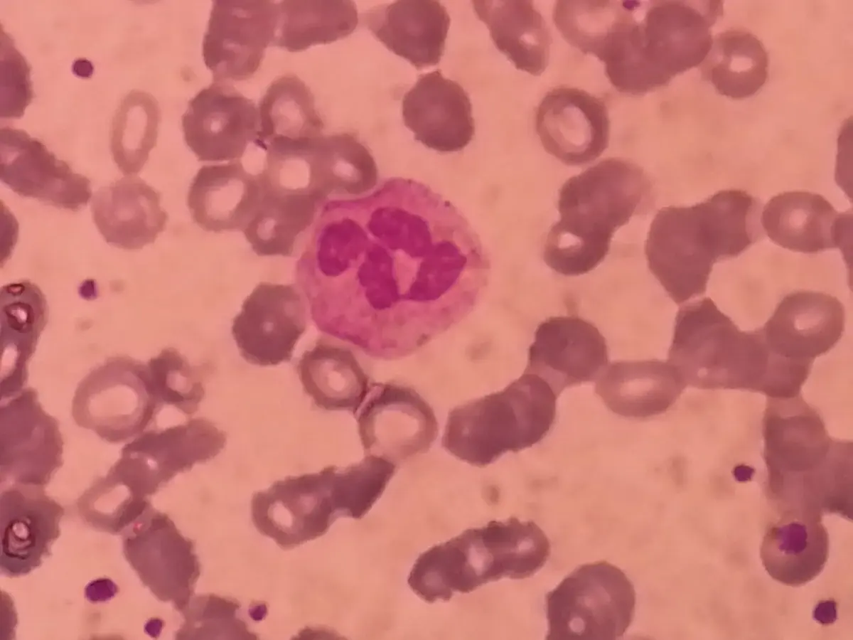
Blood looks like a red liquid, but it is a tissue: a connective tissue whose cells float in a liquid. About 55 % of it is the liquid, plasma, and 45 % is blood cells (also called blood corpuscles). There are three kinds of blood cells: red blood cells, white blood cells and platelets.
Plasma
Have you seen the blood collected when a goat is cut for a feast? After a while it sets into a dark red lump (a clot), and a pale, watery liquid separates around it. That liquid is close to plasma.
Plasma is a pale yellow, clear liquid, about 55 % of the blood. It is about 90 % water, with about 10 % dissolved substances: glucose, amino acids, fats, salts, hormones, wastes such as urea, and three important proteins: albumin, globulin and fibrinogen.
Functions of plasma
- It keeps the right amount of water in the blood.
- It carries digested food and wastes from one place to another.
- It carries enzymes and hormones made by glands to the parts of the body that need them.
- Its protein fibrinogen helps the blood to clot.
- It spreads heat around the body, keeping the temperature even.
- It keeps the chemical make-up and pH of the blood steady.
- Plasma with the fibrinogen removed is called serum. (The pale liquid around the goat’s blood clot is in fact serum: the fibrinogen has been used up making the clot.) Serum is tested in laboratories to diagnose many diseases.
What is serum?
Pick an answer to see why.
Blood corpuscles
a. Red blood cells
Red blood cells (erythrocytes, RBCs) are small, round discs, dented in on both sides (biconcave), and they have no nucleus. They are red because they are full of a red pigment called hemoglobin.
Hemoglobin is a protein (globin) joined to an iron-containing part (heme). The iron is what grabs oxygen.
- In the lungs, hemoglobin joins with oxygen to make oxyhemoglobin (bright red).
- In the body, it lets the oxygen go to the cells. Some of the cells’ carbon dioxide joins it to make carbaminohemoglobin, which is carried back to the lungs. (Most carbon dioxide, though, travels dissolved in the plasma.)
| About red blood cells | |
|---|---|
| Number | about 45 to 50 lakh (4.5 to 5 million) in each cubic millimeter of blood |
| Life | about 120 days |
| Made | in the red bone marrow of spongy bones, about 20 lakh every second |
| Destroyed | in the liver and spleen; their iron is kept and used again |
Making and destroying red cells goes on all through life, at the same rate, so the number stays steady.
- Too few red cells (or too little hemoglobin) is anemia. Less oxygen reaches the cells, so an anemic person gets tired and out of breath after even a short walk, and looks pale. Lack of iron in food is the commonest cause.
- Too many red cells is polycythemia. The blood becomes thick and flows slowly.
b. White blood cells
White blood cells (leucocytes, WBCs) have no fixed shape and are bigger than red cells. They have a nucleus and other cell parts, but no hemoglobin, so they are colorless.
They come in two kinds:
- Granular leucocytes have small grains (granules) in their cytoplasm: neutrophils, eosinophils and basophils.
- Non-granular leucocytes have none: lymphocytes and monocytes.
| About white blood cells | |
|---|---|
| Number | about 4,000 to 11,000 in each cubic millimeter of blood (one for every 500 or so red cells) |
| Life | from a few hours or days (most neutrophils) to many years (some lymphocytes) |
| Made | in the bone marrow (lymphocytes also grow up in the lymph nodes) |
| Destroyed | in the spleen, the liver, the lymph nodes, and at the site of an infection |
White blood cells fight germs that get into the body. Some swallow bacteria whole (neutrophils and monocytes do this); lymphocytes make antibodies, proteins that stick to a germ and mark it for destruction. That’s why they are called the soldiers of the body: they give us immunity.
- An abnormal fall in white cells is leucopenia: the person catches infections easily.
- Leukemia is a cancer of the blood. The bone marrow makes huge numbers of abnormal white cells that don’t work properly and crowd out the healthy red cells, white cells and platelets.
c. Platelets
Platelets (thrombocytes) are the smallest blood corpuscles: tiny, round or oval, with no nucleus. They are not complete cells but pieces broken off a giant cell in the bone marrow.
| About platelets | |
|---|---|
| Number | about 1.5 to 4.5 lakh in each cubic millimeter of blood |
| Life | about 7 to 10 days |
| Made | in the red bone marrow |
| Destroyed | in the spleen (and liver) |
Platelets stop bleeding. When you cut yourself:
- The cut blood vessel narrows, so less blood flows out.
- Platelets stick to the torn edge and to each other, making a plug.
- They release chemicals that, step by step, turn the soluble plasma protein fibrinogen into long threads of fibrin.
- The fibrin threads make a net that traps red cells: a clot. It dries into a scab, and the skin heals underneath.
Too few platelets, and even a small cut bleeds for a long time. Too many (thrombocytosis) can make clots form inside the blood vessels, where they may block an artery and cause a heart attack or a stroke.
A blood test shows that a girl has fewer red blood cells than normal. Which of these is she most likely to feel?
Pick an answer to see why.
Which blood cells are called the “soldiers of the body”?
Pick an answer to see why.
Functions of blood
- Transport. Blood carries oxygen from the lungs to the cells and carbon dioxide from the cells to the lungs. It also carries digested food, enzymes, hormones, medicines and wastes.
- Regulation. Blood spreads heat evenly, keeping the body’s temperature steady (that’s why your face goes red when you’re hot: more blood flows near the skin to lose heat). It also keeps the amounts of water and salts in the body steady.
- Protection. White blood cells fight germs, and antibodies in the blood keep us immune to diseases we’ve met before. Platelets stop bleeding from cuts and wounds.
Draw it: Blood cells
5.3 Blood group
On the surface of every red blood cell are special molecules called antigens. Two of them, antigen A and antigen B, decide a person’s ABO blood group:
| Blood group | Antigens on the red cells | Antibodies in the plasma |
|---|---|---|
| A | A | anti-B |
| B | B | anti-A |
| AB | both A and B | neither |
| O | neither A nor B | both anti-A and anti-B |
The last column is the important one for transfusions. The plasma carries antibodies against the antigens that your own cells don’t have. Anti-A antibodies stick to any red cell carrying antigen A, glue those cells together into clumps (agglutination), and destroy them.
Group O is the most common blood group in the world. (In Nepal, A and O are about equally common, each about a third of people.)
There is one more antigen that matters a lot: the D antigen, also called the Rhesus factor or Rh factor.
- If the red cells carry the Rh factor, the blood is Rh positive (+ve).
- If not, it is Rh negative (−ve).
So a person who is A+ve has antigen A and the Rh factor on their red cells. A person who is O−ve has none of the three: not A, not B, not D. With four ABO groups, each positive or negative, there are eight blood groups in all.
Your blood group is inherited from your parents, through the genes you met in Unit 4, and it never changes.
Testing a blood group
To find a blood group, a drop of blood is mixed with each of three antisera: anti-A, anti-B and anti-D (each is a liquid full of one kind of antibody). Where the antiserum finds its antigen, the blood clumps; where it doesn’t, the drop stays smooth.
Why the blood group must match
If a patient is given blood whose red cells carry an antigen the patient’s own cells don’t have, the patient’s antibodies attack those cells. They clump inside the blood vessels, burst, and block small vessels and the kidneys. This transfusion reaction can kill. So before every transfusion, the patient’s and the donor’s blood are tested and cross-matched to make sure they are compatible.
Rohan’s blood clumps with anti-B and anti-D, but not with anti-A. What is his blood group?
Pick an answer to see why.
A patient with blood group A+ve needs blood. Which of these donors is not safe?
Pick an answer to see why.
5.4 Heart
The heart is the central organ of the circulatory system: it pumps blood to every cell and tissue. It is made of cardiac muscle, a special muscle that contracts and relaxes rhythmically all your life without ever getting tired.
- Where: in the chest (the thoracic cavity), between the two lungs, behind the breastbone. It is tilted slightly to the left: about two-thirds of it lies left of the middle.
- Size and shape: about the size of its owner’s closed fist, cone-shaped (the pointed end, the apex, points down and to the left), and about 300 g in an adult.
- Covering: a double layer of membrane, the pericardium. The narrow space between the layers holds pericardial fluid, which lets the heart move without rubbing and protects it from shocks.
- Its own blood supply: the heart muscle works so hard that it needs its own blood vessels. The coronary arteries branch over its surface and bring oxygenated blood to the muscle; a large vein at the back, the coronary sinus, takes its deoxygenated blood back into the right auricle.
Heart: traced from BodyParts3D, © The Database Center for Life Science, CC BY 4.0
Inside the heart
The human heart has four chambers:
- the two upper ones, the auricles (also called atria): the right auricle and the left auricle;
- the two lower ones, the ventricles: the right ventricle and the left ventricle.
A thick muscular wall, the septum, runs down the middle and divides the heart into a right side and a left side. It keeps the deoxygenated blood of the right side from mixing with the oxygenated blood of the left side. You can think of the heart as two pumps side by side: the right pump sends blood to the lungs, the left pump sends it to the whole body.
Heart: traced from OpenStax College, Anatomy & Physiology, CC BY 3.0, via Wikimedia Commons
The auricles receive blood (from the body or the lungs); the ventricles send it out (to the lungs or the body). Pumping out needs much more force than receiving, so:
- the walls of the ventricles are thicker than those of the auricles;
- the wall of the left ventricle is the thickest of all, because it pumps blood to the whole body, from your head to your toes, while the right ventricle pumps only to the lungs next door.
The blood vessels of the heart
| Vessel | Joins | Carries |
|---|---|---|
| superior vena cava | upper body → right auricle | deoxygenated blood from the head, neck and arms |
| inferior vena cava | lower body → right auricle | deoxygenated blood from the trunk and legs |
| pulmonary artery | right ventricle → lungs | deoxygenated blood, to pick up oxygen |
| pulmonary veins (four) | lungs → left auricle | oxygenated blood |
| aorta | left ventricle → whole body | oxygenated blood; it is the largest artery |
The four valves
Blood must flow one way only. Four valves, flaps that open only in one direction like a one-way door, make sure it does.
| Valve | Where | Lets blood go |
|---|---|---|
| tricuspid valve (three flaps) | between the right auricle and right ventricle | right auricle → right ventricle |
| bicuspid or mitral valve (two flaps) | between the left auricle and left ventricle | left auricle → left ventricle |
| pulmonary valve | at the start of the pulmonary artery | right ventricle → pulmonary artery |
| aortic valve | at the start of the aorta | left ventricle → aorta |
The pulmonary and aortic valves are called semilunar valves (their three flaps are half-moon shaped).
One heartbeat, step by step
- Deoxygenated blood from the body pours into the right auricle through the superior and inferior vena cava. At the same time, oxygenated blood from the lungs pours into the left auricle through the pulmonary veins.
- Both auricles contract together. The tricuspid and bicuspid valves are open, and blood flows into the two ventricles. (In fact most of the blood flows down into the ventricles as soon as they relax, before the auricles squeeze; the squeeze tops them up.)
- Then both ventricles contract together. The tricuspid and bicuspid valves are pushed shut, so blood can’t go back into the auricles.
- In the right ventricle, the pressure opens the pulmonary valve, and deoxygenated blood goes up the pulmonary artery to the lungs.
- In the left ventricle, the pressure opens the aortic valve, and oxygenated blood goes out through the aorta to the whole body.
- The ventricles relax; the pulmonary and aortic valves shut, so the blood can’t fall back in. The auricles fill again, and it all repeats.
Why is the wall of the left ventricle thicker than that of the right ventricle?
Pick an answer to see why.
Which valve stops blood from flowing back from the right ventricle into the right auricle?
Pick an answer to see why.
Draw it: The human heart from the front, The inside of the human heart
5.5 Heart attack
Normally, blood keeps flowing into the heart from the body, out of the heart to the body, and through the coronary arteries into the heart muscle itself. But sometimes the flow of blood to part of the heart muscle is suddenly reduced or blocked. That part of the muscle gets no oxygen, and it starts to die within minutes. The heart can’t work properly, and the person feels great pain and difficulty. This is a heart attack, called myocardial infarction by doctors (myocardium: heart muscle; infarction: tissue dying for lack of blood). It is an emergency.
How it happens
Over many years, fat and cholesterol can build up inside the wall of a coronary artery. This fatty deposit is called plaque. It makes the artery narrower and stiffer, so less blood gets through. One day a plaque may crack. The body treats the crack like a wound: platelets rush in and a blood clot (a thrombus) forms on it. The clot can block the artery completely, and the heart muscle beyond it is starved.
Heart: traced from BodyParts3D, © The Database Center for Life Science, CC BY 4.0
Risk factors
These make a heart attack more likely:
- Age: men over 45 and women over 55 are at more risk than young people.
- Tobacco: smoking cigarettes and bidi, and chewing tobacco, gutkha and khaini, damage the arteries.
- High blood pressure (hypertension) for a long time.
- High cholesterol and triglycerides (fats) in the blood: “bad” cholesterol (LDL) builds up as plaque in the arteries.
- Diabetes: high blood sugar damages the walls of the arteries.
- Family history: a parent or grandparent who had a heart attack, especially at a young age.
- Little exercise and an unhealthy diet: too much trans fat (in reused frying oil, vanaspati ghee), salt, sugar, animal fat and processed foods.
- Stress: long-lasting anger, worry or negative thinking.
- Drugs such as cocaine and amphetamines, which can make the coronary arteries clamp shut.
Symptoms
Symptoms differ from person to person. The common ones:
- A sudden pain, pressure or tightness in the center of the chest, which may spread to the shoulders, arms (especially the left), neck, jaw, back or upper stomach.
- A cold sweat, tiredness, shortness of breath.
- Dizziness or light-headedness, nausea.
- Some heart attacks come suddenly. But most people have warning signs hours, days or weeks before: a discomfort or pressure in the chest that comes with effort and doesn’t go away with rest. It means the heart muscle is already short of blood. Never ignore it.
Preventing a heart attack
- Live a healthy, active life.
- Don’t smoke or chew tobacco; avoid alcohol.
- Keep a healthy body weight.
- Eat a balanced diet: more vegetables, fruit, whole grains and pulses; less salt, sugar and fried food.
- Exercise regularly: walking, cycling, games, at least 30 minutes on most days.
- Learn to manage stress.
- Treat and control high blood pressure and diabetes, which raise the risk.
- Have regular health check-ups, especially after the age of 40.
Diagnosis and treatment
To find out whether someone has had a heart attack, doctors first check the blood pressure, pulse and temperature, and then do tests of the heart:
- ECG (electrocardiogram): small metal pads on the chest record the heart’s electrical signals as a wavy line. A heart attack changes the shape of the waves. It is quick and is usually the first test.
- Blood test: damaged heart muscle leaks proteins (such as troponin) into the blood.
- Echo (echocardiogram): an ultrasound picture of the heart beating, showing which part of the wall isn’t moving well.
- Angiography, a CT coronary angiogram, and MRI: pictures of the arteries and the heart.
Angiography
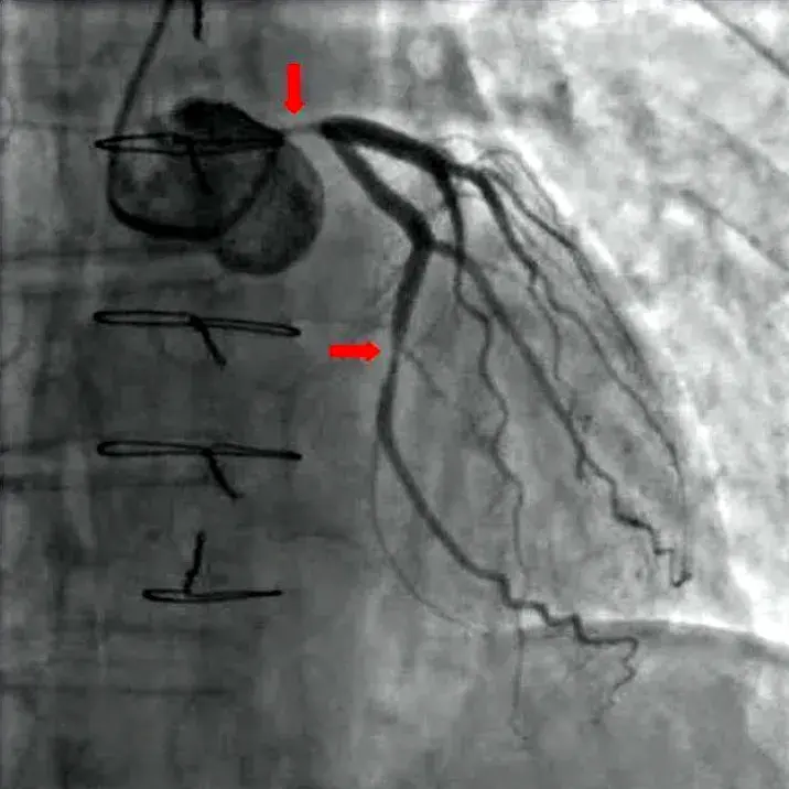
Blood vessels don’t show up clearly on an ordinary X-ray. In angiography, a special dye that X-rays can’t pass through (a contrast medium) is put into the blood, through a thin tube that the doctor guides into the coronary arteries. Then a live X-ray video (fluoroscopy) is taken. The arteries show up as dark branching lines, and any narrowing or blockage can be seen exactly. The picture is an angiogram.
Treatment
During a heart attack, heart muscle is dying every minute, so treatment must start fast to restore the blood flow. The patient is taken to hospital at once and given oxygen if their blood is short of it, and medicines. What comes next depends on whether the artery is partly or completely blocked, and on what the hospital has.
- Where angiography and angioplasty aren’t available, doctors give blood thinners and clot-dissolving medicines.
- Where they are, the blocked artery is opened with a procedure:
Coronary angioplasty and stenting (also called PCI, percutaneous coronary intervention). The cardiologist puts a long, thin, flexible tube, a catheter, into an artery in the wrist or the groin, and guides it up to the narrowed coronary artery, watching on the X-ray screen. A tiny balloon at its tip is blown up to squash the plaque against the wall. Usually a stent, a small tube of metal mesh, is left there to hold the artery open. Many stents are coated with a medicine that stops the artery from narrowing again. The patient is awake during it, and often goes home in a few days.
Coronary artery bypass surgery (open-heart surgery). The surgeon opens the chest and takes a healthy blood vessel from elsewhere in the body, usually a vein from the leg or an artery from inside the chest wall. One end is sewn onto the aorta and the other onto the coronary artery beyond the blockage. Blood now flows round the blockage by a new road, just as a bypass road takes traffic round a crowded bazaar. It is a big operation; it is usually planned, but is sometimes done as an emergency.
Heart: traced from BodyParts3D, © The Database Center for Life Science, CC BY 4.0
5.6 Heartbeat, pulse rate or heart rate
The regular, rhythmic contraction and relaxation of the heart makes a sound: the heartbeat. A doctor listens to it with a stethoscope.
The number of heartbeats in a minute is the heart rate. In a healthy adult at rest, it is 60 to 100 beats a minute. (Children’s hearts beat a little faster; a newborn baby’s about 120 to 160 times a minute.)
- A resting heart rate below 60 a minute is bradycardia (a slow heart). It is normal in fit athletes, whose strong hearts pump more blood with each beat.
- A resting heart rate above 100 a minute is tachycardia (a fast heart).
The heart rate changes with what the body is doing: it rises with exercise, fear, excitement and fever, and falls in sleep.
Each time the left ventricle squeezes, a wave of pressure runs along the arteries and makes their walls bulge. Where an artery lies near the skin, you can feel this bulge with your fingers: it is the pulse. Each heartbeat makes one pulse, so the pulse rate equals the heart rate, and the two words are used for the same thing. In a healthy adult, it is 60 to 100 a minute.
Blood vessels
Blood vessels are flexible, muscular tubes that carry blood to every cell and tissue of the body. There are three kinds: arteries, veins and capillaries.
Arteries
An artery carries blood away from the heart to the parts of the body. Arteries have thick, muscular and elastic walls, which can stand the high pressure of the blood the ventricles push into them. The pressure is high enough to keep the blood moving forward, so arteries have no valves.
The aorta is the largest artery. It branches into smaller arteries, these into still smaller arterioles, and each arteriole into a fine network of capillaries. Arteries mostly lie deep inside the body, protected by muscle, because a cut artery loses blood very fast.
Capillaries
Capillaries are the finest blood vessels, thinner than a hair. Their wall is only one cell thick, so substances can pass through it easily. Here, the real work of the circulatory system is done:
- from the blood to the cells: digested food, oxygen, hormones, enzymes and other substances;
- from the cells to the blood: carbon dioxide, urea and other wastes, which the blood takes to the excretory organs.
Capillaries reach so close to every cell that almost no cell in your body is more than a hair’s width from one. After collecting the wastes, capillaries join to form venules, which join to form veins.
Veins
A vein carries blood from the parts of the body back to the heart. Its wall has the same three layers as an artery’s, but with much less muscle, so veins are thin-walled and have a wider space inside. Most of them carry deoxygenated blood rich in carbon dioxide and wastes.
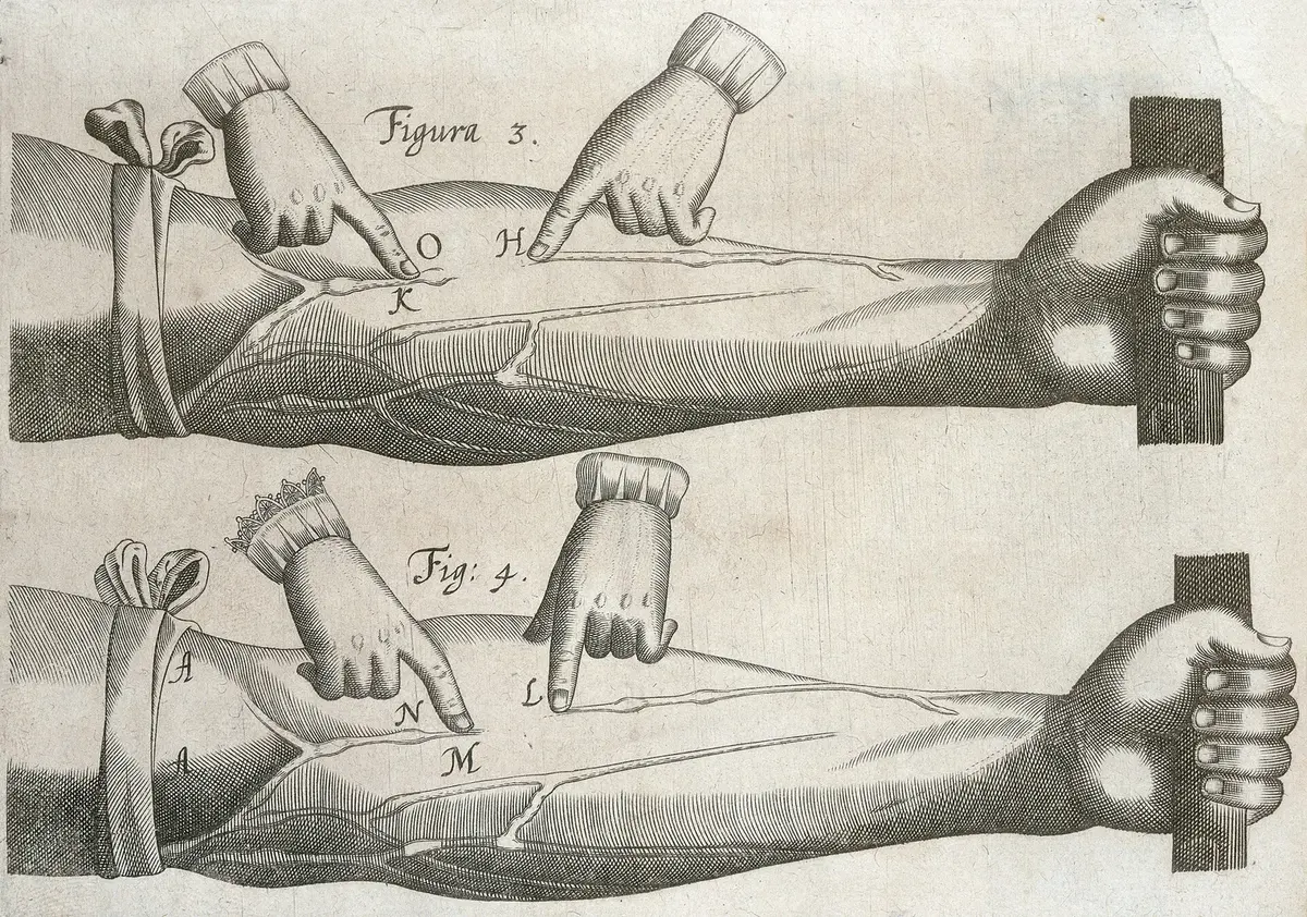
By the time blood reaches the veins, it has lost most of its pressure, so it flows slowly and could easily slip backwards, especially in the legs, where it has to climb up against gravity. To stop this, veins have valves at intervals that let blood flow only towards the heart. Squeezing muscles (when you walk) press on the veins and push the blood along, from valve to valve.
Venules, veins and the vena cavae all do the same job; they differ only in size and number. Many veins lie near the skin: the blue-green lines on your wrist are veins.
Why do veins have valves but arteries don’t?
Pick an answer to see why.
5.7 Blood circulation
The movement of blood from the heart to all parts of the body and back to the heart is blood circulation. An adult’s heart pumps about 5 to 6 liters of blood a minute at rest (and up to five times as much during hard exercise).
Look again at Figure 5.1. In one full trip round, blood passes through the heart twice: once on the right side and once on the left. That is why our circulation is called a double circulation. It has two parts:
- Systemic circulation (heart → body → heart)
- Pulmonary circulation (heart → lungs → heart)
Systemic circulation
When the left ventricle contracts, oxygenated blood flows into the aorta. From the aorta it goes into smaller arteries and arterioles, and through the capillaries it reaches the cells. There it gives the cells food, oxygen and other substances, and takes in the carbon dioxide and other wastes they have made. Then the capillaries join into venules and veins, the veins join to form the vena cavae, and the impure blood reaches the right auricle.
The circulation of blood from the left ventricle to all parts of the body through arteries, and back to the right auricle through veins, is called systemic circulation.
Pulmonary circulation
When the right ventricle contracts, the pulmonary valve opens and deoxygenated blood flows up the pulmonary artery to the lungs. There it runs through the network of capillaries around the millions of tiny air sacs, the alveoli. Carbon dioxide leaves the blood and goes into the alveoli, to be breathed out; oxygen from the air in the alveoli goes into the blood. The blood is now oxygenated, and the pulmonary veins carry it to the left auricle.
The circulation of blood from the right ventricle to the lungs and back to the left auricle is called pulmonary circulation.
| Systemic circulation | Pulmonary circulation |
|---|---|
| between the heart and the whole body | between the heart and the lungs |
| starts at the left ventricle, ends at the right auricle | starts at the right ventricle, ends at the left auricle |
| oxygenated blood goes out in the aorta; deoxygenated comes back in the vena cavae | deoxygenated blood goes out in the pulmonary artery; oxygenated comes back in the pulmonary veins |
| a long route at high pressure | a short route at lower pressure |
| gives oxygen to the body’s cells | takes oxygen from the lungs |
Blood leaves the left ventricle. Which of these does it pass through next on its way back to the heart?
Pick an answer to see why.
5.8 Blood pressure
When the left ventricle contracts and pumps blood into the arteries, the blood pushes hard on their walls. The pressure exerted by the blood on the walls of the blood vessels, especially the arteries, is called blood pressure.
Blood pressure depends on:
- how hard the ventricle contracts,
- how much blood there is, and how much flows,
- how wide or narrow the arteries are (narrow, stiff arteries raise the pressure).
Blood pressure is measured in millimeters of mercury (mmHg) and is written as two numbers, for example 120/80 mmHg, read “120 over 80”.
- The upper number is the systolic pressure: the highest pressure, when the ventricles contract (systole). In an adult it is normally about 90 to 120 mmHg.
- The lower number is the diastolic pressure: the lowest pressure, when the ventricles relax and fill again (diastole). In an adult it is normally about 60 to 80 mmHg.
The usual healthy reading for an adult is about 120/80 mmHg. Blood pressure changes with a person’s age, sex, physical activity and state of mind: it rises with exercise, anger or fear and falls in sleep.
The instrument that measures blood pressure is the sphygmomanometer (sphygmos: pulse in Greek). An inflatable cuff is wrapped round the upper arm and pumped up until it squeezes the artery shut. Then the air is let out slowly, while the health worker listens below the cuff with a stethoscope. The pressure when the first tapping sounds are heard is the systolic pressure; the pressure when the sounds disappear is the diastolic pressure. Today, digital meters do the same thing automatically, sensing the pulses in the cuff.
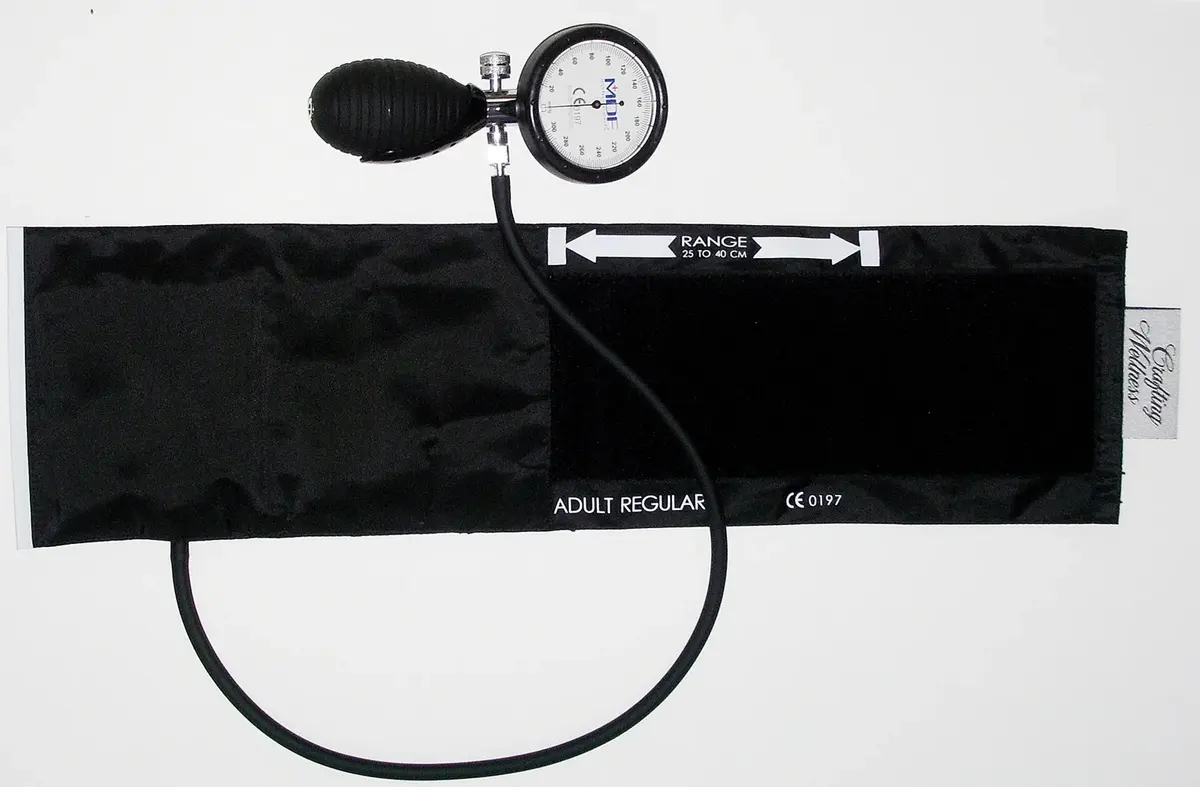
| Reading (adult) | What it means |
|---|---|
| below 90/60 mmHg | low blood pressure (hypotension): may cause dizziness and fainting |
| about 90/60 to 120/80 mmHg | normal |
| 120/80 to 139/89 mmHg | a little raised: time to look at diet and exercise |
| 140 or more, or 90 or more (measured more than once) | high blood pressure (hypertension) |
(Some countries, such as the USA, now count 130/80 and above as high. Nepal, like the WHO, uses 140/90.)
High blood pressure (hypertension)
If a person’s blood pressure stays at 140/90 mmHg or more, they have high blood pressure or hypertension. The heart has to work harder against it, and over the years it damages the arteries, the heart, the kidneys, the eyes and the brain: it is a major cause of heart attacks and strokes.
Symptoms some people notice: headache, shortness of breath, nose bleeding, sweating and tiredness, a red face, sleeplessness and anxiety.
Causes:
- Lack of regular physical exercise.
- A family history of high blood pressure.
- Smoking and drinking alcohol regularly.
- Being overweight.
- A stressful life, in body and mind.
- Too much salty and fatty food (achar, chips, instant noodles and pickled meat are very salty).
Prevention: high blood pressure leads to many problems in body and mind, but it can often be avoided by paying attention in time.
- Watch what you eat every day: less salt, less oily food.
- Don’t smoke or drink alcohol.
- Eat a balanced diet, with plenty of vegetables and fruit.
- Exercise every day.
- Do yoga, meditation and pranayama.
- Check your blood pressure regularly.
- Live a calm, stress-free life as far as you can.
- Take part in games and recreational activities.
A man’s blood pressure is 160/100 mmHg, but he says he feels fine and needs no treatment. What is the best advice?
Pick an answer to see why.
Diabetes
The glucose from our food is used by the cells for energy. A hormone made by the pancreas, insulin, lets glucose pass from the blood into the cells. If the body makes too little insulin, or the cells stop responding to it, the glucose stays in the blood and the blood sugar level rises. A condition in which the blood has more glucose than normal is called diabetes (more exactly, diabetes mellitus). Too much glucose in the blood is hyperglycemia.
(For reference: a normal fasting blood sugar is about 70 to 100 mg/dL; 126 mg/dL or more, measured twice, means diabetes.)
Symptoms of high blood glucose:
- Great thirst and hunger.
- Frequent dizziness, and sometimes fainting.
- Frequent urination (the kidneys pass the extra sugar out in the urine, with a lot of water).
- Blurred vision.
- Losing weight and feeling tired.
- Muscle cramps, and numbness or tingling in the hands and feet.
- Wounds that heal slowly.
Causes:
- Too little insulin made by the pancreas.
- Being overweight, and an inactive life.
- An unbalanced diet.
- Too much fatty and carbohydrate-rich food (sweets, sugary drinks, white rice and refined flour in large amounts).
- Heredity: it runs in families.
Prevention:
- Eat plenty of green vegetables and fruit.
- Exercise regularly.
- Keep a healthy weight.
- Live an active life.
- Live a stress-free life.
- Go for a walk every morning.
- Eat a balanced diet.
Uric acid
Many foods contain substances called purines; the body also makes purines itself, because they are part of DNA and RNA. When purines are broken down, mainly in the liver (and a little in the intestine), they become a waste called uric acid. The blood carries uric acid to the kidneys, which pass it out in the urine.
If too much uric acid is made, or the kidneys can’t remove it fast enough, its level in the blood rises. Too much uric acid in the blood is hyperuricemia. The extra uric acid can form tiny, needle-sharp crystals in the joints, which cause the painful disease gout, and in the kidneys, where they form stones.
Symptoms of high uric acid:
- Pain in the joints.
- Deep pain in the muscles.
- A greater chance of kidney stones.
- Redness, swelling and a burning feeling over the joints.
- Sharp pain in the joints of the toes, especially the big toe.
- Difficulty in walking and moving.
Causes:
- The kidneys can’t remove uric acid properly, because of diabetes or another disease.
- Eating too much purine-rich food.
- Drinking too little water.
Control and prevention:
- Exercise regularly.
- Drink enough water, a little at a time, many times a day.
- Avoid alcohol (especially beer) and smoking.
- Eat cherries: they contain anthocyanins, antioxidants that may help lower uric acid.
- Eat less fatty red meat, organ meat (liver, kidney) and seafood. (Pulses and other plant foods are less of a worry: studies find that purines from plants raise gout risk much less than those from meat.)
- Eat less purine-rich food in general; avoid sugary drinks.
Exercise
1. Choose the correct option
a. Which of the following sets of organs belong to the circulatory system?
Pick an answer to see why.
b. Which of the following blood cells is without a nucleus?
Pick an answer to see why.
c. Which disease is caused by a lack of hemoglobin?
Pick an answer to see why.
d. How many chambers are there in the human heart?
Pick an answer to see why.
e. What causes the systolic pressure on the wall of an artery when the ventricle contracts?
Pick an answer to see why.
f. The results of Rohit’s blood test are shown in the table. What is his blood group?
| Antiserum | anti-A | anti-B | anti-D |
|---|---|---|---|
| Blood clot (clumping) | seen | seen | not seen |
Pick an answer to see why.
g. A person is thirstier and hungrier than usual, feels dizzy and sometimes faints, urinates often and has blurred vision. What is the problem?
Pick an answer to see why.
h. Which test is the right one to find a blockage in a coronary artery when someone has chest pain?
Pick an answer to see why.
i. Saambhawi hurt her leg on a stone while playing at school. Whatever was done, the bleeding didn’t stop. What is the reason?
Pick an answer to see why.
j. Rasmila’s grandfather gets swollen joints and pain in his body when he eats red meat, legumes and fried food. What is his problem?
Pick an answer to see why.
2. Write the differences
Red blood cell and white blood cell
Answer
Auricle and ventricle
Answer
Artery and vein
Answer
Pulmonary circulation and systemic circulation
Answer
Systolic and diastolic blood pressure
Answer
Anemia and hemophilia
Answer
Angiogram and open-heart surgery
Answer
Platelets and white blood cells
Answer
3. Give reasons
Blood is red.
Answer
White blood cells are like the soldiers of the body.
Answer
The wall of a ventricle is more muscular (thicker) than the wall of an auricle.
Answer
The wall of the left ventricle is thicker than the wall of the right ventricle.
Answer
Arteries do not have valves but veins do.
Answer
A person with anemia feels tired after a short walk.
Answer
Arteries lie deep inside the muscles, but many veins are near the surface.
Answer
The blood group should be tested before a transfusion.
Answer
Blood clots slowly, or not at all, in a person with hemophilia.
Answer
4. Answer the following questions
Describe the main parts of the human circulatory system.
Answer
Write the main functions of blood.
Answer
When any part of the body is injured, the bleeding stops after a while. Why?
Answer
Ramila gets tired even after walking a short distance and finds it hard to breathe. What is the reason? Explain.
Answer
A person’s blood pressure is 130/90 mmHg. What does this mean?
Answer
Which device is used to measure blood pressure?
Answer
Before a blood transfusion, the doctor checks the blood groups of the donor and the patient. Why?
Answer
Sarita’s grandmother urinates often, feels tired, has muscle cramps, numbness in her hands and legs, and is always thirsty. What is her problem?
Answer
Mention the effects of high blood pressure, and how to prevent it.
Answer
The respiratory, digestive and circulatory systems are interrelated. Explain with reasons.
Answer
How is high or low blood pressure detected?
Answer
Four pictures show (1) a person swallowing a tablet, (2) the digestive system, (3) the circulatory system, and (4) a painful knee. What process do they show? Which systems take part? If the system in picture 3 were absent, how could the medicine swallowed in picture 2 still reach the knee? Explain.
Answer
Answer the questions using the table.
| Cell | Shape | Nucleus | Made in | Destroyed in |
|---|---|---|---|---|
| X | biconcave | absent | bone marrow | liver and spleen |
| Y | irregular | present | bone marrow, lymph nodes | liver, spleen, site of infection |
| Z | round | absent | bone marrow | spleen |
- i.Which blood cell’s shortage causes anemia?
- ii.What problem arises when the number of Y cells rises far above normal?
- iii.If a person’s blood clots slowly or not at all, which cells are below normal? What is the condition called?
Answer
While walking on the road, Sarita saw an old man with chest pain. She rushed him to hospital. The first examination suggested he might have a heart problem. Which test would be right to find out his heart’s condition? Explain with a reason.
Answer
Explain the blood circulation in the human body with a diagram.
Answer
Write the preventive measures for hyperuricemia.
Answer
Explain the internal structure of the human heart with a neat, labeled diagram.
Answer
Explain the main causes of a heart attack.
Answer
Introduce the angiogram and its importance.
Answer
Give a brief introduction to open-heart surgery.
Answer
When Samip’s blood was tested, his white blood cell count was outside the normal range. Which symptoms may appear in his body? Give a reason.
Answer
Harish’s blood test showed a high level of fibrinogen in his plasma. Does this affect his blood circulation? Explain with a reason.
Answer
Study the table and answer.
| Person | Injury | Flow of blood from the injured part |
|---|---|---|
| X | superficial | bled continuously; ordinary first aid didn’t stop it |
| Y | deep | bled with high pressure; ordinary first aid stopped it |
| Z | superficial | bled for a while and stopped |
- i.Which person has hemophilia?
- ii.In which person is an artery injured?
- iii.Which injury, Y’s or Z’s, is more dangerous? Give a reason.
Answer
Study the concept map: Left ventricle → Kidney → Left auricle.
- i.What type of circulation does it show?
- ii.In which parts of the map do arteries and veins play a role?
Answer
Look at the diagram and answer.
- i.Write two differences between blood vessels 15 and 18.
- ii.What are the functions of valves 11 and 13?
- iii.What happens if part 4 is absent?
- iv.Name parts 6, 7, 8 and 9.
Heart: traced from OpenStax College, Anatomy & Physiology, CC BY 3.0, via Wikimedia Commons
Answer
Draw neat, labeled diagrams of:
- i.the external structure of the human heart
- ii.the internal structure of the human heart
- iii.arteries, veins and capillaries
- iv.pulmonary circulation
- v.systemic circulation
Answer
Quiz yourself: Life Processes and the Human Body, Grades 5–10 →
Practice every question in the book, and keep track of the ones you miss →
A companion to your textbook, not a replacement for it: study from the book your school uses, and where the two differ, follow your teacher. Found a mistake? Please tell us.