
In the coal mines of Britain in the 1890s, an explosion underground killed some miners outright, but many more were found dead afterwards in passages the blast had never reached, without a mark on them. John Scott Haldane, a physiologist from Oxford, went down into the mines after such disasters, collected the air, and analyzed the blood of the dead. He showed that most had been killed by carbon monoxide in the “afterdamp”, a gas that has no smell, which grips hemoglobin far more tightly than oxygen does and so starves the body of oxygen. He breathed it himself, in measured doses, to record what it did, and he suggested that rescuers carry small animals, such as mice or canaries, which feel the gas sooner than people do. Haldane went on to measure the gases in the lungs and blood, to study breathing high on mountains and deep under water, and to treat soldiers poisoned by gas with oxygen. He is called the father of oxygen therapy.
This unit follows the air you breathe: the passages it takes into your lungs, how the chest moves it in and out, how oxygen crosses into the blood and carbon dioxide leaves it, and how the blood carries both.
8.2.1 The respiratory system and respiration
The respiratory system is the set of organs and structures that work together to exchange gases between the body and the air outside: they take in oxygen and give out carbon dioxide. It serves respiration, the oxidation of food inside the cells to release energy. The word covers two things:
- i.Breathing (external respiration): fresh air is moved from outside into the lungs, and used air from the lungs back out; gases are then exchanged between the air in the lungs and the blood.
- ii.Cellular (internal, tissue) respiration: the release of energy in the tissues by the oxidation of food.
Types of respiration
On the basis of whether oxygen is needed, cellular respiration is of two kinds.
Aerobic respiration uses oxygen to break glucose down completely, to carbon dioxide and water. It begins in the cytoplasm (glycolysis) and is finished in the mitochondria. It is efficient: one molecule of glucose gives about 38 ATP by the classic count (about 30–32 by today’s measurements: see Unit 4). Most organisms respire this way: humans and most animals, plants and many bacteria.
Anaerobic respiration (fermentation) breaks glucose down only partly, without oxygen, in the cytoplasm alone, and gives only 2 ATP per glucose. Animals’ cells make lactic acid; yeast and some bacteria make ethyl alcohol and carbon dioxide. It goes on in our muscles during a sprint, in fruits and germinating seeds short of air, and in gut parasites such as Taenia and Ascaris, which live where there is hardly any oxygen.
Which product is made when your leg muscles respire without enough oxygen in a sprint?
Pick an answer to see why.
8.2.2 The organs: nose and pharynx
The respiratory organs are, in the order the air passes them: the nose (nostrils and nasal cavity), pharynx, larynx, trachea, bronchi and bronchioles, and the lungs with their alveoli, moved by the respiratory muscles. The passages down to the finest bronchioles only carry the air (the conducting zone); gases are exchanged only in the alveoli and the smallest bronchioles that carry them (the respiratory zone).
Servier Medical Art (smart.servier.com), CC BY 4.0
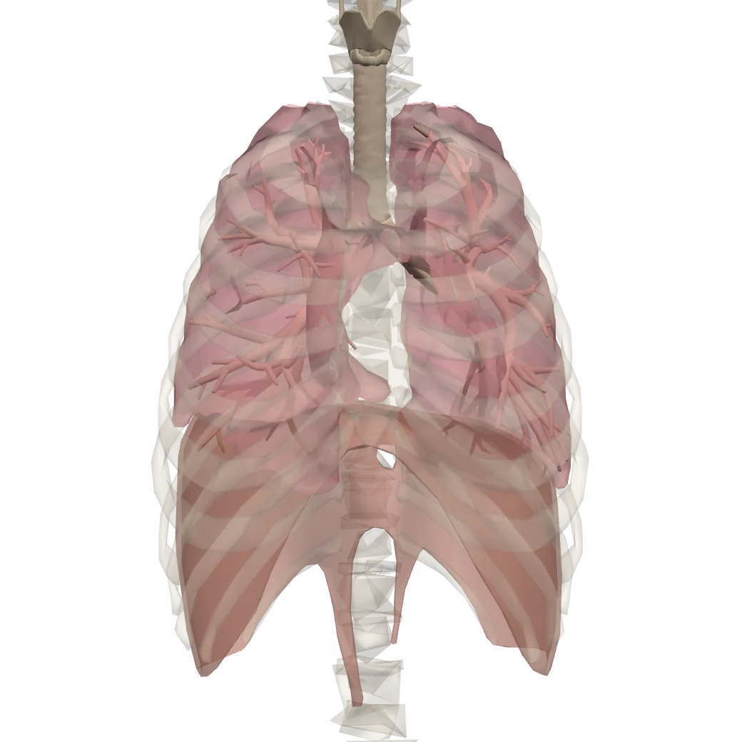
A model of a real body. Tap Show in 3D to turn it and tap its parts.
Voice box (larynx)Sits at the top of the windpipe, in the neck. Inside it are the vocal cords: air from the lungs makes them vibrate when you speak, and that is your voice.
Windpipe (trachea)A tube held open by rings of cartilage; you can feel them at the front of your neck. It carries air down into the chest.
BronchiAt its lower end the windpipe divides into two bronchi (one is a bronchus), one going to each lung.
BronchiolesInside the lung the bronchi branch again and again into thinner and thinner tubes, the bronchioles. Each ends in a bunch of tiny air sacs, the alveoli.
Right lungA soft, spongy organ in the chest, protected by the ribs and full of millions of alveoli. The right lung has three lobes and is a little bigger than the left.
Left lungIt has only two lobes and a hollow on its inner side where the heart sits, so it is a little smaller than the right.
DiaphragmA dome-shaped sheet of muscle under the lungs, between the chest and the belly. It moves down to draw air into the lungs and up again to push it out.
Body and organs: BodyParts3D, The Database Center for Life Science (CC BY 4.0)
Nose (nostrils and nasal cavity)
The nose is where fresh air comes in and used air goes out. Its outer part is a triangular frame of bone above and cartilage below, covered by skin and small muscles. It opens to the outside by two oval openings, the external nostrils (nares), and inside, at the back, into the pharynx by the internal nostrils (choanae). A vertical wall, the nasal septum, divides its cavity into a right and a left nasal chamber.
The nasal cavity has three regions:
- Vestibule, the lowest part, just inside the nostrils, lined by skin with stiff hairs, the vibrissae.
- Respiratory region, the middle part, the largest. From its outer wall three pairs of curled, scroll-like shelves of thin bone stick out into the cavity: the nasal conchae (turbinates), superior, middle and inferior. They make the air swirl over a large, warm, wet surface. This region is lined by pseudostratified ciliated columnar epithelium with goblet cells, which make mucus, over a thick bed of blood vessels.
- Olfactory region, the upper part, in the roof. Its lining carries the olfactory receptors, nerve cells whose fibers pass up through the bone to the olfactory bulb of the brain.
Servier Medical Art (smart.servier.com), CC BY 4.0
Paranasal sinuses. Air-filled hollows in the bones of the skull round the nose open into the nasal cavity. There are four pairs, named after their bones: frontal (in the forehead), ethmoidal (between the eyes), maxillary (in the cheeks, the largest) and sphenoidal (deep behind the nose). They are lined by mucous membrane, whose mucus drains into the nasal cavity. They make the skull lighter and give the voice its resonance; when their lining swells in a cold, their openings block and they ache (sinusitis).
Functions of the nose:
- i.It stops dust and other particles from entering the airways.
- ii.It filters, warms and moistens the air: by the time air reaches the pharynx it is close to body temperature and nearly saturated with water vapor.
- iii.Its olfactory region detects smells.
- iv.Its vibrissae and sticky mucus trap dust and microorganisms, and the cilia sweep the mucus back to the throat to be swallowed.
- v.With the sinuses, it gives the voice its resonance (try speaking with your nose pinched).
Pharynx
The pharynx is a short vertical tube behind the nasal cavity and the mouth, about 12 cm long, the common passage for food and air. It has three regions:
- Nasopharynx, behind the nasal cavity: the internal nostrils open into it, and so do the two Eustachian tubes from the middle ears. Air passes through it only.
- Oropharynx, behind the mouth: the part you see when you open your mouth wide. Food and air both pass.
- Laryngopharynx, the lowest part: it leads into two tubes, the trachea (windpipe) in front and the esophagus (food pipe) behind.
During swallowing the soft palate rises to close the nasopharynx, and the larynx rises under the epiglottis, so food goes to the esophagus and not into the nose or the windpipe.
Where do the paths of food and air cross?
Pick an answer to see why.
8.2.3 The larynx (voice box)
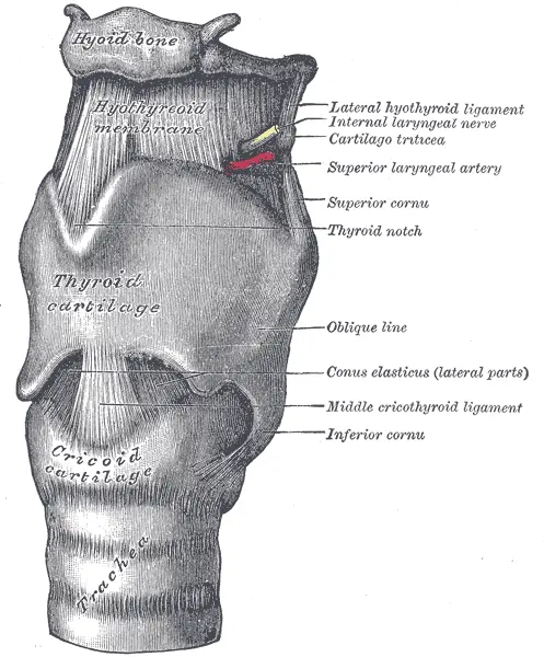
The larynx is a hollow box of cartilage at the top of the trachea, in the front of the neck, at the level of the third to sixth cervical vertebrae (C3–C6). It is the voice box, and it guards the entrance to the lower airways.
Its framework is nine cartilages: three single and three paired.
| Single (unpaired) | Paired |
|---|---|
| thyroid cartilage: the largest, two plates meeting in front in a ridge | arytenoid: small pyramids on the back of the cricoid; the vocal cords are fixed to them |
| cricoid cartilage: a ring like a signet ring, with its broad part at the back, the only complete ring of cartilage in the airway | corniculate: small horns on the tips of the arytenoids |
| epiglottis: a leaf-shaped flap of elastic cartilage above the opening | cuneiform: small rods in the folds beside them |
They support the larynx, protect the vocal cords and, moved by small muscles, change the tension of the cords to make sound. At puberty, under the influence of testosterone, the larynx of a boy grows, and the ridge of his thyroid cartilage juts forward in the neck as the Adam’s apple; his vocal cords lengthen and his voice deepens (it “breaks”).
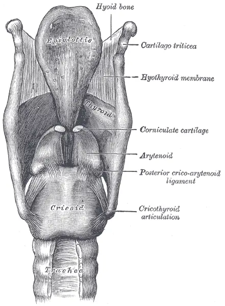
The epiglottis closes over the opening of the larynx, the glottis, when you swallow. Inside, two pairs of folds stretch from the thyroid cartilage in front to the arytenoids behind: the upper false vocal cords, which take no part in making sound, and the lower true vocal cords. Air breathed out between the true vocal cords, drawn close together, sets them vibrating, and the vibration is the sound of your voice; tighter, thinner cords give a higher note.
8.2.4 Trachea, bronchi and bronchioles
Trachea (windpipe)
The trachea is a tube about 12 cm long and 2.5 cm across that runs from the larynx down into the chest, from the sixth cervical to about the fifth thoracic vertebra. It lies in front of, and parallel to, the esophagus. Its wall is held open by 16 to 20 C-shaped rings of cartilage, the tracheal rings, which stop it collapsing when the pressure inside falls as you breathe in hard. The open part of each C faces backwards, towards the esophagus, and is closed by smooth muscle (the trachealis), so that a swallowed mouthful can bulge the esophagus forward as it passes. It is lined by pseudostratified ciliated columnar epithelium with goblet cells.
Servier Medical Art (smart.servier.com), CC BY 4.0
Bronchi and bronchioles
At the level of the fifth thoracic vertebra the trachea divides into the right and left primary (main) bronchi, one to each lung. The right bronchus is wider, shorter and more upright (about 2.5 cm long) than the left (about 5 cm), which has to pass under the arch of the aorta and over the heart; so a peanut or a coin breathed in by mistake usually lodges in the right one.
Inside the lungs they branch again and again, like a tree upside down:
- the right bronchus into three secondary (lobar) bronchi, one for each lobe of the right lung; the left into two;
- these into tertiary (segmental) bronchi;
- then into bronchioles, under 1 mm across, with no cartilage in their walls;
- terminal bronchioles, the last that only carry air;
- respiratory bronchioles, with a few alveoli in their walls;
- alveolar ducts, which end in alveolar sacs, clusters of alveoli.
The bronchi are held open by plates and incomplete rings of cartilage and lined, like the trachea, by pseudostratified ciliated epithelium. The bronchioles have smooth muscle instead, which can narrow them (as in asthma), and are lined by ciliated columnar epithelium that becomes cuboidal in the smallest.
Why does a peanut breathed in by mistake usually go into the right bronchus rather than the left?
Pick an answer to see why.
8.2.5 The lungs
The lungs are a pair of soft, spongy, pinkish, cone-shaped organs that fill most of the chest (thoracic cavity), one on each side of the heart. Each has a broad, hollowed base resting on the diaphragm and a narrow apex reaching just above the collarbone.
- The right lung is slightly larger and is divided by two fissures into three lobes (upper, middle and lower).
- The left lung is a little smaller and has two lobes; its front edge has a hollow, the cardiac notch, where the heart lies.
Each lung is wrapped in a double membrane, the pleura: the inner visceral pleura covers the lung itself, and the outer parietal pleura lines the chest wall and the diaphragm. Between them is a very thin pleural space (pleural cavity) holding a little pleural fluid. The fluid lets the two layers slide over each other without friction as the lungs swell and shrink, and, like a film of water between two glass plates, it holds the lungs against the chest wall, so that when the chest grows the lungs grow with it.
Inside, each lung is the bronchial tree ending in the alveoli. There are some 300 to 500 million alveoli in the two lungs (a careful count in 2004 found 480 million on average), each about 0.2 mm across, wrapped in a net of capillaries. Spread out, their surface is about 70 square meters, roughly the floor of a classroom.
Servier Medical Art (smart.servier.com), CC BY 4.0
The alveolar wall is made of two kinds of cells, the pneumocytes:
- Type I pneumocytes: very thin, flat (squamous) cells covering about 95% of the surface, across which gases diffuse.
- Type II pneumocytes: fewer, rounded (cuboidal) cells that secrete surfactant, a film of fat and protein that lowers the surface tension of the water lining the alveoli. Without it the tiny wet alveoli would tend to collapse at the end of each breath and be hard to blow up again. Babies born very early often haven’t made enough surfactant yet and struggle to breathe.
The lungs get two blood supplies. The pulmonary arteries bring deoxygenated blood from the heart to be loaded with oxygen in the alveoli’s capillaries, and the pulmonary veins take it back. The bronchial arteries, from the aorta, bring oxygenated blood to nourish the lung’s own tissue: its bronchi, their walls and the pleura. The lungs’ capillaries also carry, on their inner lining, angiotensin-converting enzyme (ACE), which converts angiotensin I in the passing blood to angiotensin II, a hormone that raises blood pressure.
8.2.6 The respiratory muscles
The chief muscles of breathing are the diaphragm and the intercostal muscles.
- The diaphragm is a dome-shaped sheet of skeletal muscle and tendon separating the chest from the abdomen. When it contracts it pulls down and flattens; when it relaxes it rises back into its dome.
- The intercostal muscles lie between the ribs, in two layers: the external intercostals, whose fibers slope down and forward, lift the ribs; the internal intercostals, sloping the other way, pull them down.
Other muscles, of the neck (sternocleidomastoid, scalenes), the shoulders and the abdomen, join in only when breathing is hard: the accessory respiratory muscles.
8.2.7 Pulmonary ventilation (breathing)
The physiology of respiration covers how oxygen is taken in from the air and carbon dioxide given out, how the gases are exchanged between the air and the blood and between the blood and the tissues, how they are carried, and how food is oxidized in the cells. It is described under five heads: ventilation, exchange of gases, transport of oxygen, internal respiration and transport of carbon dioxide.
Pulmonary ventilation (breathing) is the movement of air into the lungs and out again, brought about by the respiratory muscles. An adult at rest breathes about 12 to 16 times a minute, about half a liter each time. It has two steps, inspiration and expiration. Air, like any gas, flows from where its pressure is higher to where it is lower: the muscles don’t pull or push air, they change the size of the chest, and with it the pressure in the lungs.
Inspiration (inhalation)
Air is drawn in when the pressure in the lungs (intrapulmonary pressure) falls below the atmospheric pressure. It is always active:
- The external intercostal muscles contract, pulling the ribs and the sternum upwards and outwards (like the handle of a bucket being lifted).
- At the same time the diaphragm contracts and flattens, moving down.
- The chest grows from front to back, side to side and top to bottom, and the lungs, held to it by the pleural fluid, expand with it.
- The air in the lungs now has more room, so its pressure falls, by about 1 mmHg in quiet breathing.
- Air from outside rushes in until the pressures are equal again.
Expiration (exhalation)
Air leaves when the pressure in the lungs rises above the atmospheric pressure.
- In quiet breathing expiration is passive: the external intercostals and the diaphragm simply relax. The ribs fall downwards and inwards, the diaphragm rises back into its dome, and the stretched elastic lungs recoil, like a balloon letting go.
- The chest and lungs get smaller, the pressure in the lungs rises above the air’s, and air flows out.
- In forced breathing (running, coughing, blowing up a balloon) expiration becomes active: the internal intercostal muscles contract and pull the ribs down harder, and the abdominal muscles squeeze the belly’s organs up against the diaphragm, pushing air out fast.
What makes air flow into the lungs during inspiration?
Pick an answer to see why.
8.2.8 Exchange of gases
The alveoli are the sites of gas exchange: tiny air sacs at the ends of the alveolar ducts, each wrapped in capillaries. Their wall is extremely thin, and the blood flows very close to the air. Gases cross by simple diffusion, each down its own partial pressure gradient, from where its partial pressure is higher to where it is lower. (The partial pressure of a gas in a mixture is its share of the total pressure: oxygen is 21% of the air, so at sea level, out of 760 mmHg, its partial pressure, PO₂, is about 160 mmHg. In blood it is the pressure the dissolved gas would be in balance with.)
| PO₂ (mmHg) | PCO₂ (mmHg) | |
|---|---|---|
| air in the alveoli | about 100 | 40 |
| blood coming to the lungs (deoxygenated) | 40 | about 45 |
| blood leaving the lungs (oxygenated) | about 100 | 40 |
| tissues | 40 or less | 45 or more |
So in the lungs oxygen diffuses from the alveoli into the blood, and carbon dioxide diffuses from the blood into the alveoli, to be breathed out. (Books give the PCO₂ of deoxygenated blood as 44, 45 or 46 mmHg: it varies a little.) Carbon dioxide’s gradient is much smaller than oxygen’s, but it is about twenty times more soluble, so it diffuses just as fast.
The diffusion (respiratory) membrane the gases cross is made of three layers: the squamous epithelium of the alveolus, the endothelium of the capillary, and the basement membrane between them, together less than a thousandth of a millimeter (about 0.5 µm) thick. A huge area, a very thin membrane, steep gradients kept up by breathing and by the blood flowing on, and a moist surface: these make exchange fast. A red blood cell takes about three-quarters of a second to pass through an alveolar capillary at rest, and it is fully loaded with oxygen in about a third of that.
Why does oxygen move from the alveoli into the blood?
Pick an answer to see why.
8.2.9 Transport of oxygen and internal respiration
Transport of oxygen
Oxygen that has diffused into the blood is carried to the tissues. 100 mL of arterial blood carries about 20 mL of oxygen, in two forms:
- a.Dissolved in the plasma: only about 1–3%, because oxygen dissolves poorly in water.
- b.As oxyhemoglobin: about 97–99%. Oxygen joins loosely with the hemoglobin (Hb) of the red blood cells, each molecule of which can carry four molecules of oxygen, one on the iron of each of its four heme groups.
Oxyhemoglobin is an unstable compound: it forms where PO₂ is high and breaks apart again where PO₂ is low. Its forming is helped by the conditions in the lungs: a low PCO₂, a lower temperature and a higher pH (less acid).
The oxygen–hemoglobin dissociation curve shows how full of oxygen hemoglobin is (its saturation) at each PO₂. It is S-shaped: once one heme group has taken an oxygen, the others take it more easily. So hemoglobin is almost full over a wide range of high PO₂ (it loads well even if the air is thinner), and on the steep middle part a small fall in PO₂ makes it give up a lot.
Internal (tissue) respiration
In the tissue capillaries the PO₂ is low, so oxyhemoglobin dissociates, and the freed oxygen diffuses out into the tissue fluid and into the cells:
Its dissociation is helped by a high PCO₂, a higher temperature and a low pH: exactly the conditions in a working muscle, which is making carbon dioxide, acid and heat. This shift of the curve to the right, so that hemoglobin gives up more oxygen where carbon dioxide is high, is the Bohr effect. In the cells, the oxygen oxidizes food (glucose) in the mitochondria to release energy as ATP:
8.2.10 Transport of carbon dioxide
Respiring tissues make a great deal of carbon dioxide, and it would be harmful if it built up: it forms an acid in water. The blood carries it to the lungs in three ways.
Dissolved in the plasma (about 7%). Carbon dioxide is far more soluble than oxygen. A very little of the dissolved gas joins with water to form carbonic acid, slowly, because plasma has hardly any of the enzyme that speeds this up.
As bicarbonate ions (about 70%). Most of the carbon dioxide diffuses into the red blood cells, which contain the enzyme carbonic anhydrase. It joins carbon dioxide and water very fast into carbonic acid, which splits into a hydrogen ion and a bicarbonate ion:
The H⁺ is taken up by hemoglobin (which has just given up its oxygen and takes up H⁺ more readily), so the blood hardly becomes more acid. Most of the HCO₃⁻ diffuses out of the red cell into the plasma, and is carried there. To keep the charges balanced, chloride ions (Cl⁻) move from the plasma into the red cell in exchange: the chloride shift (Hamburger phenomenon). Books often write the bicarbonate as joined to potassium in the red cell (KHCO₃) and to sodium in the plasma (NaHCO₃): in the blood these are dissolved ions, the bicarbonate balanced by the K⁺ and Na⁺ around it.
As carbaminohemoglobin (about 23%). Carbon dioxide joins directly with the amino (–NH₂) groups of hemoglobin (and a little of the plasma proteins):
In the lungs all three reverse: the PCO₂ in the alveoli is lower, so bicarbonate comes back into the red cells (chloride goes out), carbonic acid re-forms and is split by carbonic anhydrase into carbon dioxide and water, and carbon dioxide leaves hemoglobin; the gas diffuses into the alveoli and is breathed out.
Where is most of the bicarbonate in the blood made?
Pick an answer to see why.
Exercise
A. Tick the correct alternatives
- Which of the following cartilages of the larynx is unpaired?
Pick an answer to see why.
- Which epithelium lines the inside of the trachea?
Pick an answer to see why.
- Pneumocytes are modified ……………. epithelium.
Pick an answer to see why.
- What happens to the intercostal muscles during inspiration?
Pick an answer to see why.
- The most numerous and narrowest tubes in the lungs are the
Pick an answer to see why.
- The passages of food and air separate at the
Pick an answer to see why.
- Which of the following statements is true?
Pick an answer to see why.
- What happens to the diaphragm during expiration?
Pick an answer to see why.
- In which of these are bicarbonate ions mostly made?
Pick an answer to see why.
- Into how many lobes is the left lung of a human divided?
Pick an answer to see why.
- Which statement best describes the role of surfactant in the lungs?
Pick an answer to see why.
- Which best explains why oxygen moves from the alveoli into the blood and carbon dioxide from the blood into the alveoli?
Pick an answer to see why.
- The open part of the C-shaped cartilage rings of the trachea is on its
Pick an answer to see why.
B. Answer the following questions in brief
Describe the structure of the larynx. What causes the prominent Adam’s apple in the neck of males?
Answer
What is pulmonary ventilation? How does it occur in the human body? Explain.
Answer
What is the primary respiratory organ of the human body? Describe its morphology.
Answer
What mechanism is mainly responsible for the exchange of oxygen and carbon dioxide in the alveoli of the lungs? Elaborate with a diagram.
Answer
How are oxygen and carbon dioxide transported by the blood? Describe briefly.
Answer
Study the diagram and answer the questions.
Servier Medical Art (smart.servier.com), CC BY 4.0
- a.Label A to I.
- b.What are the functions of D and H?
- c.Give the differences between G and I.
Answer
C. Long answer questions
Inhaling oxygen and exhaling carbon dioxide is called breathing, or external respiration. Which organs take part in it? Describe them briefly, with a neat, labeled diagram.
Answer
Explain the physiological processes of respiration in terms of pulmonary ventilation, gas exchange, transport of gases and internal respiration.
Answer
Printable PDFs
- Study notes 4 pages
- Worksheet 4 pages Answer key
Quiz yourself: Life Processes and the Human Body, Grades 5–12 →
Practice every question in the book, and keep track of the ones you miss →
A companion to your textbook, not a replacement for it: study from the book your school uses, and where the two differ, follow your teacher. Found a mistake? Please tell us.