
Put two fingers on the side of your neck, just under the jaw, and you can feel it: a push, a pause, a push. That push started a fraction of a second earlier in a small patch of special muscle, about as long as your fingernail, high in the wall of your heart. It has been firing about 72 times a minute since months before you were born, more than 100,000 times a day, and it will not stop for a rest until the day you die. Each push sends about 70 ml of blood into a network of tubes so long that, laid end to end, the smallest of them, the capillaries, would stretch for thousands of kilometers.
This unit is about that network, the circulatory system: the heart that drives it, how each beat starts and spreads, how blood pressure is made and measured, why blood from one person can kill another, and the arteries and veins that reach every part of the body.
The circulatory system
The circulatory system is the group of organs that carries useful substances to the tissues and takes unwanted ones away. Useful substances include oxygen, nutrients and hormones. Unwanted ones are metabolic wastes such as urea, carbon dioxide and toxins, which are carried to the organs that get rid of them.
In humans the circulation is closed: blood always stays inside the heart and blood vessels, and only fluid leaks out through the thin walls of the capillaries to bathe the cells. (In an insect it is open: the blood is pumped out into the body cavity and soaks the organs directly.)
The circulatory system has two parts:
- The blood vascular system: the heart, the blood vessels and the blood.
- The lymphatic system: lymph vessels, lymph nodes and lymph, which collect the fluid that leaked from the capillaries and return it to the veins.
This unit is about the blood vascular system. Blood itself, its plasma and cells, you met as a fluid connective tissue in Unit 14.
Heart
Location and size
The human heart is a red, hollow, muscular organ shaped roughly like a cone, with its broad base at the top and its narrow apex at the bottom. It lies in the chest (thoracic cavity) between the two lungs, in the space called the mediastinum, tilted so that its apex points to the left. It is about the size of its owner’s clenched fist: in an adult about 12 cm long and about 300 g in weight. Like all muscle and blood, it develops from the embryo’s mesoderm.
Turn the heart and see where it sits between the lungs, tipped to the left, with the great vessels at its top.
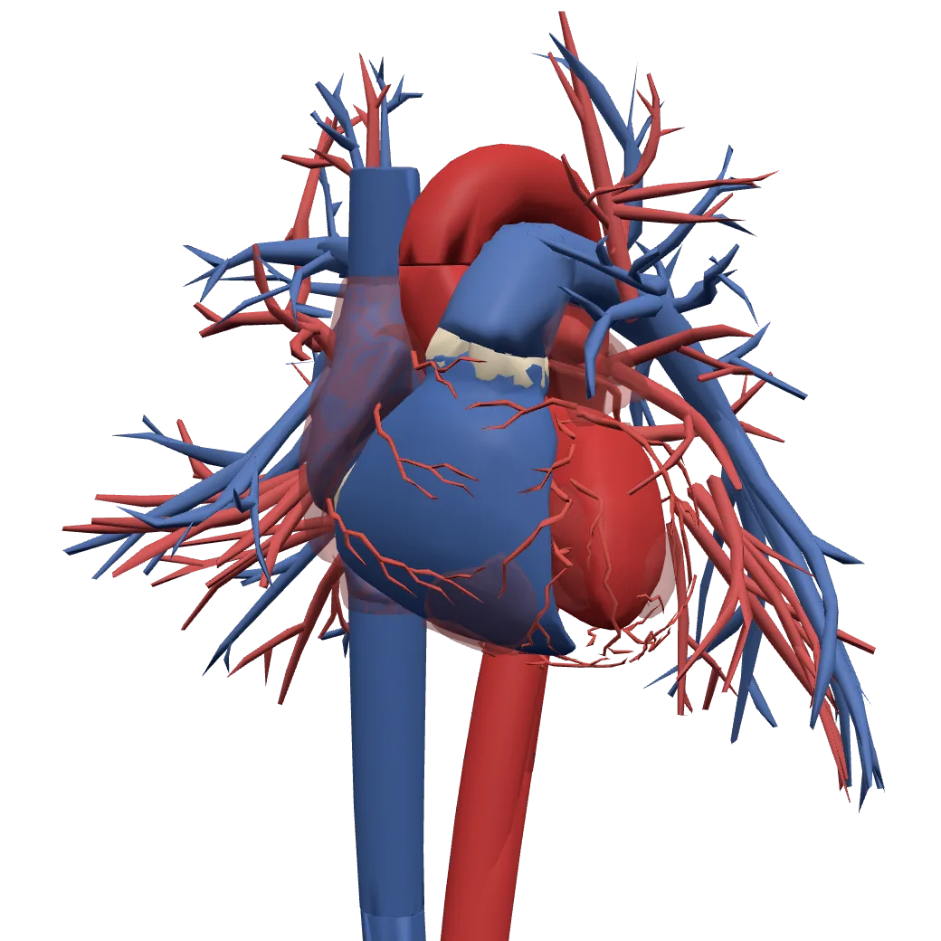
A model of a real body. Tap Show in 3D to turn it and tap its parts.
Wall of the right auricleA thin wall of cardiac muscle. An auricle only receives blood and passes it down, so it needs little force.
Wall of the right ventricleThicker than an auricle’s, since it pumps the blood out, but thinner than the left ventricle’s: it only pumps to the lungs next door.
Wall of the left auricleThin, like the right auricle’s: it receives the blood coming back from the lungs and passes it down to the left ventricle.
Wall of the left ventricleThe thickest wall of the heart, because the left ventricle pumps blood to the whole body, from the head to the toes.
Right auricle (atrium)Receives the deoxygenated blood coming back from the whole body through the superior and inferior vena cava.
Right ventricleTakes the blood from the right auricle and pumps it up the pulmonary artery to the lungs.
Left auricle (atrium)Receives the oxygenated blood coming back from the lungs through the four pulmonary veins.
Left ventricleTakes the oxygenated blood from the left auricle and pumps it out through the aorta to the whole body.
Heart valvesFour flaps that open one way only, like one-way doors, so the blood can’t flow back: the tricuspid, bicuspid (mitral), pulmonary and aortic valves.
Coronary arteriesThe heart’s own blood supply: they branch over its surface and bring oxygenated blood to the hard-working heart muscle.
AortaThe largest artery. It carries oxygenated blood from the left ventricle and branches into the arteries of the whole body.
Venae cavaeThe two largest veins. The superior vena cava brings deoxygenated blood from the head, neck and arms, the inferior vena cava from the trunk and legs, both into the right auricle.
Pulmonary arteriesCarry deoxygenated blood from the right ventricle to the lungs: the only arteries that carry deoxygenated blood.
Pulmonary veinsFour veins that bring oxygenated blood from the lungs to the left auricle: the only veins that carry oxygenated blood.
Body and organs: BodyParts3D, The Database Center for Life Science (CC BY 4.0)
Pericardium (protective covering)
The heart sits inside a double-layered sac, the pericardium, which also wraps the bases of the great vessels. It has two parts:
- The fibrous pericardium, the outer, tough bag of dense connective tissue. It anchors the heart in place and stops it from over-stretching when it fills.
- The serous pericardium, a thin, smooth, double layer inside it:
- its parietal layer lines the inside of the fibrous bag;
- its visceral layer is stuck to the surface of the heart itself. It is a single layer of flattened epithelium, and it is also the outermost layer of the heart wall, the epicardium.
Between the two serous layers is a narrow space, the pericardial cavity, holding a small amount (about 15–50 ml) of pericardial fluid made by the serous layers. The fluid lets the heart beat without friction against its bag, and the whole sac helps protect the heart from shocks and injury.
The wall of the heart itself has three layers:
- The epicardium, outermost: the visceral pericardium.
- The myocardium, the middle and thickest layer, made of cardiac muscle fibers and rich in blood vessels.
- The endocardium, innermost: a smooth lining of endothelium (squamous epithelium) on a basement membrane, continuous with the lining of the blood vessels, so blood flows over a slippery surface.
Which of these is not a layer of the heart wall itself?
Pick an answer to see why.
What is the job of the pericardial fluid?
Pick an answer to see why.
External features
The human heart has four chambers: two upper atria (or auricles) and two lower ventricles. Its outside shows:
Sulci (grooves). The two atria are separated outside by a shallow interatrial groove. The atria are marked off from the ventricles by a groove running round the heart, the atrioventricular groove or coronary sulcus. The two ventricles are marked off by the anterior and posterior interventricular sulci. The coronary arteries and veins run in these grooves, bedded in fat.
Atria. The two thin-walled, upper receiving chambers:
- the right atrium, the larger, receives deoxygenated blood through the superior vena cava (from the head, neck and arms), the inferior vena cava (the larger, from the lower body) and the coronary sinus (from the heart wall itself);
- the left atrium, with a thicker wall, receives oxygenated blood from the lungs through four pulmonary veins (two pairs).
Each atrium has a small ear-shaped pouch, the auricle, on its front.
Ventricles. The two lower, thick-walled pumping chambers. The left ventricle is the larger and has a wall about three times thicker than the right’s: it pumps blood to the whole body, while the right ventricle pumps only as far as the lungs. Together the ventricles make most of the heart’s front and its apex.
Pulmonary trunk and aorta. The pulmonary trunk leaves the right ventricle and divides into the right and left pulmonary arteries, to the lungs. The aorta leaves the left ventricle; it is described in three parts:
- the ascending aorta, which gives off the right and left coronary arteries to the heart’s own muscle;
- the arch of the aorta, which gives off the brachiocephalic (innominate) artery, the left common carotid and the left subclavian;
- the descending aorta, running down through the thorax and abdomen.
The pulmonary trunk is joined to the arch of the aorta by a fibrous cord, the ligamentum arteriosum: the remains of the ductus arteriosus, a short vessel that let the fetus’s blood skip the lungs it did not yet use.
BodyParts3D, © The Database Center for Life Science, CC BY 4.0
Internal structure
The inside of the heart is seen in a section cut lengthwise, or by removing its front wall.
i. Atria
The two atria are completely separated by a muscular wall, the interatrial septum.
- The right atrium has the openings of the superior vena cava, the inferior vena cava and the coronary sinus.
- The opening of the inferior vena cava has a small, rudimentary fold, the Eustachian valve. (Before birth it steered blood towards the foramen ovale.)
- The opening of the coronary sinus has the Thebesian valve (coronary valve).
- On the interatrial septum is an oval hollow, the fossa ovalis. It marks the place of the foramen ovale, an opening through which, in the fetus, blood passed from the right atrium into the left atrium. It closes soon after birth.
- The left atrium has the openings of the four pulmonary veins.
The inner wall of each auricle and of the right atrium’s front is ridged with parallel bands of muscle, the musculi pectinati (pectinate muscles, “comb muscles”), while the rest of the atrial wall is smooth.
ii. Valves
The heart has four valves, which let blood flow forward only:
- The tricuspid valve, with three flaps (cusps), guards the opening between the right atrium and right ventricle.
- The bicuspid or mitral valve, with two flaps, guards the opening between the left atrium and left ventricle. (Its two flaps looked to anatomists like a bishop’s hat, a miter.)
- The pulmonary semilunar valve, at the base of the pulmonary trunk, and
- the aortic semilunar valve, at the base of the aorta, each with three half-moon-shaped pockets.
The flaps of the tricuspid and bicuspid valves are tied by tendon-like cords, the chordae tendineae, to papillary muscles in the ventricle walls.
iii. Ventricles
The atria open into the thick, muscular ventricles, which are separated by a thick, oblique interventricular septum. The left ventricle is longer and thicker-walled.
- The inner wall of each ventricle has a few cone-shaped papillary muscles. From their tips the chordae tendineae run to the flaps of the tricuspid and bicuspid valves. When the ventricles contract hard, the papillary muscles contract too and the cords hold the flaps, so they cannot be pushed back up into the atria (like a sail held by its ropes).
- The rest of the ventricle wall is criss-crossed by irregular muscular ridges, the columnae carneae (“fleshy columns”), with small spaces, or fissures, between them.
Heart: traced from OpenStax College, Anatomy & Physiology, CC BY 3.0, via Wikimedia Commons
The chordae tendineae are attached at one end to the valve flaps. What are they attached to at the other end?
Pick an answer to see why.
A baby is born and takes its first breaths. What happens to the foramen ovale?
Pick an answer to see why.
How is blood supplied to the heart muscle itself?
The heart works without a rest, so its muscle needs a steady supply of oxygen and food. It cannot use the blood inside its chambers: the wall is too thick for that to soak through. Instead the ascending aorta, just above the aortic valve, gives off the right and left coronary arteries, which run in the grooves and branch through the myocardium. The deoxygenated blood is collected by coronary veins into the coronary sinus, a wide vein in the groove at the back of the heart, which empties into the right atrium.
A clot in a coronary artery cuts off the blood to the muscle beyond it, which begins to die: a heart attack (myocardial infarction).
Course of blood circulation in the heart
- The right atrium receives deoxygenated blood from the upper body through the superior vena cava, from the lower body through the inferior vena cava, and from the heart wall through the coronary sinus.
- The right atrium contracts, and blood passes through the open tricuspid valve into the right ventricle.
- The right ventricle contracts. The tricuspid valve shuts, the pulmonary valve opens, and blood is pumped through the pulmonary arteries to the lungs, where it gives up carbon dioxide and takes up oxygen.
- Oxygenated blood returns from the lungs to the left atrium through the four pulmonary veins.
- The left atrium contracts, and blood passes through the open bicuspid (mitral) valve into the left ventricle.
- The left ventricle contracts. The bicuspid valve shuts, the aortic valve opens, and blood is pumped into the aorta and on to every part of the body.
In fact the two sides work at the same time: both atria contract together, then both ventricles together.
Origin and conduction of heartbeat
The heartbeat is the heart’s rhythmic contraction (systole) and relaxation (diastole). A healthy adult’s heart at rest beats about 72 times a minute.
The human heart is myogenic: each beat starts in the heart muscle itself, in a group of specialized cardiac muscle cells (nodal tissue), not in a nerve. (A heart taken out for a transplant, with every nerve cut, goes on beating when it is put into the new body.) Nerves and hormones only speed it up or slow it down. (The heart of some invertebrates, such as many crustaceans, is neurogenic: each beat is started by a nerve.)
The parts that start and carry the beat are:
- The sinoatrial node (SA node), the pacemaker: a small mass of specialized cardiac muscle in the wall of the right atrium, near the opening of the superior vena cava. Its cells fire on their own, about 70–75 times a minute, faster than any other part of the heart, so they set the pace.
- The atrioventricular node (AV node): another small mass of nodal tissue in the wall of the right atrium, low on the interatrial septum, near the opening of the coronary sinus.
- The bundle of His (atrioventricular bundle): a bundle of specialized fibers running from the AV node down into the interventricular septum, where it divides into a right and a left bundle branch, one for each ventricle.
- The Purkinje fibers: a network of fine fibers from the bundle branches, spreading through the muscle of the ventricle walls.
How a beat travels:
- The SA node fires. The impulse spreads across the walls of both atria, which contract together (atrial systole), pushing blood into the ventricles.
- The impulse reaches the AV node, the only way through from the atria to the ventricles: elsewhere a ring of fibrous tissue between them blocks it. The AV node delays the impulse by about 0.1 s, so the atria finish emptying before the ventricles begin to contract.
- From the AV node the impulse races down the bundle of His, its branches and the Purkinje fibers, which carry it to all the ventricle muscle almost at once, starting from the apex.
- Both ventricles contract together, from the apex upwards (ventricular systole), squeezing blood up into the aorta and pulmonary trunk, like toothpaste squeezed from the bottom of the tube.
In short:
SA node → atrial walls → AV node (delay) → bundle of His → right and left bundle branches → Purkinje fibers → ventricle muscle
Servier Medical Art (smart.servier.com), CC BY 4.0
What is the job of the AV node’s short delay?
Pick an answer to see why.
Why is the human heart called myogenic?
Pick an answer to see why.
Control of heartbeat
The SA node sets a basic rhythm, but the body changes the rate to suit its needs in two ways.
a. Nervous control. The cardiac center lies in the medulla oblongata of the brain. It has two parts:
- the cardio-inhibitory center, which slows the heart, through parasympathetic fibers (in the vagus nerve) to the SA and AV nodes;
- the cardio-acceleratory center, which speeds up the heart and makes each beat stronger, through sympathetic nerve fibers of the autonomic nervous system.
b. Hormonal control. The adrenal medulla releases adrenaline (epinephrine) and noradrenaline (norepinephrine), which speed up the heart and make it beat more strongly. Adrenaline is poured out above all in emergencies: fear, anger, excitement (the “fight or flight” response). Noradrenaline is also the chemical released by the sympathetic nerves themselves. Thyroxine, from the thyroid gland, raises the heart rate by raising the body’s basal metabolic rate (BMR); people with an overactive thyroid often have a fast pulse.
A signal from the cardio-inhibitory center reaches the heart through:
Pick an answer to see why.
Working mechanism of the human heart
The right atrium receives deoxygenated blood from the body through the venae cavae and the coronary sinus, and the left atrium receives oxygenated blood from the lungs through the pulmonary veins.
Because the heart is myogenic, its chambers contract (systole) and relax (diastole) in a regular rhythm, the atria first, then the ventricles. A wave of excitation starting at the SA node spreads across both atria, which contract and fill the ventricles. The wave is picked up by the AV node and, after its short delay, passed through the bundle of His and the Purkinje fibers to the whole ventricle wall. The ventricles then contract: the left pumps oxygenated blood through the aorta to the body, and the right pumps deoxygenated blood through the pulmonary arteries to the lungs. Then the whole heart relaxes and fills again, ready for the next beat.
Concept of double circulation
In humans, as in all mammals and birds, blood passes through the heart twice in each complete round of the body: once in the systemic circulation and once in the pulmonary circulation. This is double circulation.
- Systemic circulation (greater or peripheral circulation): between the heart and all parts of the body.
- Oxygenated blood leaves the left ventricle through the aorta, is carried by arteries to every organ, gives up oxygen in the capillaries, and returns as deoxygenated blood through the superior and inferior venae cavae (and from the heart wall through the coronary sinus) to the right atrium.
- The blood travels a long way, against high resistance.
- Pulmonary circulation (lesser circulation): between the heart and the lungs.
- Deoxygenated blood leaves the right ventricle through the pulmonary trunk and the right and left pulmonary arteries to the lungs, picks up oxygen, and returns as oxygenated blood through the pulmonary veins to the left atrium.
- The blood travels only a short way.
Significance of double circulation:
- Oxygenated and deoxygenated blood never mix, because the heart is fully divided into a right and a left side, so the blood sent to the body carries as much oxygen as possible.
- The heart pumps twice: the blood returning from the lungs, slowed by their fine capillaries, gets a second push, so it reaches the body at a high pressure while the delicate lungs receive it at a low one.
- A steady, rich supply of oxygen (and removal of carbon dioxide) allows the high rate of metabolism that keeps mammals and birds warm-blooded.
The pulmonary artery carries deoxygenated blood, yet it is called an artery. Why?
Pick an answer to see why.
Pacemakers
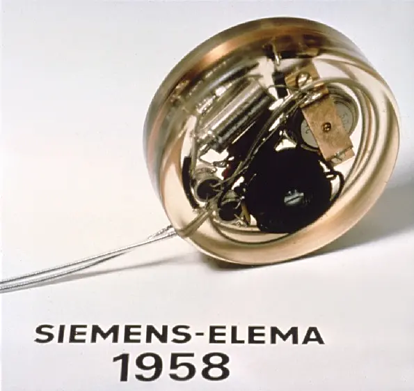
The SA node is the heart’s natural pacemaker: it starts the electrical impulse for each beat. If it fails to fire properly, or if the impulse cannot get through to the ventricles because of a block in the AV node or the bundle of His (heart block), the heart beats too slowly and the person feels faint, tired and breathless. They can be given an artificial pacemaker.
An artificial pacemaker is a small medical device that starts electrical impulses in the heart to keep it beating at a proper rate. Most modern ones watch the heart and fire only when its own beat is missing or too slow. It is put under the skin below the collarbone in a short operation, usually under local anesthetic. It has three parts:
- A pulse generator: a small, sealed metal case holding a lithium battery (lasting about 7–10 years) and the circuit that makes the electrical impulses.
- Leads: insulated wires that carry the impulses, threaded through a vein into the right side of the heart.
- Electrodes at the ends of the leads, touching the heart muscle, which deliver the impulses.
Cardiac cycle
The cardiac cycle is the sequence of events in one heartbeat: one round of contraction and relaxation of the heart. Contraction is systole and relaxation is diastole. At 72 beats a minute, one cycle lasts about 0.8 s (60 ÷ 72). It has three main phases:
| Phase | What happens | Duration |
|---|---|---|
| Atrial systole | Both atria contract together, topping up the ventricles (which have already filled about three-quarters by themselves). | about 0.1 s |
| Ventricular systole | Both ventricles contract together. The AV valves shut (lubb), the semilunar valves open, and blood is pumped into the pulmonary trunk and aorta. | about 0.3 s |
| Joint (complete) diastole | Atria and ventricles all relax. The semilunar valves shut (dub). Blood returning through the veins fills the atria and flows on through the open AV valves into the ventricles. | about 0.4 s |
So in each cycle the atria rest for 0.7 s and the ventricles for 0.5 s: the heart spends more of its life resting than working. During ventricular systole the atria are already relaxing and filling again.
While blood is being pushed out of the ventricles into the arteries, which valves are open?
Pick an answer to see why.
Cardiac output
The number of heartbeats in a minute is the heart rate (HR), about 72 at rest. It rises during exercise, fever, fear and anger.
The volume of blood pumped out by each ventricle in one beat is the stroke volume (SV), about 70 ml at rest.
The volume of blood pumped out by each ventricle in one minute is the cardiac output (CO):
At rest: CO = 72 × 70 ml = 5040 ml per minute, about 5 liters: roughly all the blood in the body, every minute.
Heart sounds and heart murmur
Each heartbeat makes sounds, which a doctor hears by putting a stethoscope (or an ear) on the chest. They are made mainly by the closing of the valves and the sudden stop and vibration of the blood behind them. Heart sounds tell a doctor a great deal about how the heart is working.
There are four heart sounds, but only the first two are normally heard in everyone:
- First heart sound (S1), “lubb”: made by the closing of the atrioventricular (tricuspid and bicuspid) valves at the start of ventricular systole, with the ventricles’ contraction. It is longer (about 0.15 s) and lower-pitched, a dull sound.
- Second heart sound (S2), “dub”: made by the closing of the semilunar valves at the end of ventricular systole. It is shorter (about 0.1 s), higher-pitched and sharper.
- Third heart sound (S3): a soft, low-pitched sound just after S2, made as blood rushes into the ventricles at the start of their filling. It is normal in children and young people, and in athletes; in an older adult it can be a sign of heart failure.
- Fourth heart sound (S4): a soft, low-pitched sound just before S1, made as the atria contract and push blood into a stiff ventricle. It is usually a sign of a stiffened ventricle (for example after years of high blood pressure).
A heart murmur is an unusual sound heard during a heartbeat, a whooshing or hissing made when blood flows turbulently through a valve or vessel.
- A damaged tricuspid or bicuspid valve changes the first heart sound.
- A damaged semilunar valve that does not shut properly lets blood leak back into the ventricle, and a soft hissing is heard after the second sound, “lubb-dub-shhh”.
Valves are damaged by diseases such as rheumatic fever (after an untreated throat infection by Streptococcus bacteria), syphilis and other infections. Some murmurs, in children, are harmless (“innocent murmurs”) and go away.
Blood pressure
As blood is pumped through the vessels, above all the arteries, it pushes on their walls. Blood pressure is the pressure exerted by the blood on the walls of the vessels it flows through. Measured in an artery, it is the arterial blood pressure.
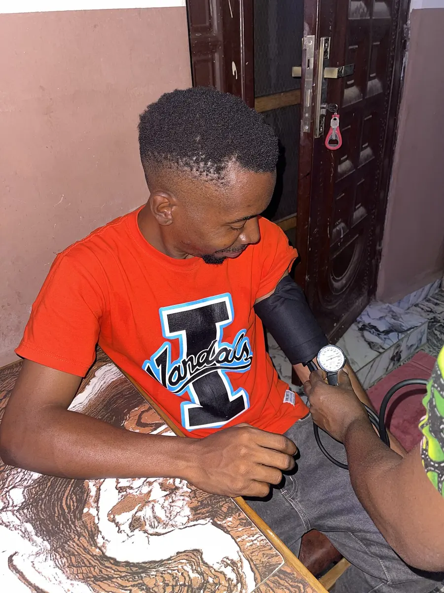
It is measured with a sphygmomanometer by the auscultatory method (listening with a stethoscope): a cuff round the upper arm is pumped up until it squeezes the brachial artery shut, then slowly let down while the doctor listens below it. The pressure at which the first tapping sounds are heard is the systolic pressure; the pressure at which they disappear is the diastolic pressure. The brachial artery is used because it is a large artery in the upper arm, at about the level of the heart, and easy to press against the bone.
In a healthy adult, blood pressure is about 120/80 mmHg (said “120 over 80”). It rises and falls in each cardiac cycle:
- Systolic blood pressure: the highest pressure in the arteries, during ventricular systole: about 120 mmHg (the upper limit).
- Diastolic blood pressure: the lowest pressure in the arteries, while the ventricles are relaxed: about 80 mmHg (the lower limit).
- Pulse pressure: the difference between the two, about 40 mmHg.
Blood pressure changes with exercise, excitement and other emotions, meals, sex, posture and time of day; it also tends to rise with age.
Problems related to blood pressure
Hypertension (high blood pressure) is blood pressure that stays at 140/90 mmHg or above (some newer guidelines take 130/80 as the line).
- Causes and risk factors: long-term stress, obesity, too much salt and fatty food, smoking, too much alcohol, lack of exercise, kidney disease (such as nephritis), and the narrowing and hardening of arteries (atherosclerosis), which also makes it worse. Often no single cause is found, and it runs in families.
- Harm: it overworks the heart and damages the walls of arteries, leading to heart attack, stroke (bleeding or a clot in the brain), kidney failure and damage to the eyes. It usually causes no symptoms for years, so it is called the “silent killer”: the only way to know is to measure it.
Hypotension (low blood pressure) is blood pressure that stays below about 90/60 mmHg.
- Causes: loss of blood (hemorrhage), loss of fluid by vomiting and diarrhea, heart problems, widening of blood vessels (as in a severe allergic reaction or infection), and some medicines.
- Effects: too little blood reaches the brain, so the person feels dizzy and may faint, especially on standing up suddenly.
| Hypertension | Hypotension | |
|---|---|---|
| Blood pressure | stays at or above 140/90 mmHg | stays below about 90/60 mmHg |
| Common causes | stress, obesity, salty and fatty food, smoking, kidney disease, narrowed arteries | blood loss, vomiting and diarrhea, heart problems, widened vessels |
| Effects | heart attack, stroke, kidney failure; often no symptoms | dizziness, fainting, tiredness |
A person’s blood pressure is 130/85 mmHg. What is the pulse pressure?
Pick an answer to see why.
Blood grouping
Blood grouping is the sorting of human blood into types by the antigens on the surface of the red blood cells and the antibodies in the plasma. The two best-known systems are the ABO system and the Rh system.
The ABO system was discovered in 1900–1901 by the Austrian doctor Karl Landsteiner, who found three groups, A, B and O (he called O “C”). The fourth, rarer group, AB, was found in 1902 by his colleagues in Vienna, Alfred von Decastello and Adriano Sturli. Landsteiner is called the father of blood grouping, and he received the Nobel Prize in Physiology or Medicine in 1930.
ABO blood grouping system
The ABO system depends on two antigens (agglutinogens), A and B, which may be present on the surface of the red blood cells, and two antibodies (agglutinins), anti-A (written a) and anti-B (b), which may be present in the plasma. A person never has the antibody against their own antigen. So there are four blood groups:
| Blood group | Antigen on the red blood cells | Antibody in the plasma |
|---|---|---|
| A | A | anti-B (b) |
| B | B | anti-A (a) |
| AB | A and B | none |
| O | none | anti-A and anti-B (a and b) |
When red blood cells carrying an antigen meet the antibody against it, the antibodies bind the cells together in clumps: agglutination. The clumped cells then burst (hemolysis).
Blood transfusion
Blood transfusion is the medical procedure of giving blood or its parts (red cells, plasma, platelets) from one person, the donor, into the bloodstream of another, the recipient. Before a transfusion the blood of both is grouped, and then cross-matched (a little of each mixed in the lab), to make sure they are compatible. An incompatible transfusion makes the recipient’s antibodies clump the donor’s red cells: they block small vessels, burst, and can cause clotting inside the vessels, kidney failure, circulatory failure and even death.
What matters is the donor’s antigens and the recipient’s antibodies:
- Group A blood can be given only to people without anti-A antibodies: A and AB.
- Group B blood can be given only to B and AB.
- Group AB blood can be given only to AB, since everyone else has anti-A, anti-B or both.
- Group O blood has no A or B antigens, so it can be given to all four groups.
| Recipient (plasma antibodies) ↓ / Donor (red cell antigens) → | A | B | AB | O |
|---|---|---|---|---|
| A (anti-B) | ✓ | clumps | clumps | ✓ |
| B (anti-A) | clumps | ✓ | clumps | ✓ |
| AB (none) | ✓ | ✓ | ✓ | ✓ |
| O (anti-A and anti-B) | clumps | clumps | clumps | ✓ |
So group AB is called the universal recipient (its plasma has no anti-A or anti-B), and group O the universal donor (its red cells have no A or B antigen).
Rabina’s blood clumps with anti-B serum but not with anti-A serum. Which group is she, and which antibody is in her plasma?
Pick an answer to see why.
Inheritance of blood group
The ABO blood group is set by one gene with three alleles: Iᴬ (makes antigen A), Iᴮ (makes antigen B) and i (sometimes written Iᴼ; makes neither). Each person carries two of these alleles, one from each parent. Iᴬ and Iᴮ are both dominant over i, and codominant with each other. So there are six genotypes and four phenotypes (blood groups):
| Genotype | Blood group | Antigen |
|---|---|---|
| IᴬIᴬ | A | A |
| Iᴬi | A | A |
| IᴮIᴮ | B | B |
| Iᴮi | B | B |
| IᴬIᴮ | AB | A and B |
| ii | O | none |
The blood groups of the parents tell which groups their children can and cannot have:
| Parents’ blood groups | Children: possible | Children: impossible |
|---|---|---|
| O × O | O | A, B, AB |
| O × A | O, A | B, AB |
| A × A | O, A | B, AB |
| O × B | O, B | A, AB |
| B × B | O, B | A, AB |
| A × B | O, A, B, AB | none |
| O × AB | A, B | O, AB |
| A × AB | A, B, AB | O |
| B × AB | A, B, AB | O |
| AB × AB | A, B, AB | O |
Rh grouping
Rh grouping sorts blood by the presence or absence of another antigen on the red blood cells, the Rh factor (Rhesus factor, the D antigen). It was discovered in 1940 by Karl Landsteiner and Alexander Wiener, who injected red blood cells of the rhesus monkey into rabbits and found that the antibodies the rabbits made also clumped the red cells of most people.
- Blood with the Rh antigen is Rh-positive (Rh+); blood without it is Rh-negative (Rh−). Rh-positive is dominant over Rh-negative.
- How common each is depends on the people: about 85% of Europeans are Rh-positive, and in most of Asia, including South Asia, the great majority are.
- Unlike anti-A and anti-B, anti-Rh antibodies are not present from birth. An Rh-negative person makes them only after Rh-positive blood enters their body.
So the first transfusion of Rh-positive blood into an Rh-negative person usually does little harm at once, but it makes the person sensitized: they start making anti-Rh antibodies. A second transfusion of Rh-positive blood is then attacked by these antibodies, which clump the cells, and it can be serious, even fatal. Rh-positive and Rh-negative blood are therefore never mixed in transfusion.
The same thing can happen in pregnancy: an Rh-negative mother carrying an Rh-positive baby can be sensitized by the baby’s blood at birth, and her antibodies may attack the red cells of her next Rh-positive baby (erythroblastosis fetalis, hemolytic disease of the newborn). An injection of anti-D given to the mother after each birth prevents this.
Identifying blood groups
A blood group is found with antisera: serum containing known antibodies.
- Antiserum A (anti-A) clumps red cells carrying antigen A.
- Antiserum B (anti-B) clumps red cells carrying antigen B.
- Antiserum D (anti-D) clumps red cells carrying the Rh antigen.
A drop of blood is mixed with a drop of each antiserum on a clean slide or tile. Clumping (agglutination) means the antigen is there; a smooth, evenly red drop means it is not.
| Blood group | Anti-A | Anti-B | Anti-D |
|---|---|---|---|
| A+ | clumps | smooth | clumps |
| A− | clumps | smooth | smooth |
| B+ | smooth | clumps | clumps |
| B− | smooth | clumps | smooth |
| AB+ | clumps | clumps | clumps |
| AB− | clumps | clumps | smooth |
| O+ | smooth | smooth | clumps |
| O− | smooth | smooth | smooth |
A sample of blood clumps with anti-A and anti-D, but not with anti-B. What is its group?
Pick an answer to see why.
Arterial system
The arterial system is the system of arteries and their branches that carry blood away from the heart to every part of the body. All carry oxygenated blood except the pulmonary arteries, which carry deoxygenated blood to the lungs. It can be studied as:
A. Pulmonary arteries. The pulmonary trunk leaves the right ventricle and divides into the right and left pulmonary arteries, which carry deoxygenated blood to the two lungs.
B. Systemic aorta, the largest artery of the body, about 2.5–3 cm wide at its start. It has three parts: the ascending aorta, the arch of the aorta and the descending aorta.
- The ascending aorta gives off the right and left coronary arteries, supplying oxygenated blood to the heart muscle.
- The arch of the aorta gives off three branches: the brachiocephalic (innominate) artery, the left common carotid artery and the left subclavian artery.
- The brachiocephalic artery is short; it divides into the right common carotid and the right subclavian arteries.
- Each common carotid artery (right and left) runs up the neck and, at the level of the upper border of the voice box, divides into:
- the external carotid, supplying its side of the face, scalp, neck, jaw and tongue;
- the internal carotid, entering the skull to supply the brain, eyes, forehead and nose.
- Each subclavian artery (right and left) gives off:
- the vertebral artery, which runs up through holes in the neck vertebrae to the base of the brain;
- and then continues into the armpit as the axillary artery (supplying the shoulder and chest wall), which continues down the upper arm as the brachial artery. Below the elbow the brachial divides into the radial and ulnar arteries of the forearm and hand. (The pulse felt at the wrist is in the radial artery.)
- The descending aorta runs down through the thorax (thoracic aorta), passes through the diaphragm, and continues through the abdomen (abdominal aorta). Its main branches, from top to bottom, are:
- Posterior intercostal arteries: to the ribs and the muscles between them (intercostal muscles).
- Esophageal arteries: to the esophagus (and small bronchial arteries to the lungs’ own tissue).
- Inferior phrenic arteries: to the underside of the diaphragm.
- The celiac trunk: a short, single artery that divides at once into three:
- the left gastric artery, to the stomach (and the lower esophagus);
- the splenic artery, to the spleen, pancreas and part of the stomach;
- the common hepatic artery, to the liver, gall bladder, stomach, duodenum and pancreas.
- The superior mesenteric artery: a long, single branch to the small intestine, part of the pancreas, the cecum, and the ascending and transverse colon.
- Renal arteries (a pair): to the kidneys.
- Gonadal arteries (a pair): to the testes in a man (testicular arteries) or the ovaries in a woman (ovarian arteries).
- Lumbar arteries (four pairs): to the body wall and the vertebrae at the back of the abdomen.
- The inferior mesenteric artery: a single branch to the rest of the large intestine (the descending and sigmoid colon and the rectum).
- At the level of the fourth lumbar vertebra the aorta ends by dividing into the right and left common iliac arteries.
- Each common iliac artery divides into:
- the internal iliac artery, short, supplying the organs of the pelvis (bladder, rectum, reproductive organs) and the buttock;
- the external iliac artery, which runs down into the thigh as the femoral artery. Behind the knee the femoral becomes the popliteal artery, which divides just below the knee into the anterior tibial and posterior tibial arteries of the leg and foot.
BodyParts3D, © The Database Center for Life Science, CC BY 4.0
Which of these arteries is not a branch of the descending aorta?
Pick an answer to see why.
A doctor feels your pulse at the wrist, on the thumb side. Which artery is it?
Pick an answer to see why.
Venous system
The venous system is the system of veins and their branches that collect blood from every part of the body and carry it back to the heart. All carry deoxygenated blood except the pulmonary veins. It consists of:
- Pulmonary veins: two from each lung (four in all), bringing oxygenated blood from the lungs to the left atrium.
- Coronary veins: collecting deoxygenated blood from the heart muscle into the coronary sinus, which opens into the right atrium.
- The superior vena cava (precaval vein): collecting blood from the head, neck, upper limbs and chest wall and pouring it into the right atrium. It is formed by the union of the right and left brachiocephalic veins. Each brachiocephalic vein is formed, behind the collarbone, by the union of:
- the internal jugular vein, collecting blood from the brain, the inside of the skull, the face and the neck;
- the subclavian vein, collecting blood from the upper limb (it is the continuation of the axillary vein, which receives the arm’s deep veins and the cephalic and basilic veins just under the skin). The external jugular vein, collecting blood from the scalp, face and jaw, joins the subclavian vein.
- The inferior vena cava (postcaval vein), the largest vein: collecting blood from the parts of the body below the diaphragm and opening into the right atrium. It is formed, at the level of the fifth lumbar vertebra, by the union of the two common iliac veins, and receives on its way up:
- Common iliac veins, each formed by the union of:
- the external iliac vein, which carries the blood of the whole lower limb. It is the continuation of the femoral vein, which receives the deep veins of the leg (the anterior and posterior tibial veins, joining as the popliteal vein behind the knee) and the great saphenous vein, the longest vein in the body, running under the skin from the inner side of the foot to the groin;
- the internal iliac (hypogastric) vein, collecting blood from the pelvic organs: the rectum, urinary bladder, uterus and vagina, or the prostate gland, seminal vesicles and penis, and the buttock.
- Lumbar veins: from the back wall of the abdomen.
- Gonadal (testicular or ovarian) veins: from the testes or ovaries. The right one enters the inferior vena cava; the left one enters the left renal vein.
- Renal veins: from the kidneys.
- Suprarenal (adrenal) veins: from the adrenal glands. The right one is short and empties into the inferior vena cava; the left one is longer and empties into the left renal vein.
- Hepatic veins: from the liver, just below the diaphragm.
- Inferior phrenic veins: from the underside of the diaphragm.
- Common iliac veins, each formed by the union of:
BodyParts3D, © The Database Center for Life Science, CC BY 4.0
A surgeon needs a length of vein to bypass a blocked coronary artery. Which vein is usually taken from the leg?
Pick an answer to see why.
Hepatic portal system
A portal system is a vein that begins in capillaries in one organ and ends in capillaries in another, instead of going straight to the heart: blood passes through two capillary beds in a row.
The hepatic portal vein collects blood from the alimentary canal (stomach and intestines), the pancreas, the spleen and the gall bladder, and carries it to the liver. It is formed behind the pancreas by the union of the superior mesenteric and splenic veins, and receives:
- the cystic vein, from the gall bladder;
- the splenic vein, from the spleen, pancreas and part of the stomach (the inferior mesenteric vein usually joins the splenic vein);
- the gastric veins, from the stomach and the lower end of the esophagus;
- duodenal veins, from the duodenum;
- the mesenteric veins, from the intestines.
These veins bring nutrient-rich, deoxygenated blood into the hepatic portal vein, which divides into capillaries (sinusoids) inside the liver. Only after passing through the liver does the blood leave by the hepatic veins into the inferior vena cava and so to the heart. This is the hepatic portal system.
Significance of the hepatic portal system:
- Nutrients absorbed in the intestine (glucose, amino acids, vitamins, minerals) are taken first to the liver, which stores them (glucose as glycogen, iron, vitamins A and B₁₂), changes them, or sends them on as the body needs: so the blood leaving for the body has a steady level of glucose, whether you have just eaten or not.
- Toxins absorbed from the gut (and many drugs and alcohol) are detoxified in the liver before they reach the rest of the body.
- Germs that slip through the gut wall are destroyed by the liver’s Kupffer cells.
- Ammonia, made when amino acids are broken down, is turned into the less toxic urea in the liver, to be removed by the kidneys.
Why does blood from the intestines go to the liver before it goes back to the heart?
Pick an answer to see why.
Exercise
A. Tick the correct alternatives
- Which of the following layers of the pericardium is closest to the heart?
Pick an answer to see why.
- Which of the following layers consists of cardiac muscle?
Pick an answer to see why.
- What are the muscular ridges on the inner surface of the atria called?
Pick an answer to see why.
- The tricuspid valve is situated between the:
Pick an answer to see why.
- In the embryo, the foramen ovale is the opening between the right atrium and the:
Pick an answer to see why.
- Which of the following statements best describes the role of the SA node in the heartbeat?
Pick an answer to see why.
- Rashmi’s grandfather, 65 years old, has been living with a dangerously slow heartbeat. If he consults a cardiologist, he is most likely to be advised to:
Pick an answer to see why.
- Which of the following events is responsible for the “lubb dub” sound of the heartbeat?
Pick an answer to see why.
- To which of the following does the bundle of His carry the impulse of contraction?
Pick an answer to see why.
- Which of the following statements is incorrect?
Pick an answer to see why.
- Sushant, who doesn’t know his blood group, is hurt in an accident and needs a blood transfusion at once. A friend who has a valid certificate of his own blood group offers blood at once, and it is accepted without testing Sushant’s blood. What was the donor’s blood group?
Pick an answer to see why.
- Through which blood vessel does the left atrium receive oxygenated blood?
Pick an answer to see why.
- Which of the following valves is present at the opening of the coronary sinus?
Pick an answer to see why.
- From which of the following does the hepatic portal vein bring blood to the liver?
Pick an answer to see why.
- Which of the following is associated with a decreased cardiac output?
Pick an answer to see why.
- Which of the following is used to detect the presence or absence of the Rh antigen on red blood cells?
Pick an answer to see why.
- Which of the following arteries supplies oxygenated blood to the diaphragm?
Pick an answer to see why.
- Which of the following is the longest vein in the human body?
Pick an answer to see why.
- Which phase of the cardiac cycle involves the flow of blood from the ventricles to the different parts of the body?
Pick an answer to see why.
- Mesenteric veins collect deoxygenated blood from the:
Pick an answer to see why.
B. Answer the following questions in brief
Describe the external structure of the human heart.
Answer
Draw a well-labeled diagram of the internal structure of the human heart.
Answer
Describe the mechanism of the origin and conduction of the heartbeat.
Answer
What is blood pressure? Differentiate between hypotension and hypertension. What are the causes of hypertension?
Answer
Observe the diagram given and answer the following questions.
Servier Medical Art (smart.servier.com), CC BY 4.0
- i.Identify the parts A and B.
- ii.What are their functions?
- iii.What is the remedy when part A fails to work?
Answer
Describe in brief the arterial system of humans.
Answer
Write a brief note on human blood groups.
Answer
Draw a neat, labeled diagram of the venous system of the human body.
Answer
C. Long answer questions
What is a myogenic heart? Explain the structure and working mechanism of the human heart.
Answer
Describe the venous system of the human body. Which type of portal system is found in humans? Explain.
Answer
Printable PDFs
- Study notes 5 pages
- Worksheet 4 pages Answer key
Quiz yourself: Life Processes and the Human Body, Grades 5–12 →
Practice every question in the book, and keep track of the ones you miss →
A companion to your textbook, not a replacement for it: study from the book your school uses, and where the two differ, follow your teacher. Found a mistake? Please tell us.