
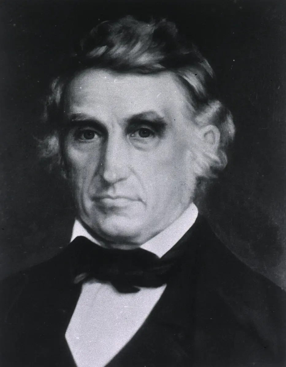
On 6 June 1822, on Mackinac Island in North America, a shotgun went off by accident a meter from a young fur trader, Alexis St. Martin. The blast tore open his side and his stomach. The army surgeon, William Beaumont, did not expect him to live. He lived, but the wound never closed: it healed into a hole, a little window from the skin into the stomach. Over the next ten years Beaumont lowered pieces of meat, bread and cabbage on silk threads through the window and pulled them out again to see what had happened to them, and drew off the stomach’s juice to watch it digest food in a glass jar at body warmth. He showed that digestion in the stomach is chemical, done by an acid juice, and not just grinding or rotting. He is remembered as the father of gastric physiology.
This unit follows a meal through your own body: the long tube it travels down, the glands that pour their juices into it, the wall of the tube, and what happens to each kind of food on the way, until what is useful is in your blood and the rest leaves as feces.
8.1.1 Nutrition and digestion
Nutrition is the process by which living things obtain the substances they need for growth, development and energy. Food is anything taken in to supply them, and nutrients are the chemical substances in food that the body uses: for growth and repair of tissues, for making gametes, for protection against disease, and for energy. They are of two kinds:
- organic nutrients: carbohydrates, lipids (fats), proteins and vitamins;
- inorganic nutrients: minerals and water.
Vitamins, minerals and water are small enough to be absorbed as they are. But carbohydrates, proteins and fats are mostly large molecules (starch, proteins, fats) that cannot pass through the wall of the gut or be used by the cells as they are eaten. They must first be broken down into small, soluble, diffusible molecules: starch into glucose, proteins into amino acids, fats into fatty acids and glycerol (or monoglycerides). This is digestion: the catabolic process that turns complex food into simple, absorbable forms. It happens in two ways, together:
- Mechanical (physical) digestion breaks large pieces of food into smaller ones, by chewing, churning and mixing, so that enzymes have a much larger surface to act on. It doesn’t change the molecules.
- Chemical (enzymatic) digestion breaks the large molecules into small ones by hydrolysis, with water, in reactions sped up by enzymes (hydrolases): starch to glucose, proteins to amino acids, and so on.
The organs that do this make up the digestive system: the set of organs concerned with the ingestion of food, its digestion, the absorption of the products, their assimilation into the body, and the egestion of what can’t be digested, as feces. It has two parts, the alimentary canal and the digestive glands; how they work together is the physiology of digestion.
Which of these needs to be digested before it can be absorbed?
Pick an answer to see why.
8.1.2 The alimentary canal: mouth and buccal cavity
The alimentary canal is a long, coiled tube, open at both ends (a complete gut), running from the mouth to the anus. It is about 9 meters long in a body after death; in a living person, whose muscles are partly tensed, it is shorter. Its width changes from part to part, and each part is shaped for its own job. In order, its parts are the mouth, vestibule, buccal cavity, pharynx, esophagus, stomach, small intestine, large intestine, and anal canal with the anus.
Servier Medical Art (smart.servier.com), CC BY 4.0
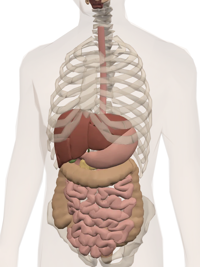
A model of a real body. Tap Show in 3D to turn it and tap its parts.
TongueTurns the food as you chew and pushes it back to be swallowed.
Salivary glandsMake saliva while you chew. Food wet with saliva is easy to swallow, and its amylase starts turning starch into sugar.
Food pipe (esophagus)Carries the swallowed food from the mouth down to the stomach.
StomachIts gastric juice has hydrochloric acid, which kills most germs in the food, and pepsin, which starts digesting protein. The food is churned into a thick paste.
LiverMakes bile, which breaks fat into tiny drops so it digests easily.
Gall bladderStores the bile the liver makes and sends it into the duodenum.
PancreasSends pancreatic juice into the duodenum: trypsin digests protein, amylase starch and lipase fat.
DuodenumThe first part of the small intestine, where bile and pancreatic juice join the half-digested food.
Small intestineIntestinal juice finishes digestion here, and millions of tiny finger-like villi on its inside wall pass the digested food into the blood.
Large intestineTakes most of the water back into the body from what is left.
AppendixA small closed tube off the start of the large intestine.
RectumHolds the waste until it leaves the body through the anus.
Body and organs: BodyParts3D, The Database Center for Life Science (CC BY 4.0)
1. Mouth
The mouth is a slit across the face just below the nose, bounded by two movable lips, the upper and the lower. It takes in food (ingestion); the lips hold it in while it is chewed.
2. Vestibule
Behind the lips is a narrow space, the vestibule, between the lips and cheeks on the outside and the teeth and gums on the inside. (Run your tongue round the outside of your teeth: it is moving in your vestibule.)
3. Buccal (oral) cavity
The vestibule opens into the large buccal cavity, lined by stratified squamous epithelium, a lining many cells thick that stands up to rubbing by hard food. It holds the palate as its roof, the tongue on its floor, and the teeth round its edge.
Servier Medical Art (smart.servier.com), CC BY 4.0
a. Palate. The palate is the roof of the buccal cavity. Its front part, the hard palate, is made by the palatine bones and the maxillae (upper jaw bones); it is arched and carries ridges across it, the palatine rugae, against which the tongue presses and grips food. The back part, the soft palate, is smooth and fleshy, made of muscle; it helps food to be swallowed smoothly. From its back edge hangs a small cone of muscle, the uvula (velum palati). When you swallow, the soft palate and uvula rise and close off the passage from the internal nostrils, so that food and drink don’t go up into the nose.
b. Tongue. The tongue is a flat, roughly triangular, muscular organ on the floor of the buccal cavity. It is made of striated (voluntary) muscle, in bundles that run lengthwise, across and up and down, so it can change its shape and position in almost any way. Its front is free; under it a fold of mucous membrane, the lingual frenulum, ties it to the floor of the mouth; its back is fixed to the hyoid bone and the mandible (lower jaw). A shallow groove shaped like an upside-down V, the terminal sulcus, divides its upper surface into the front oral part (about two-thirds) and the back pharyngeal part (about one-third).
The upper surface is rough with small projections, the papillae. Many of them carry taste buds, the small groups of cells that detect taste. Humans have three kinds of papillae:
| Papillae | Where | Shape and number | Taste buds |
|---|---|---|---|
| Circumvallate (vallate) | at the back of the oral part, in a V just in front of the terminal sulcus | the largest, each sunk in a circular trench; only 7–12 | many, in the walls of the trench |
| Fungiform | on the tip and edges, scattered among the filiform | like small mushrooms, red dots you can see; a few hundred | a few on each |
| Filiform | all over the front two-thirds | the smallest and most numerous, thin and pointed like threads | none: they give the tongue its rough grip on food |
Servier Medical Art (smart.servier.com), CC BY 4.0
The tongue also has glands of two kinds: von Ebner’s glands, which open into the trenches of the circumvallate papillae and secrete a watery fluid containing lingual lipase, and Weber’s glands, along its edges, which secrete mucus.
The tongue detects five basic tastes: sweet, salty, sour, bitter and umami (the savoury taste of meat broth, tomatoes, mushrooms and the monosodium glutamate in instant noodles). Each taste bud holds receptor cells for all five, and taste buds sit on the tip, edges and back of the tongue, so every taste can be detected by every part of the tongue that has taste buds. The middle of the top surface has few taste buds and tastes weakly.
The tongue’s jobs: it tastes food; it moves food between the teeth and mixes it with saliva during chewing; it rolls the chewed food into a ball, the bolus, and pushes it back for swallowing; and it shapes sounds in speech.
Which papillae of the human tongue carry no taste buds?
Pick an answer to see why.
8.1.3 Teeth
Teeth are hard structures fixed in the upper and lower jaws. They develop from two germ layers: the enamel from the ectoderm, the rest from the mesoderm. Human teeth are:
- thecodont: each tooth sits in a socket in the jaw bone;
- diphyodont: two sets grow in a lifetime, the milk (deciduous) teeth and the permanent teeth;
- heterodont: the teeth are of different kinds, shaped for different jobs.
The milk teeth come through from about 6 months to 2½ years of age. From about 6 years they fall out one by one and permanent teeth replace them, a process that lasts until about 12. The permanent molars have no milk teeth before them: the first comes at about 6, the second at about 12, and the third molars, the wisdom teeth, between about 17 and 25, if at all.
By shape and job there are four kinds:
- Incisors, at the front: flat with a sharp, chisel-like edge, for cutting and biting.
- Canines, one each side of the incisors: pointed like a dagger, for tearing and piercing.
- Premolars, behind the canines: broad with two cusps, for crushing and grinding. They have one root or two.
- Molars, at the back: the broadest, with four or five cusps, for grinding. Upper molars have three roots, lower molars two.
A dental formula shows how many teeth of each kind there are in half of the upper jaw (above the line) and half of the lower jaw (below), in the order incisors, canines, premolars, molars; multiplying by 2 gives both sides.
A child has no premolars: the two milk molars on each side are replaced by the two permanent premolars, and the permanent molars come in behind them.
Structure of a tooth
A tooth cut lengthwise (an L.S.) shows three regions:
- Crown: the shiny part above the gum. It is covered by enamel, made by cells called ameloblasts before the tooth comes through. Enamel is about 96% mineral (calcium phosphate as hydroxyapatite), the hardest substance in the body; once the tooth is through, the ameloblasts are gone, so enamel can never be remade.
- Neck: the short part where crown meets root, at the gum line.
- Root: the part fixed in the jaw bone, covered by a thin bony layer, the cement (cementum). The periodontal ligament (periodontal membrane), with its fibers and blood vessels, ties the cement to the bone of the socket and cushions the tooth.
The bulk of the tooth, under the enamel and cement, is dentine (ivory), made by odontoblasts lining the inside of the tooth. In the middle is the pulp cavity, filled with pulp: loose connective tissue with blood vessels and nerves, which enter through a small hole at the root’s tip, and odontoblasts round its edge.
Servier Medical Art (smart.servier.com), CC BY 4.0
How many premolars does a full set of adult human teeth have in all?
Pick an answer to see why.
8.1.4 Pharynx, esophagus and stomach
4. Pharynx
The pharynx (throat) is a short tube, about 12 cm long, behind the nasal cavity, the buccal cavity and the larynx. It is the common passage for food and air, and it is partly divided into three regions:
- the nasopharynx, at the top, behind the nose: the internal nostrils and the two Eustachian tubes from the middle ears open into it;
- the oropharynx, in the middle, behind the mouth: food and air both pass through it;
- the laryngopharynx, at the bottom: it leads into the larynx and windpipe through the glottis, in front, and into the esophagus through the gullet, behind.
A leaf-shaped flap of elastic cartilage, the epiglottis, stands above the glottis. When you swallow, the larynx rises and the epiglottis folds back over the glottis, so that food goes into the esophagus and not into the windpipe.
The pharynx is guarded by patches of lymphoid tissue, the tonsils: the pharyngeal tonsil (adenoids) in the roof of the nasopharynx, the tubal tonsils round the openings of the Eustachian tubes, the palatine tonsils on each side of the oropharynx, and the lingual tonsil on the back of the tongue. Together they make a ring round the entrance to the throat, Waldeyer’s ring, which catches germs coming in with food and air.
5. Esophagus
The esophagus (food pipe, gullet) is a narrow muscular tube about 25 cm long. It runs down behind the windpipe, through the chest, and passes through the diaphragm at the level of the 10th thoracic vertebra, through an opening called the esophageal hiatus. It opens into the stomach at the cardiac orifice, kept closed between swallows by a ring of muscle, the cardiac sphincter (lower esophageal sphincter), which stops food and acid flowing back up.
Swallowed food travels down it as a ball, the bolus, pushed by peristalsis: a wave of contraction of the smooth muscle in the wall, squeezing the tube shut behind the food while the muscle just ahead of it relaxes. Peristalsis moves food along the whole alimentary canal, which is why you can swallow lying down, or even upside down.
The esophagus is lined by non-keratinized stratified squamous epithelium, many cells thick, to stand up to rubbing by hard food. Its inner lining lies in long folds, so the tube can stretch to let a bolus through. It makes no digestive enzymes; it is kept slippery by mucus from small glands in its submucosa. Its outer layer is not a serosa but a fibrous coat, the tunica adventitia, because most of it lies in the chest, outside the body cavity of the abdomen.
6. Stomach
The stomach is the widest part of the alimentary canal: a J-shaped muscular bag that can stretch to hold a liter or two and more. It lies in the upper left of the abdomen, just below the diaphragm, slanting across the body, about 25–30 cm long and 15 cm wide when full. It is held in place by folds of peritoneum, the omenta; in the embryo it hangs from the back wall of the abdomen by one fold, the mesogaster (dorsal mesogastrium), which grows into the greater omentum. Its inner, concave edge is the lesser curvature; its outer, convex edge is the greater curvature. From the greater curvature hangs a large apron of peritoneum, the greater omentum, which covers the intestines in front and stores fat. It is called the “policeman of the abdomen”, because it moves to wrap an inflamed organ (a burst appendix, say), walling off the infection.
The stomach has four regions:
- Cardia: the narrow upper part, round the cardiac orifice, where the esophagus opens in.
- Fundus: the dome that bulges up beside the cardia, to the left and above it; it usually holds swallowed air (what you see as a bubble on an X-ray).
- Body (corpus): the large middle part. Its lining lies in folds, the gastric rugae, which flatten out as the stomach fills.
- Pylorus: the narrower last part, the wide pyloric antrum and then the narrow pyloric canal, ending at the pyloric sphincter, which lets food out into the duodenum a little at a time.
Servier Medical Art (smart.servier.com), CC BY 4.0
The stomach’s lining is dotted with millions of tiny pits, the openings of the gastric glands (8.1.6). Its muscle coat has three layers instead of two, an extra oblique layer inside the circular one, so it can churn food thoroughly. The stomach stores a meal for a few hours, churns it and mixes it with gastric juice, starts the digestion of proteins, and absorbs a little: water, alcohol, and some drugs such as aspirin.
Which part of the stomach usually holds a bubble of swallowed air?
Pick an answer to see why.
8.1.5 Small and large intestine
7. Small intestine
The small intestine is a narrow, coiled, muscular tube, the longest part of the canal: about 6–7 m measured after death, but only about 3 m in a living person, measured by X-ray or with a tube passed down it, because its muscle is partly contracted. Folds of peritoneum, the mesenteries, hold its coils in place and carry its blood vessels, nerves and lymph vessels. Its lining is raised into permanent ring-shaped folds, the plicae circulares (valves of Kerckring), which slow the food and give a bigger surface. It has three parts (their lengths, like the 6–7 m, are measured after death):
i. Duodenum. The first, shortest and widest part, a C-shaped loop about 25–30 cm long round the head of the pancreas. It receives bile from the liver and pancreatic juice from the pancreas through a common opening, the ampulla of Vater (hepatopancreatic ampulla, or hepatopancreatic duct), guarded by the sphincter of Oddi. Its wall holds two kinds of glands: the simple tubular crypts of Lieberkühn in the mucosa, as all along the small intestine, and the branched tubular Brunner’s glands, found only in the duodenum, in the submucosa. Its villi are broad and leaf-like. Most chemical digestion happens here.
ii. Jejunum. The middle part, about 2.5 m long. Its wall is thicker and has more blood vessels than the ileum’s, its plicae are large and close together, and its villi are tall. It is where most of the digested food is absorbed.
iii. Ileum. The last and longest part, about 3.5 m long and much coiled. Its villi are thinner and fewer. It absorbs what the jejunum leaves, and it is the only part that absorbs vitamin B₁₂ (with intrinsic factor) and the bile salts, which go back to the liver to be used again. Its wall holds patches of lymphoid tissue, Peyer’s patches, with white cells that check the gut’s contents for germs; they are called the intestinal tonsils, and are part of the MALT (mucosa-associated lymphoid tissue). The ileum opens into the cecum of the large intestine through the ileocecal opening, guarded by the ileocecal valve, which stops the contents of the large intestine from coming back.
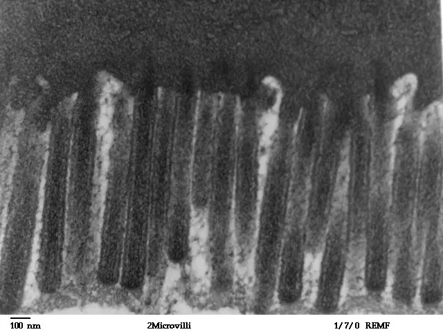
All along the small intestine the lining is covered with millions of tiny finger-like projections, the villi (about 1 mm long), each with blood capillaries and a lymph vessel, the lacteal, inside it. The cells covering each villus carry thousands of even tinier projections, the microvilli, which look like the bristles of a brush under the microscope: a brush border. Folds, villi and microvilli together give the small intestine an absorbing surface of about 30 m², nearly half a classroom’s floor.
Servier Medical Art (smart.servier.com), CC BY 4.0
8. Large intestine
The small intestine leads into the large intestine, a wider muscular tube about 1.5 m long, which frames the coils of the small intestine. It has no villi, though its cells still carry short microvilli. It absorbs most of the water and salts left in the food, stores the waste for a time, and passes it out as feces. It has three parts:
- Cecum: a small pouch, closed at its lower end, into which the ileum opens through the ileocecal valve. From it hangs a thin tube like a worm, about 8 cm long, the vermiform appendix. It is rich in lymphoid tissue and seems to work as a store of helpful gut bacteria that can re-seed the colon after an infection.
- Colon: the main part, an upside-down U round the small intestine, in four parts: the ascending colon up the right side, the transverse colon across, the descending colon down the left side, and the S-shaped sigmoid colon into the pelvis. Its outer longitudinal muscle is gathered into three ribbons running along it, the teniae coli; being shorter than the tube, they pucker it into a row of pouches, the haustra, which give it its bulging look. Its bacteria, such as Escherichia coli, make vitamin K and some B vitamins (such as biotin), and part of them is absorbed.
- Rectum: the last part, about 12–15 cm long, a slightly wider tube with folds inside and large blood vessels in its wall. It stores feces until defecation.
Servier Medical Art (smart.servier.com), CC BY 4.0
9. Anal canal and anus
The rectum leads into the anal canal, about 3–4 cm long, whose lining is raised into 6–10 lengthwise folds, the anal columns. It opens to the outside at the anus, between the buttocks. Two rings of muscle keep it closed: the internal anal sphincter, of smooth muscle, not under our control (involuntary), and the external anal sphincter, of striated muscle, which we control (voluntary). That is why toilet training is possible.
Which part of the small intestine absorbs vitamin B₁₂?
Pick an answer to see why.
8.1.6 Digestive glands: salivary and gastric glands
The juices that digest food come from digestive glands: some in the wall of the canal itself (the gastric and intestinal glands), others outside it, pouring their juice in through ducts (the salivary glands, liver and pancreas).
1. Salivary glands
Three pairs of salivary glands lie outside the buccal cavity and pour saliva into it through ducts:
| Gland | Where | Duct, and where it opens | Share of saliva | Its saliva |
|---|---|---|---|---|
| Parotid (the largest) | in front of and below each ear | Stensen’s duct, into the vestibule opposite the upper second molar | about 25% | watery, rich in salivary amylase |
| Submandibular (medium) | under the angle of the lower jaw, below the floor of the mouth | Wharton’s duct, into the floor of the mouth beside the frenulum | about 70% | both watery and mucous: amylase and mucus |
| Sublingual (the smallest) | under the tongue, in the floor of the mouth | several small ducts of Rivinus, into the floor of the mouth | about 5% | mostly mucus, a little amylase |
Servier Medical Art (smart.servier.com), CC BY 4.0
Mumps is an infection of the parotid glands by a virus: they swell, so the face puffs out in front of the ears. The MMR (measles, mumps and rubella) vaccine protects against it; Nepal’s routine program gives children the MR vaccine instead, against measles and rubella only.
Saliva is a clear, slightly sticky fluid, about 1–1.5 liters a day, nearly neutral (pH about 6.8). It contains water (over 99%), salts (Na⁺, K⁺, Cl⁻, HCO₃⁻), salivary amylase (ptyalin), lingual lipase, lysozyme (which kills bacteria), mucus, and antibodies (immunoglobulin A). Its jobs:
- i.It moistens food and, with its mucus, makes the bolus slippery to swallow.
- ii.It keeps the mouth clean and moist, and lets us speak easily.
- iii.Its lysozyme and antibodies kill or hold back germs.
- iv.Its enzymes start digestion: salivary amylase begins splitting starch, and lingual lipase fats.
- v.It dissolves food, so the taste buds can taste it.
2. Gastric glands
The lining of the stomach holds millions of microscopic gastric glands: branched tubular glands opening into the bottoms of the gastric pits. Their secretion is gastric juice. They are made of several kinds of cells:
- Chief (peptic, zymogen) cells, deep in the gland, secrete the inactive enzyme pepsinogen, a little gastric lipase, and in some young mammals prorennin.
- Parietal (oxyntic) cells, large cells on the gland’s walls, secrete hydrochloric acid (HCl) and Castle’s intrinsic factor.
- Mucous cells, lining the gastric pits and the necks of the glands (mucous neck cells), secrete thick mucus with bicarbonate, which forms a protective coat on the lining and keeps the acid and pepsin from digesting the stomach itself.
- Enteroendocrine (argentaffin) cells, scattered among them, secrete hormones into the blood: the G cells, mostly in the pyloric antrum, secrete gastrin.
Servier Medical Art (smart.servier.com), CC BY 4.0
Gastric juice is a thin, clear, nearly colorless liquid, about 2–3 liters a day. It is strongly acidic: about pH 1.5–2.5 in adults, and less acidic in babies, whose milk feeds buffer the acid. It contains water, HCl, pepsinogen, gastric lipase, mucus and intrinsic factor (and, in calves and other young mammals, prorennin). It has no amylase of its own worth the name: the starch in a bolus goes on being digested by swallowed salivary amylase until the acid reaches it. The jobs of gastric juice:
- i.Its acid makes the stomach’s contents acidic, which activates pepsinogen into pepsin and gives pepsin the pH it works best at.
- ii.Its acid kills most germs swallowed with food.
- iii.Its acid dissolves the minerals in small pieces of bone and loosens connective tissue in meat.
- iv.Its pepsin starts the digestion of proteins.
- v.Its mucus makes the food slippery and protects the stomach’s lining.
Which cells of the gastric gland make hydrochloric acid?
Pick an answer to see why.
8.1.7 The liver
The liver is the largest gland and the largest organ inside the body, reddish-brown and weighing about 1.5 kg. It lies in the upper right of the abdomen, just under the diaphragm, protected by the lower ribs. A fold of peritoneum, the falciform ligament, divides it from the front into a large right lobe and a smaller left lobe; on its underside two small lobes, the quadrate and the caudate lobes, are counted with the right lobe.
Tucked under the right lobe is the gall bladder, a thin-walled, pear-shaped, greenish sac that stores bile and makes it more concentrated.
The ducts
Bile leaves the liver through the right and left hepatic ducts, which join into the common hepatic duct. This joins the cystic duct from the gall bladder to form the common bile duct, which runs down behind the duodenum. There it meets the main pancreatic duct (duct of Wirsung), and the two open together through the hepatopancreatic ampulla (ampulla of Vater) into the duodenum. Two rings of muscle control the flow: the sphincter of Boyden, round the end of the common bile duct, and the sphincter of Oddi, round the ampulla’s opening into the duodenum. Between meals they are closed, so bile backs up the cystic duct into the gall bladder and is stored; after a fatty meal they relax and the gall bladder contracts.
Inside the liver
The liver is wrapped in a thin coat of connective tissue, Glisson’s capsule, which also runs into the liver along the branches of its vessels and ducts. Under the microscope the liver is made of about a hundred thousand small units, the hepatic lobules, each about 1 mm across and roughly six-sided:
- In the middle of each lobule is a central vein (intralobular vein).
- From it, rows of liver cells, the hepatocytes, run outwards like the spokes of a wheel: the hepatic cords.
- Between the cords run wide, leaky blood vessels, the sinusoids, in which blood flows inwards to the central vein. Their walls hold Kupffer cells, phagocytes that eat bacteria, old red blood cells and debris. Beside them lie Ito cells (stellate cells), which store fat and vitamin A.
- Between the cells of each cord run tiny channels, the bile canaliculi, into which the cells secrete bile. Bile flows outwards, the opposite way to the blood, through the short canals of Hering into the bile ductules, which join into bile ducts.
- At the corners where lobules meet lie the portal canals, each with a portal triad: a branch of the hepatic portal vein (bringing blood rich in absorbed food from the gut), a branch of the hepatic artery (bringing oxygen-rich blood), and an interlobular bile duct (taking bile away).
Servier Medical Art (smart.servier.com), CC BY 4.0
What the liver does
The liver does hundreds of jobs. The main ones:
- It makes bile. Its cells secrete bile, a yellow-green alkaline liquid (pH 7.6–8.6), 0.5–1 liter a day, stored and concentrated in the gall bladder. Bile contains bile salts (sodium glycocholate and sodium taurocholate), bile pigments, cholesterol, lecithin and inorganic salts, including sodium bicarbonate; it has no enzymes. Its jobs:
- Its bile salts emulsify fats: they break large fat drops into tiny droplets, giving lipase a far larger surface to work on (as soap does to grease on a plate).
- Its bicarbonate helps to neutralize the acid chyme coming from the stomach, so the enzymes of the duodenum can work.
- Its bile salts help to absorb fats and the fat-soluble vitamins A, D, E and K.
- It slows the growth of some bacteria and helps to keep the intestine moving.
- It controls blood glucose. It turns extra glucose into glycogen and stores it (glycogenesis), turns glycogen back into glucose when blood glucose falls (glycogenolysis), and makes new glucose from amino acids, lactic acid and glycerol (gluconeogenesis).
- It works on fats. It turns extra glucose and amino acids into fats, makes cholesterol and the proteins that carry fats in the blood.
- It deals with extra amino acids and poisons. It removes the amino group (–NH₂) from amino acids the body doesn’t need (deamination), making poisonous ammonia, which it turns at once into much less poisonous urea (in the ornithine cycle) for the kidneys to remove. It also makes alcohol, drugs and other poisons harmless (detoxification).
- It cleans the blood. Its Kupffer cells eat bacteria, dead cells and foreign particles from the blood coming from the gut.
- It breaks down old red blood cells. Kupffer cells (with the spleen) break down worn-out red blood cells; the heme of their hemoglobin becomes the bile pigments biliverdin (green) and bilirubin (yellow), which leave in bile and color the feces. The iron is kept.
- It makes blood cells before birth. In the fetus the liver makes red blood cells: it is a hemopoietic organ.
- It makes plasma proteins: albumin, and the clotting factors fibrinogen and prothrombin.
- It stores iron and copper, the fat-soluble vitamins A, D, E and K, and vitamin B₁₂.
- It makes heat. With so much chemistry going on, the liver is one of the body’s main sources of heat at rest.
In a liver lobule, which way do blood and bile flow?
Pick an answer to see why.
8.1.8 The pancreas and the intestinal glands
4. Pancreas
The pancreas is a soft, lobed, pinkish-gray gland, about 12–15 cm long and about 80 g (60–100 g). It lies behind the stomach, across the back wall of the abdomen, with its head in the C of the duodenum, then its neck, body, and tail reaching to the spleen. Small ducts join into the main pancreatic duct (duct of Wirsung), which runs its whole length and joins the common bile duct at the ampulla of Vater. An accessory pancreatic duct (duct of Santorini), branching from it, drains the head and opens into the duodenum by itself, a little above.
The pancreas is a mixed (heterocrine) gland: it is both exocrine, pouring a digestive juice into a duct, and endocrine, releasing hormones into the blood.
- The exocrine part (about 98–99% of the gland) is made of countless small sacs, the acini. Each acinus is a ring of wedge-shaped secretory cells round a tiny central space, which leads into a ductule. The cells make the pancreatic juice; the acini lie in loose connective tissue with blood vessels, nerves and ducts.
- The endocrine part is about a million small, pale clusters of cells scattered among the acini, the islets of Langerhans. Their α cells secrete glucagon, their β cells (most of them) insulin, and their δ cells somatostatin; these hormones control blood glucose, and come again in the unit on the endocrine glands.
Servier Medical Art (smart.servier.com), CC BY 4.0
Pancreatic juice is clear and alkaline (about pH 8.4), 1–1.5 liters a day. Its sodium bicarbonate neutralizes the acid chyme. It contains:
- four inactive enzymes (proenzymes) for proteins: trypsinogen, chymotrypsinogen, procarboxypeptidase and proelastase;
- pancreatic amylase (amylopsin), for starch;
- pancreatic lipase (steapsin), for fats;
- nucleases (DNase and RNase), for nucleic acids.
5. Intestinal glands
The lining of the small intestine has glands of two kinds:
- Brunner’s glands: branched tubular glands found only in the duodenum, in its submucosa. They secrete thick alkaline mucus, rich in bicarbonate, which protects the duodenum’s wall from the acid chyme, and a little enzyme.
- Crypts of Lieberkühn: simple tubular glands, pits between the villi, all along the small intestine’s mucosa. They secrete the watery intestinal juice, and their bottoms hold the stem cells that replace the lining every 3–5 days.
Intestinal juice (succus entericus), 1.5–2 liters a day, is slightly alkaline (about pH 7.8). With it work the enzymes that finish digestion: enterokinase (enteropeptidase), peptidases (aminopeptidase and dipeptidase), maltase, isomaltase, sucrase, lactase, intestinal lipase, nucleotidases and nucleosidases. Most of these, in fact, are not poured out into the juice: they are fixed to the surface of the microvilli, the brush border, so the last splits happen right on the surface that absorbs the products.
Which of these is an endocrine part of the pancreas?
Pick an answer to see why.
8.1.9 The wall of the alimentary canal
From the esophagus to the anus the wall of the alimentary canal is built on one plan of four layers, the same as in the frog Grade 11: Grade 11, 8.4.4, changed a little in each part. From the outside in:
- Serosa: the outermost layer, a thin sheet of flat cells (mesothelium, part of the visceral peritoneum) on a little loose connective tissue. Its wet, smooth surface lets the organs slide over each other. (The esophagus has no serosa: it has a fibrous tunica adventitia instead.)
- Muscularis (muscularis externa): two layers of smooth muscle, an outer longitudinal and an inner circular layer, which make the movements of peristalsis and mixing. Between them lies a network of nerves and nerve cell bodies, the myenteric plexus (Auerbach’s plexus), which controls these movements. (The stomach has a third, oblique, layer inside the circular.)
- Submucosa: loose connective tissue with blood vessels, lymph vessels and nerves, and a second network of nerves, the submucous plexus (Meissner’s plexus), which controls the glands and the blood flow of the lining. In the duodenum it holds Brunner’s glands.
- Mucosa: the innermost layer, folded into pits, glands and (in the small intestine) villi. It has three layers of its own:
- the muscularis mucosae, a thin sheet of smooth muscle (inner circular, outer longitudinal) that moves the lining;
- the lamina propria, loose connective tissue with blood capillaries, lacteals, nerves and lymphoid tissue;
- the epithelium, the lining itself: a single layer of columnar cells, many of them glandular, in the stomach and intestines; and stratified squamous in the mouth, esophagus and the end of the anal canal, where food or feces rub hard on it.
Between which two layers does Auerbach’s (myenteric) plexus lie?
Pick an answer to see why.
8.1.10 Physiology of digestion
The physiology of digestion is the set of processes by which complex, insoluble food is broken down into simple, soluble substances, absorbed, and the waste expelled. Food contains carbohydrates, proteins, fats, vitamins, minerals and water. Carbohydrates, proteins and fats are hydrolyzed by enzymes (hydrolases) into glucose and other simple sugars, amino acids, fatty acids and glycerol; vitamins, minerals and water need no digestion. The whole process is kept running at the right pace by hormones of the gut (8.1.12) and by nerves, both the gut’s own plexuses and the vagus nerve from the brain (the sight or smell of food makes your mouth water and starts your stomach’s juice flowing before you have eaten anything).
Follow one kind of food through the canal here:
Digestion in the mouth
Food is chewed (masticated) by the teeth, helped by the tongue, cheeks and lips: mechanical digestion. The tongue turns the food over between the teeth and mixes it with saliva; the mucus of saliva wets it and binds it into a slippery bolus. Saliva also starts chemical digestion:
Up to about 30% of the starch in a meal is digested in the mouth and in the bolus before stomach acid stops the amylase. Lingual lipase starts on a little fat (it does most of its work later, in the acid stomach), and lysozyme kills bacteria. The bolus is then swallowed through the pharynx and carried down the esophagus by peristalsis. No digestion happens in the pharynx and esophagus themselves.
Digestion in the stomach
Food stays in the stomach for about 2–5 hours, longer for a fatty meal, churned and mixed with gastric juice by the stomach’s muscles. Its HCl kills most of the germs in the food and activates the protein enzymes:
Pepsin itself then activates more pepsinogen, so once a little pepsin is made the change speeds up: pepsin is an autocatalyst.
In calves and other young mammals the stomach also makes prorennin, activated by HCl into rennin (chymosin), which curdles milk so that it stays longer in the stomach to be digested:
Gastric lipase begins on fats, splitting a little of them:
By the end, the food is a thick, semi-liquid, acid mixture, the chyme. The pyloric sphincter lets it into the duodenum in small squirts.
Digestion in the small intestine
In the duodenum the chyme meets three juices: bile, pancreatic juice and intestinal juice.
Bile contains no enzymes. Its bicarbonate (with the pancreas’s) neutralizes the acid chyme and makes the duodenum’s contents slightly alkaline, which the enzymes there need. Its bile salts emulsify fats.
Pancreatic juice: its proenzymes are switched on in a chain. Enterokinase, an enzyme on the brush border of the duodenum, activates a little trypsinogen; the trypsin made then activates more trypsinogen and all the other proenzymes:
Then the pancreatic enzymes digest the food:
Intestinal juice and the brush border finish the job:
What is left, undigested and unabsorbed, passes into the large intestine, where no digestive enzymes are secreted. Its bacteria, though, ferment some of the fiber that our enzymes can’t touch, making gases and short fatty acids that the colon’s cells use as food.
Which enzyme starts the chain that activates the pancreas’s protein enzymes?
Pick an answer to see why.
8.1.11 Absorption, assimilation and egestion
Absorption
Absorption is the passage of the end products of digestion through the lining of the alimentary canal into the blood and lymph. About 90% of it takes place in the small intestine, where digestion is finished right against its lining. They are taken up by the cells covering the villi, by four mechanisms: simple diffusion, facilitated diffusion (through carrier proteins, down a concentration gradient), active transport (with carriers, using energy) and osmosis (water).
- Glucose and galactose are taken in by active transport, carried in together with sodium ions (Na⁺); Na⁺ is pumped out again on the other side of the cell, using ATP. When there is a lot of glucose in the gut, some also enters by facilitated diffusion.
- Fructose enters by facilitated diffusion.
- Amino acids (and short peptides) are taken in by active transport, mostly with Na⁺ too.
- Water follows the absorbed food and salts by osmosis.
- Water-soluble vitamins (the B group and C) are absorbed mostly by carrier proteins, some by diffusion; vitamin B₁₂ only with intrinsic factor, in the ileum.
- Minerals are absorbed by carriers, some by active transport (iron and calcium, mostly in the duodenum).
Sugars and amino acids pass from the cells into the blood capillaries of the villi, which drain into the hepatic portal vein and so go to the liver first.
Fats take a different road:
- Fatty acids and monoglycerides gather with bile salts and lecithin into tiny water-soluble balls, the micelles, which carry them through the watery contents of the gut to the surface of the cells.
- There the fatty acids and monoglycerides leave the micelles and diffuse through the cell membrane, which is itself made of lipids. The bile salts stay behind, to be used again, and are reabsorbed in the ileum.
- In the smooth endoplasmic reticulum of the cells they are joined back into fats (triglycerides), coated with protein and cholesterol into tiny droplets, the chylomicrons.
- Chylomicrons are too big to enter the blood capillaries; they pass into the lacteal of the villus.
- The lacteals drain into larger lymph vessels and the thoracic duct, which empties into the blood at the left subclavian vein, near the neck.
The fat-soluble vitamins A, D, E and K travel with the fats.
In the large intestine most of the remaining water and salts are absorbed, with some vitamins made by its bacteria (vitamin K and biotin) and some drugs (which is why some medicines are given as suppositories).
Assimilation
Assimilation is the use of the absorbed food by the body’s cells. The blood carries it to every tissue. Glucose is respired for energy or stored as glycogen and fat; amino acids are built into the cell’s own proteins (enzymes, muscle, antibodies, hormones); fatty acids and glycerol build cell membranes, or are stored as fat or respired.
Egestion (defecation)
Egestion is getting rid of the undigested remains of food as feces through the anus. Feces contain undigested fiber (cellulose), water, dead cells from the lining, huge numbers of bacteria (dead and alive, a third to a half of their dry mass), a little fat, salts and mucus. The mucus of the large intestine and rectum makes them slippery. Their brown color comes from stercobilin, made from bile pigment by bacteria; their smell from indole, skatole and sulphur compounds made by the bacteria. When the rectum fills, its stretching gives the urge to go; the rectum contracts, and the anal sphincters relax, the internal one by reflex and the external one when we choose.
Why do the products of fat digestion enter the lacteals instead of the blood capillaries?
Pick an answer to see why.
8.1.12 Hormones that control digestion
Several parts of the alimentary canal secrete hormones into the blood, which tell other parts when to secrete their juices and how fast to move food on. The main ones:
| Hormone | Made by | Acts on | Action |
|---|---|---|---|
| Gastrin | G cells in the stomach (pyloric antrum), when food arrives | stomach | makes the gastric glands secrete gastric juice, especially acid; stirs the stomach’s muscle |
| Gastric inhibitory peptide (GIP), one of the enterogastrones | duodenum, when fats and sugars arrive | stomach | slows gastric secretion and emptying (it also makes the pancreas release insulin) |
| Secretin | duodenum, when acid chyme arrives | pancreas, liver, stomach | makes the pancreas release a juice rich in bicarbonate; increases the flow of bile; reduces gastric secretion |
| Cholecystokinin (CCK) | small intestine (duodenum), when fats and proteins arrive | gall bladder, pancreas | makes the gall bladder contract and release bile; makes the pancreas secrete a juice rich in enzymes |
| Enterocrinin | small intestine | small intestine | said to stimulate the crypts of Lieberkühn |
| Duocrinin | duodenum | duodenum | said to stimulate Brunner’s glands |
Secretin, found in 1902 by William Bayliss and Ernest Starling, was the first hormone ever discovered; Starling coined the word hormone, from the Greek for “to set in motion”. Enterocrinin and duocrinin were named between the 1930s and 1950s from experiments with gut extracts, but were never purified, and they are left out of most newer books; the glands they were said to control are now known to be stirred mostly by nerves and by the food itself.
A meal rich in fat reaches the duodenum. Which hormone makes the gall bladder squeeze out bile?
Pick an answer to see why.
Exercise
A. Tick the correct alternatives
- Which type of gland is present in the stomach?
Pick an answer to see why.
- What is the hardest substance of the human body?
Pick an answer to see why.
- Which part of the alimentary canal is chiefly responsible for the absorption of nutrients?
Pick an answer to see why.
- Which of the following is the main function of the esophagus?
Pick an answer to see why.
- Which of the following changes trypsinogen into trypsin?
Pick an answer to see why.
- Which of the following ducts is also called the duct of Santorini?
Pick an answer to see why.
- The phagocytic cells of the liver are called…
Pick an answer to see why.
- What is the duct of the submandibular salivary gland called?
Pick an answer to see why.
- Which of the following best describes the effect of HCl in the stomach on enzyme activity?
Pick an answer to see why.
- A person with chronic diarrhea is losing a large amount of water and salts in watery feces. Which part of the digestive system normally takes back the last of the water, turning the liquid that reaches it into solid feces?
Pick an answer to see why.
- Where is bile produced in the body?
Pick an answer to see why.
- Which of the following is characteristic of human teeth?
Pick an answer to see why.
B. Answer the following questions in brief
What is the spacious cavity immediately behind the mouth? List the structures present in it and describe them briefly.
Answer
Observe the diagram and answer the questions.
Servier Medical Art (smart.servier.com), CC BY 4.0
- a.Label the parts A, B and C.
- b.What do A, B and C secrete? What is the function of their secretion?
- c.Name their ducts.
Answer
Describe the structure of the L.S. of a human tooth.
Answer
Identify the gland that produces bile juice. Describe its morphology, anatomy and functions.
Answer
Describe the process of digestion in the small intestine.
Answer
What is absorption? How are the end products of carbohydrate, protein and fat digestion absorbed?
Answer
Describe the digestion of protein throughout the gastrointestinal tract.
Answer
Observe the diagram and answer the questions.
Servier Medical Art (smart.servier.com), CC BY 4.0
- a.Label the parts A, B and C.
- b.Write any three differences between A and C.
- c.What are the functions of A and C?
Answer
C. Long answer questions
What is the digestive system? Describe the various organs of the alimentary canal involved in the digestion of food.
Answer
Elucidate the biochemical processes involved in the digestion of carbohydrates, proteins and fats, including the enzymes and their regulation.
Answer
Printable PDFs
- Study notes 6 pages
- Worksheet 4 pages Answer key
Quiz yourself: Life Processes and the Human Body, Grades 5–12 →
Practice every question in the book, and keep track of the ones you miss →
A companion to your textbook, not a replacement for it: study from the book your school uses, and where the two differ, follow your teacher. Found a mistake? Please tell us.