
Every time one of your skin cells divides, it copies about six billion letters of DNA, a thread about two meters long, packed into a nucleus a few thousandths of a millimeter across. It does this in a few hours, while the cell goes on living, and it makes about one mistake in every billion letters. Then, all day long, it reads pieces of that thread, three letters at a time, and turns them into proteins: the hemoglobin in your blood, the keratin in your hair, the enzymes that digest your dal bhat.
The science of how this works began in a monastery garden. In the 1850s and 1860s Gregor Johann Mendel (1822–1884), a monk in Brünn in the Austrian Empire (now Brno, in the Czech Republic), crossed thousands of pea plants and counted how characters such as seed color and shape were passed on. He found that some characters are dominant and others recessive, and that they pass on by fixed rules. His work was hardly noticed in his lifetime, but it became the basis of modern genetics.
This unit opens the part of the book called genetics. It looks at the molecule that carries the instructions, DNA (and its working copy, RNA), at how DNA is copied so exactly, and at the code in which its instructions are written. The next unit follows those instructions from parents to children, as Mendel did with his peas.
3.1.1 Introduction to genetics
A buffalo always gives birth to a buffalo calf, never a goat, and a seed of a watermelon grows into a watermelon plant, never a pumpkin. Offspring resemble their parents. Relatives at a naming ceremony say “she has her father’s nose” or “his grandfather’s ears”. Yet the same children also differ: from their parents, and from each other.
Both facts need explaining, and three words name them.
Heredity is the passing of characters from one generation to the next. It is why offspring resemble their parents.
Variation is the differences between the individuals of a species, including between the offspring of the same parents. Some variation is inherited (from different genes); some comes from the surroundings (a plant in shade grows taller and paler than its twin in the sun).
Genetics is the branch of biology that studies heredity and variation. The word was coined in 1905 by the English biologist William Bateson, from the Greek genesis, origin.
Two sisters have the same blood group, but one is 12 cm taller than the other. Which statement is best?
Pick an answer to see why.
3.1.2 Genetic materials
The genetic material is the substance that carries the instructions for building and running an organism, and passes them from parents to offspring. It shapes everything about an organism: how it develops, what characters it has, and, over many generations, how its species changes. In almost all living things the genetic material is DNA; in some viruses it is RNA.
Both are nucleic acids: very large molecules (macromolecules), each a long chain of small units called nucleotides. Every nucleotide has three parts: a pentose sugar, a nitrogenous base and a phosphate group (from phosphoric acid).
1. Pentose sugar
A pentose is a sugar with five carbon atoms. Nucleic acids have two kinds: ribose in RNA, and deoxyribose in DNA. Deoxyribose (“de-oxy”, one oxygen less) has only an H on carbon 2′ where ribose has an –OH.
2. Nitrogenous bases
The bases are ring compounds containing nitrogen, of two classes:
a. Purines have two rings, a six-membered ring joined to a five-membered one: adenine (A) and guanine (G). Both are found in DNA and RNA.
b. Pyrimidines have one ring: cytosine (C), in both DNA and RNA; thymine (T), in DNA only; and uracil (U), in RNA only.
3. Phosphoric acid
Phosphoric acid, H₃PO₄, gives the phosphate group. In a chain, each phosphate joins carbon 3′ of one sugar to carbon 5′ of the next, by a phosphodiester bond. These bonds link nucleotides into a strand.
Which pair are both pyrimidines?
Pick an answer to see why.
3.1.3 Deoxyribonucleic acid (DNA)
DNA is the genetic material of every cellular organism and of most viruses; only the RNA viruses use RNA instead. Where it lies depends on the cell:
- In prokaryotic cells (bacteria), DNA lies in the cytoplasm, as one circular chromosome in a region called the nucleoid, often with small extra rings, the plasmids.
- In eukaryotic cells, DNA is mostly in the nucleus, in the chromosomes, and a little in the mitochondria and plastids (chloroplasts).
DNA is a very long molecule made of units called deoxyribonucleotides, each with deoxyribose, a phosphate and one of four bases (A, G, C or T). Along a strand, the units are joined by phosphodiester bonds.
Structure of DNA
In 1953 James Watson, an American biologist, and Francis Crick, a British scientist who had trained in physics, working at the University of Cambridge, proposed the double helix model of DNA. They built it from the X-ray photographs of DNA made by Rosalind Franklin and Maurice Wilkins in London, and from Erwin Chargaff’s measurements of the bases (the story is in Grade 10 Science).
The main features of the Watson and Crick model:
- DNA has two strands (polynucleotide chains) that twist round each other, and round a common axis, into a double helix. The helix is right-handed, like the thread of an ordinary screw.
- The two strands run in opposite directions: one runs 5′ → 3′, the other 3′ → 5′. They are antiparallel.
- The helix has a constant diameter of about 2 nm.
- Because the two backbones are not opposite each other, the gaps between them are of two widths: a wide major groove and a narrow minor groove, winding round the helix.
- One major groove and one minor groove together make one complete turn.
- One complete turn is about 3.4 nm long (the pitch of the helix).
- The sugar–phosphate backbone (deoxyribose and phosphate, taking turns) forms the outside of the helix, and the bases point inward, stacked flat like the steps of a spiral staircase.
- The bases of the two strands are joined in pairs by hydrogen bonds: adenine with thymine by two hydrogen bonds (A=T), and guanine with cytosine by three (G≡C). A purine always pairs with a pyrimidine, so every pair is the same width, and the helix stays 2 nm wide all along. Bases that pair like this are complementary: the order of bases on one strand fixes the order on the other.
- Neighboring base pairs are about 0.34 nm apart, so there are about 10 base pairs in one complete turn (3.4 ÷ 0.34 = 10).
- Since A always pairs with T, and G with C, a molecule of DNA has as much A as T, and as much G as C. So the total of the purines equals the total of the pyrimidines:
This is Chargaff’s rule, after Erwin Chargaff, who measured it in 1950.
Turn a real piece of DNA, every atom where X-ray crystallography put it. Find the two backbones, the base pairs between them, and the two grooves.
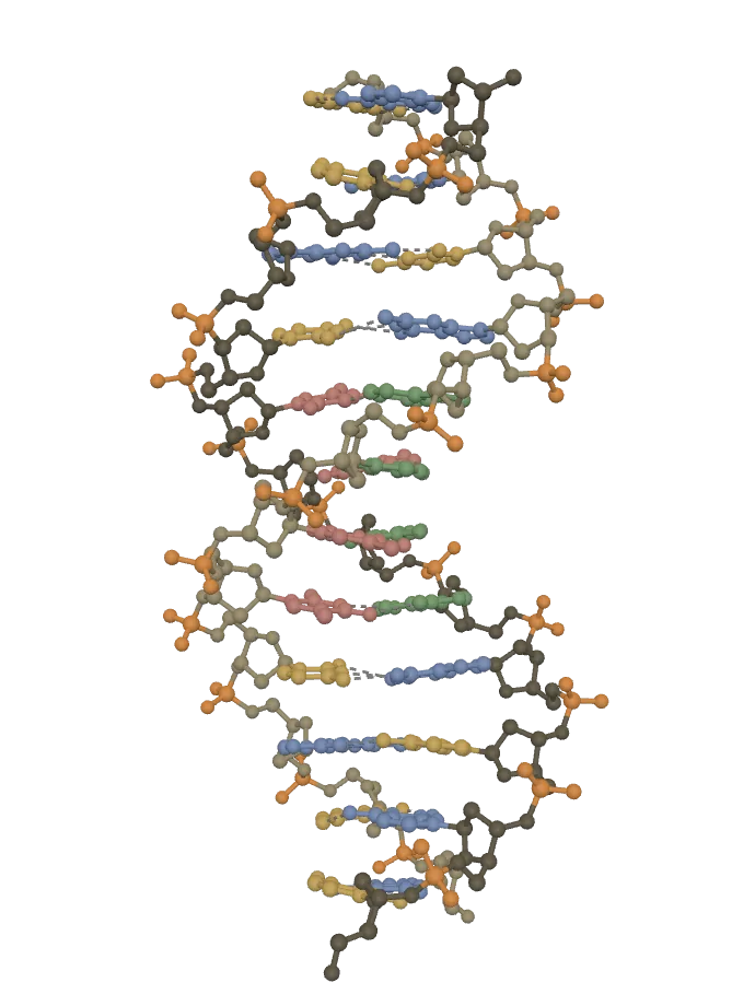
This browser can’t show the 3D model. The picture above is the same model.
Twelve base pairs of real DNA: a right-handed helix, the backbones outside, the bases stacked inside 0.34 nm apart, one turn in about 10 pairs. Turn it side-on for the wide major and narrow minor grooves. (X-rays don’t place hydrogen atoms, so none are shown.)
Sugar and phosphateThe sides of the ladder: each strand is a chain of deoxyribose sugars (brown; one strand darker) joined through phosphate groups (orange).
Adenine (A)Always paired with thymine (T) on the other strand, by two hydrogen bonds (dashed).
Thymine (T)Always paired with adenine (A) on the other strand, by two hydrogen bonds (dashed).
Guanine (G)Always paired with cytosine (C) on the other strand, by three hydrogen bonds (dashed).
Cytosine (C)Always paired with guanine (G) on the other strand, by three hydrogen bonds (dashed).
Drag to turn it. Tap a part, or a name below, to pick it out; tap it again to see the whole.
DNA: PDB 1BNA, Drew, Dickerson and others (1981), RCSB Protein Data Bank, CC0: www.rcsb.org/structure/1BNA
A stretch of DNA has 30 complete turns. About how many base pairs does it have?
Pick an answer to see why.
Functions of DNA
- It carries the hereditary characters from parents to offspring, written as the order (sequence) of its bases.
- It copies itself (replication, 3.1.5), so each new cell and each new generation gets a complete copy of the instructions.
- Crossing over between homologous chromosomes in meiosis exchanges pieces of DNA, giving new combinations of genes (genetic recombination; Unit 9).
- Changes in its bases (mutations) give rise to new variations; over many generations, with natural selection, these build new species (Unit 10).
- It is transcribed into the RNAs the cell needs for its different jobs.
- It controls the cell’s metabolism through the RNAs and proteins (enzymes) it codes for. Information flows DNA → RNA → protein, the central dogma of molecular biology, named by Crick in 1958.
3.1.4 Ribonucleic acid (RNA)
RNA is present in every living cell. It is made in the nucleus, copied from DNA, and found in the nucleus, the nucleolus, the cytoplasm, the ribosomes, mitochondria and plastids. It is a chain of ribonucleotides, each with ribose, a phosphate and one of the four bases adenine, guanine, cytosine and uracil.
- RNA is usually single-stranded. The strand can fold back on itself where its bases pair (A with U, G with C), forming short double-stranded loops, hairpins and coils (a pseudohelix).
- Like DNA, a strand has a direction: a 5′ end and a 3′ end, set by the way the sugar–phosphate backbone runs.
- Its bases can come in any order. RNA made on a DNA strand (the template) is complementary to it: A in the DNA gives U in the RNA, T gives A, G gives C, and C gives G. So if the template reads 3′-CAATGA-5′, the RNA made on it reads 5′-GUUACU-3′.
Types of RNA
There are three main kinds, all made on DNA and all working together to make proteins.
- Messenger RNA (mRNA) carries the genetic information, a copy of one gene, from DNA in the nucleus to the ribosomes, where it is read to make a protein. It is only about 3–5 % of the cell’s RNA, and short-lived (minutes to hours).
- Ribosomal RNA (rRNA) is a stable RNA that, with proteins, makes up the ribosomes. It is the most plentiful, about 80 % of the RNA in a cell. It holds the mRNA in place and joins the amino acids into a chain.
- Transfer RNA (tRNA) is the adapter. Each kind of tRNA carries one particular amino acid to the ribosome and has, at the bottom of its clover leaf, three bases, the anticodon, that pair with the matching three bases (codon) of the mRNA. It is small, about 75 to 90 nucleotides, and about 15 % of the cell’s RNA.
Functions of RNA
RNA’s jobs are mostly about using the genetic information: carrying it, and turning it into proteins.
- mRNA carries the genetic information from DNA to the ribosomes for protein synthesis.
- tRNA brings amino acids to the ribosome, in the order the mRNA’s codons ask for.
- rRNA forms part of the ribosome, where proteins are made; it is the rRNA itself that joins the amino acids by peptide bonds.
- In certain viruses RNA is the genetic material: the viruses of influenza, measles, polio, rabies, HIV and COVID-19, and the tobacco mosaic virus of plants.
- Some RNA molecules work as enzymes, called ribozymes: they catalyze reactions, such as the cleavage (cutting) of other RNA molecules. The ribosome’s rRNA is a ribozyme too.
A sample of nucleic acid from a virus contains 25 % uracil. What can you say about it?
Pick an answer to see why.
3.1.5 DNA replication
A living cell must be able to copy its whole set of genetic instructions every time it divides; and a many-celled organism must pass complete copies to the next generation. So the instructions must be kept in a form that can be copied exactly.
DNA replication is the process by which a double-stranded DNA molecule is copied into two identical DNA molecules. It happens before a cell divides, in the S phase of the cell cycle Grade 11: Interphase, so that when the cell divides, each daughter cell gets one complete copy. In outline: the two strands of DNA separate, and each acts as a template on which a new complementary strand is built.
Types of DNA replication
When Watson and Crick saw that the two strands are complementary, they suggested that each strand could serve as a template for a new one. But there were three possible ways the old and new strands could end up shared out, depending on how much of the old molecule is kept (conserved) in each copy:
- Conservative replication. The original double helix stays whole, and a completely new double helix is made beside it. Of the two copies, one has both old strands and the other both new strands.
- Semi-conservative replication. The two old strands separate, and each gets a new partner. Each of the two copies has one old strand and one new strand.
- Dispersive replication. The old molecule is broken into pieces, and the new molecules are patchworks: each strand of both copies is made of stretches of old DNA and stretches of new.
Only one of these happens. In 1958, at the California Institute of Technology, Matthew Meselson and Franklin Stahl found out which. They grew the bacterium E. coli for many generations on food in which all the nitrogen was ¹⁵N, a heavy isotope, so all its DNA was heavy. Then they moved the bacteria to ordinary ¹⁴N food, and took samples after each generation (each time the bacteria had doubled). They spun each sample’s DNA very fast in a solution of cesium chloride, in which DNA settles in a band at the height where its density matches the solution’s: heavy DNA low in the tube, light DNA higher, half-and-half DNA in between. Try each model in the widget, and compare it with what they saw.
What they saw:
- After one generation, all the DNA formed one band, halfway between heavy and light: every molecule was half old, half new. This ruled out conservative replication, which would give one heavy band and one light band.
- After two generations, there were two bands, one halfway and one light, in equal amounts. This ruled out dispersive replication, which would give a single band, a quarter heavy.
- Only semi-conservative replication predicts both. So DNA replication is semi-conservative.
Semi-conservative mode of DNA replication
Replication is called semi-conservative because each daughter DNA molecule is a hybrid: it conserves one parental strand (polynucleotide chain) and has one newly made strand. It happens in three stages: initiation, elongation and termination.
1. Initiation
- The two strands begin to separate at particular places called origins of replication (ori). A bacterium or a virus usually has one origin on its DNA; a eukaryotic chromosome has many (human DNA uses tens of thousands), so its long DNA is copied in many pieces at once.
- The enzyme helicase unwinds the double helix by breaking the hydrogen bonds between the base pairs, so the two strands separate. Each separated strand is a template for a new strand.
- Opening a helix makes two prongs, a Y-shaped structure called the replication fork. From an origin the DNA opens in both directions, so there are two forks moving apart, making a “bubble” that grows until it meets the next one.
- Two helpers: single-strand binding (SSB) proteins coat the separated strands so they don’t pair up again, and topoisomerase (in bacteria, DNA gyrase) cuts and rejoins the DNA ahead of the fork to release the twisting that unwinding causes, as a rope twists up when you pull its strands apart.
- DNA polymerase, the enzyme that builds the new strand, can’t start a strand from nothing: it can only add nucleotides to the end of a strand that is already there. So an enzyme called primase first makes a short piece of RNA, about ten nucleotides long, complementary to the template: the RNA primer. The primer gives DNA polymerase a starting point.
2. Elongation
- DNA polymerase (DNA polymerase III in E. coli) reads the template strand and adds DNA nucleotides one by one, each pairing with the base it faces (A with T, G with C). It joins each new nucleotide to the 3′ end of the growing new strand (starting with the primer’s 3′ end). So a new strand always grows in the 5′ → 3′ direction, while the polymerase moves along the template from its 3′ end towards its 5′ end.
- The two template strands are antiparallel, but both new strands must grow 5′ → 3′. As the fork opens, this works out differently on the two sides:
- On one template (the one running 3′ → 5′ towards the fork), the new strand can grow towards the fork, following it as it opens. It is made in one continuous piece, from one primer. This is the leading strand.
- On the other template (running 5′ → 3′ towards the fork), the new strand must grow away from the fork. So it is made in short pieces: each time the fork has opened a little more, primase lays a new primer near the fork, and DNA polymerase builds a short stretch back towards the last piece. These pieces are Okazaki fragments, about 1000 to 2000 nucleotides long in bacteria and 100 to 200 in eukaryotes. Because it is made in pieces and always lags behind the opening fork, this new strand is the lagging strand.
- The RNA primers are then removed (in E. coli by another polymerase, DNA polymerase I), and the gaps they leave are filled with DNA.
- Finally, DNA ligase joins neighboring pieces of the new strand by forming the last missing phosphodiester bond between them, sealing the lagging strand into one continuous chain.
Because one new strand is made continuously and the other in pieces, replication is also called semi-discontinuous.
3. Termination
Replication ends when the whole DNA molecule has been copied: in a bacterium, when the two forks moving round its circular DNA meet on the far side; in a eukaryote, when the forks from neighboring origins meet and the pieces are joined, and the ends of each chromosome (its telomeres) are finished. The result is two DNA molecules, each with one old and one new strand: semi-conservative, half of each molecule from the original DNA, half brand new. Each new molecule winds up into a double helix by itself.
DNA polymerase also proofreads: when it adds a wrong base, it usually notices the mismatch, removes the base and tries again. With proofreading and repair, a cell makes only about one mistake in a billion bases copied.
Why is one new strand made in short pieces?
Pick an answer to see why.
What is the job of DNA ligase?
Pick an answer to see why.
3.1.6 Genetic code
DNA carries genetic information from cell to cell and from generation to generation. That information is expressed as proteins: enzymes, hormones, pigments, the proteins of muscle and blood. A protein is a chain of amino acids, of 20 kinds. So the order of bases in DNA must somehow spell out the order of amino acids in a protein.
How many bases are needed for one amino acid? With four bases (A, C, G, U in the mRNA):
- one base for each amino acid would give only 4¹ = 4 words;
- two bases, 4² = 16 words: still fewer than 20;
- three bases, 4³ = 64 words: enough, with plenty to spare.
So the code is read in groups of three bases. A group of three bases on the mRNA that stands for one amino acid (or for “stop”) is a codon. The set of rules by which the order of bases in mRNA is translated into the order of amino acids in a protein is the genetic code: the instructions a cell uses to make its proteins.
The code is written for the mRNA (with U); the DNA’s coding strand has the same order with T for U, and the template strand is complementary to both.
| First two bases | third U | third C | third A | third G |
|---|---|---|---|---|
| UU | UUU Phe | UUC Phe | UUA Leu | UUG Leu |
| UC | UCU Ser | UCC Ser | UCA Ser | UCG Ser |
| UA | UAU Tyr | UAC Tyr | UAA stop | UAG stop |
| UG | UGU Cys | UGC Cys | UGA stop | UGG Trp |
| CU | CUU Leu | CUC Leu | CUA Leu | CUG Leu |
| CC | CCU Pro | CCC Pro | CCA Pro | CCG Pro |
| CA | CAU His | CAC His | CAA Gln | CAG Gln |
| CG | CGU Arg | CGC Arg | CGA Arg | CGG Arg |
| AU | AUU Ile | AUC Ile | AUA Ile | AUG Met (start) |
| AC | ACU Thr | ACC Thr | ACA Thr | ACG Thr |
| AA | AAU Asn | AAC Asn | AAA Lys | AAG Lys |
| AG | AGU Ser | AGC Ser | AGA Arg | AGG Arg |
| GU | GUU Val | GUC Val | GUA Val | GUG Val |
| GC | GCU Ala | GCC Ala | GCA Ala | GCG Ala |
| GA | GAU Asp | GAC Asp | GAA Glu | GAG Glu |
| GG | GGU Gly | GGC Gly | GGA Gly | GGG Gly |
The standard genetic code (the NCBI’s table 1). Ala alanine, Arg arginine, Asn asparagine, Asp aspartic acid, Cys cysteine, Gln glutamine, Glu glutamic acid, Gly glycine, His histidine, Ile isoleucine, Leu leucine, Lys lysine, Met methionine, Phe phenylalanine, Pro proline, Ser serine, Thr threonine, Trp tryptophan, Tyr tyrosine, Val valine.
To read a codon: find the row of its first two bases, then the column of its third. For example, GGA: row GG (the last row), column A: glycine. Notice how often the four codons of a row all mean the same amino acid.
Characteristics of the genetic code
- Triplet code. Each codon is three bases, and specifies one amino acid. For example, AUG stands for methionine.
- Non-overlapping. Neighboring codons don’t share bases: each base belongs to only one codon. For example, ACUAAG is read as ACU and AAG, not as ACU, CUA, UAA, AAG.
- Commaless (without punctuation). There are no gaps or “commas” between codons: once reading starts, the bases are read continuously, three at a time. For example, mRNA reading CCUCAGC… is read as CCU, CAG, C…, and so on. So adding or losing one base shifts every codon after it (a frameshift).
- Universal. A given codon stands for the same amino acid in almost all organisms, from bacteria to plants to people. That is why a human gene put into a bacterium makes the human protein (Unit 13). There are a few small exceptions: in the mitochondria of humans, for example, UGA means tryptophan instead of stop.
- Degenerate. Most amino acids have more than one codon. There are 64 codons; 3 are stop signals, so 61 stand for the 20 amino acids. For example, glycine has four codons, GGU, GGC, GGA and GGG. Only methionine (AUG) and tryptophan (UGG) have one each. Codons for the same amino acid usually differ only in their third base, so a change in the third base often changes nothing.
- Unambiguous. Each codon stands for only one amino acid: GGA is always glycine, never anything else. (Degenerate means one amino acid, several codons; unambiguous means one codon, one amino acid.)
- Stop (nonsense, terminator) codons. UAA, UAG and UGA don’t stand for any amino acid. They mark the end of the protein: when the ribosome reaches one, the chain is released.
- Start (initiation) codon. AUG begins the making of a protein in all organisms, and stands for methionine there (and wherever it appears inside a gene). In some bacteria GUG (and, rarely, UUG) can also serve as the start codon; at the start it too brings methionine.
- Colinearity. The order of codons in the gene (and in its mRNA) is the same as the order of amino acids in the protein: the first codon gives the first amino acid, the second the second, and so on. The mRNA is complementary to the DNA’s template strand, and identical, except for U in place of T, to its other strand, the coding strand. For example:
| Base sequence | ||
|---|---|---|
| DNA coding strand | 5′- ATG GGA CTT -3′ | |
| DNA template strand | 3′- TAC CCT GAA -5′ | read by the enzyme |
| mRNA | 5′- AUG GGA CUU -3′ | complementary to the template |
| Protein | Met – Gly – Leu | one amino acid per codon, in the same order |
How many different codons stand for amino acids?
Pick an answer to see why.
A change in a gene turns the mRNA codon GGU into GGC. What happens to the protein?
Pick an answer to see why.
Exercise
A. Tick the correct alternatives
- Which nitrogenous base is found in RNA but not in DNA?
Pick an answer to see why.
- What type of bond connects the sugar and phosphate in the RNA backbone?
Pick an answer to see why.
- Which of the following correctly pairs nitrogenous bases in DNA?
Pick an answer to see why.
- How many nucleotide bases form one codon in the genetic code?
Pick an answer to see why.
- Which of the following statements about the genetic code is true?
Pick an answer to see why.
- Which of the following enzymes unwinds the DNA helix during replication?
Pick an answer to see why.
- Which of the following best explains why one strand is made continuously and the other discontinuously during replication?
Pick an answer to see why.
- Study the figure of the types of DNA replication. Identify the types A, B and C.
Pick an answer to see why.
- If a DNA molecule has 2000 base pairs, about how long will it be?
Pick an answer to see why.
- If a drug strengthened the hydrogen bonds between DNA base pairs, which process would be most directly affected?
Pick an answer to see why.
B. Answer the following questions
Define heredity and variation.
Answer
What are the three components of a nucleotide?
Answer
Differentiate between purine and pyrimidine bases.
Answer
Describe the structure and function of DNA with a necessary diagram.
Answer
Describe the structure and function of RNA with a necessary diagram.
Answer
What are the three types of RNA? Describe them.
Answer
Define genetic code. Mention its characteristics.
Answer
Describe the semi-conservative mode of DNA replication with an appropriate diagram.
Answer
Compare the leading and lagging strands during DNA replication.
Answer
If the DNA template strand is TACGGA, what will be the RNA sequence formed?
Answer
A scientist extracts a nucleic acid and finds that it contains uracil but no thymine, and its sugar is ribose. Identify the molecule. In which organisms might it serve as the genetic material?
Answer
A student is given a nucleotide containing ribose sugar, adenine and phosphate. Is it a component of DNA or RNA? Justify your answer.
Answer
Study the diagram and answer the following questions.
- a.What is shown in the figure?
- b.Label A, B and C.
- c.Name the enzyme that adds DNA nucleotides on the template.
- d.What is an Okazaki fragment?
Answer
Printable PDFs
- Study notes 4 pages
- Worksheet 4 pages Answer key
Quiz yourself: Heredity and Evolution, Grades 5–12 →
Practice every question in the book, and keep track of the ones you miss →
A companion to your textbook, not a replacement for it: study from the book your school uses, and where the two differ, follow your teacher. Found a mistake? Please tell us.