
Drop a few fertilized frog eggs into a jar of pond water and look at them every morning. On the first day they are black-and-white dots. By the third day each dot has stretched into a little curved body with a head end and a tail end. Within a week or two, tadpoles wriggle out of the jelly. Nobody adds anything: no food, no instructions. A single cell, the zygote, builds the whole animal by itself, step after step, in the same order every time.
The last unit followed the making of a sperm and an egg. This unit follows what happens when they meet: fertilization, then the early stages of embryonic development, in which the zygote divides into thousands of cells (cleavage, the morula and the blastula), the cells move into three layers (gastrulation), and the layers begin to fold into organs (organogenesis).
7.2.1 Fertilization
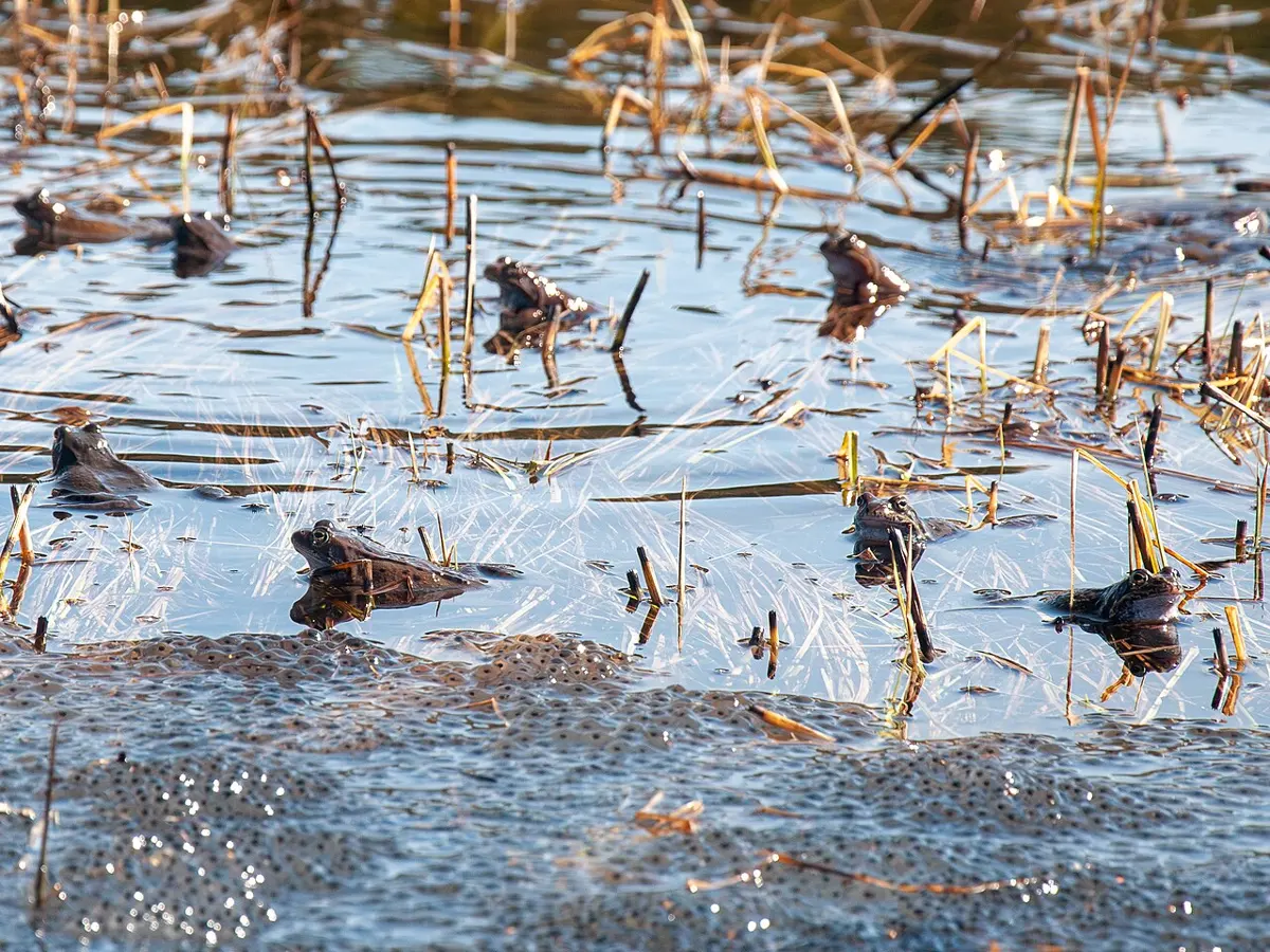
Breeding and amplexus
Frogs breed in the rainy season. The male calls with a loud, special croak (the breeding call) that females of his own species recognize; a ready female comes to him. The male climbs onto her back and holds her firmly round the body, gripping with rough nuptial pads that grow on his thumbs in the breeding season. This embrace is amplexus. It can last for hours. In amplexus the female lays her eggs, as secondary oocytes, through her cloaca into the water, and at the same time the male sheds his sperms, in seminal fluid, over them: this is insemination. A sperm then fuses with each egg: fertilization.
So in a frog, fertilization is external: it happens in the water, outside the female’s body.
The process of fertilization
The stages are in Figure 16.2.
- The sperm reaches the egg. A sperm enters the egg through its dark animal hemisphere. Only its head and middle piece enter; the tail is left outside.
- The cone of reception. Where the sperm touches the egg, the egg’s surface rises a little to meet it, as a small cone, the cone of reception (receptive cone).
- Through the coverings. The sperm’s acrosome releases its protein-digesting enzymes, the sperm lysins, which dissolve a way through the jelly and the vitelline membrane.
- The fertilization membrane. As soon as one sperm is in, the egg releases fluid under its vitelline membrane. The membrane lifts off, becomes thick and tough, and is now called the fertilization membrane; the space under it is the perivitelline space. No other sperm can get through it.
- Meiosis II is completed. The entry of the sperm makes the egg finish its second maturation division: the second polar body is pushed out, and the egg’s 13 chromosomes form the female pronucleus (egg nucleus).
- The male pronucleus. Inside the egg, the sperm’s nucleus swells into the male pronucleus. It moves inwards, dragging a little of the surface pigment with it, which leaves a dark streak, the sperm path or penetration path. Then it turns towards the female pronucleus (the copulation path).
- Fusion. The two haploid pronuclei meet and fuse. The zygote has 26 chromosomes again (2n = 26): fertilization restores the diploid number.
- Ready to divide. The sperm brought a centriole, which the egg lacks. It divides into two, the halves move apart and make the spindle, and the chromosomes line up in the middle for the first cleavage.
What stops a second sperm from entering a frog’s egg?
Pick an answer to see why.
Effects of fertilization
Fertilization changes the egg in many ways.
- i.The entry of the sperm makes the secondary oocyte complete meiosis II, and the second polar body is formed.
- ii.The egg’s cytoplasm contracts and gives out a little fluid into the perivitelline space, which lifts the vitelline membrane into the thick, tough fertilization membrane.
- iii.The fertilization membrane keeps out other sperms, which ensures monospermy (only one sperm fertilizes each egg).
- iv.Fertilization brings in a centriole, which the egg lacks, to make the spindle for cleavage.
- v.On the side opposite the sperm’s entry, a crescent-shaped gray area appears, between the dark animal hemisphere and the pale vegetal hemisphere: the gray crescent. The egg’s pigmented surface layer (cortex) turns by about 30° towards the sperm’s entry point, and on the opposite side its edge moves up, uncovering the paler cytoplasm beneath Grade 12: Unit 15, The gray crescent.
- vi.The gray crescent marks where the dorsal lip of the blastopore will form, and so the future dorsal side (back) of the embryo.
- vii.Before fertilization, the egg is radially symmetrical: it has only an animal–vegetal axis. The gray crescent gives it a front and back too, so the fertilized egg becomes bilaterally symmetrical.
So the plan of the future body is already marked on the fertilized egg: the gray crescent marks the embryo’s future dorsal surface, the region of the animal pole its future anterior (head) end, and the region of the vegetal pole its future posterior end.
Significance of fertilization
- It forms a diploid zygote, which develops into the embryo.
- It brings in the centriole (centrosome) for the zygote’s divisions.
- It prevents polyspermy, through the fertilization membrane.
- It stimulates the egg to complete its second maturation division.
- It brings variation: the zygote combines chromosomes from two parents, so each offspring is a new mixture of genes.
7.2.2 Cleavage
Cleavage is the series of rapid mitotic divisions by which the zygote becomes a many-celled embryo. The cells it makes are blastomeres. The embryo doesn’t grow in this time: its size stays the same, while the number of its cells goes up and each gets smaller. The cells don’t grow between divisions; they only copy their DNA and divide again.
In a frog, cleavage starts about 2–3 hours after fertilization. It is holoblastic: each furrow cuts right through the egg, from the animal pole to the vegetal pole. And it is unequal: the yolk in the vegetal half slows the furrows and pushes the spindles up, so the cells of the vegetal half are larger.
- i.The first cleavage is meridional (vertical): the furrow runs from the animal pole down to the vegetal pole, along the egg’s axis, usually through the middle of the gray crescent.
- ii.It gives two equal blastomeres: the 2-celled stage.
- iii.The second cleavage is also meridional, at right angles to the first, giving four equal blastomeres: the 4-celled stage.
- iv.The third cleavage is latitudinal (horizontal), but a little above the equator, giving the 8-celled stage with cells of two sizes.
- v.The four upper cells, small, pigmented and with little yolk, are micromeres; the four lower cells, large and full of yolk, are megameres (macromeres).
- vi.The fourth cleavage is meridional (vertical) again, two furrows dividing all eight cells at once: the 16-celled stage, 8 micromeres and 8 megameres.
- vii.The fifth cleavage is latitudinal (horizontal), again dividing every cell at once: the 32-celled stage, with 16 smaller micromeres above and 16 larger megameres below.
After that the divisions stop keeping time: the micromeres divide faster than the yolk-laden megameres, and the pattern becomes irregular.
Why are the megameres of a frog’s 8-celled embryo larger than its micromeres?
Pick an answer to see why.
7.2.3 Morula and blastula
Morula
After the fifth cleavage, the 32-celled embryo is a ball of cells shaped like a mulberry: the morula (Latin morum, “mulberry”), and its formation is morulation. In the textbooks’ definition, a morula is a solid ball of cells, without the cavity of a blastula. In a frog, though, the cavity has already begun: a small space has opened among the cells by the 8-celled stage. So the frog’s morula is a short stage at the start of blastula formation, not a separate, solid one.
Blastulation
From the 8-celled stage, a small fluid-filled space, the segmentation cavity, appears in the middle of the embryo, between the cells, nearer the animal pole. As the cells go on dividing, it grows, and the embryo becomes a hollow ball of cells, the blastula. Its formation is blastulation.
- The small, yolk-free micromeres of the animal half divide quickly; the large, yolky megameres of the vegetal half divide slowly, because the yolk gets in the way.
- The large cavity inside is the blastocoel (segmentation cavity). It is filled with fluid, which the cells lining it pump in: mostly water, with salts and some proteins and sugars.
- In a fully formed blastula, the blastocoel is a dome-shaped (hemispherical) cavity in the animal half. Its roof is two or three layers of small cells, pigmented black at the surface; its floor is the large yolky megameres (Figure 16.3).
The blastocoel matters because it keeps the cells of the roof and the floor apart, so that they don’t affect each other too early, and it gives cells room to move during gastrulation. It doesn’t feed the embryo: the food is the yolk inside the cells, used up as they divide and, later, as they build organs.
A blastula with a large cavity like this is a coeloblastula (from the Greek koilos, “hollow”); the name comes from its cavity. Since it is still one ball of cells, without separate germ layers, the blastula is called monoblastic (single-layered), even though in the frog its roof is a few cells thick.
What is the blastocoel of a frog’s blastula?
Pick an answer to see why.
Presumptive areas: the fate map of the blastula
Each region of a blastula has a future: it will become a particular tissue. These regions are presumptive (prospective) areas, and a drawing of them is a fate map (Figure 16.4). They were found by marking small patches of living blastulae with harmless dyes and following each patch until it became part of an organ (and later by microsurgery, and dyes injected into single cells).
- Almost the whole animal half is presumptive ectoderm: presumptive epidermis over most of it, and presumptive neural plate on its dorsal side.
- A belt round the middle, the marginal zone, is presumptive mesoderm. On the dorsal side, in the region of the gray crescent, it is widest: there it is presumptive notochord (chorda-mesoderm).
- The yolky vegetal half is presumptive endoderm.
- A small area just above the future dorsal lip, the prechordal plate, will form the endoderm of the front of the gut (the pharynx) and the mesoderm (mesenchyme) of the head.
7.2.4 Gastrulation
Gastrulation is the stage in which the cells of the blastula move, in a great rearrangement, into the layers of the body: the blastula becomes a gastrula. Cells don’t only divide; whole sheets of them spread, fold in and slide past one another (morphogenetic movements). Cells that were on the outside of the blastula, the future gut and muscles, move inside, and those that stay outside become the skin and the nervous system.
The main events of gastrulation are below (Figure 16.5).
1. Epiboly. The small, pigmented micromeres of the animal half divide fast and spread down over the megameres of the vegetal half, like a cap pulled over a ball. In the end they cover the whole embryo except a small round patch, the yolk plug.
2. Emboly or invagination. On the dorsal side, a little below the gray crescent, a few cells sink inwards, and a crescent-shaped groove appears: the blastopore. The groove deepens into a new cavity, the archenteron (the future gut). The archenteron opens to the outside through the blastopore, whose upper edge is the dorsal lip, its sides the lateral lips and its lower edge the ventral lip. As the archenteron grows, the blastocoel is squeezed and shrinks until it is gone. The floor and sides of the archenteron are endoderm (megameres); its roof is chorda-mesoderm, the future notochord and mesoderm.
3. The lips of the blastopore close in. The groove spreads sideways and round until it is a ring, and the ring then narrows from all sides, dorsal, ventral and lateral. The blastopore becomes small and round, and yolky megameres show through it as the yolk plug: this is the yolk plug stage.
4. Involution. Cells of the marginal zone roll in over the dorsal lip, and later over the lateral and ventral lips too, and spread forwards on the inside, under the ectoderm. Those over the dorsal lip form the roof of the archenteron, the chorda-mesoderm; those over the other lips form the rest of the mesoderm.
5. Rotation. As the heavy yolky cells shift and the archenteron fills with fluid, the embryo’s center of gravity changes, and it turns through about 90° inside its fertilization membrane. Its long axis then lies parallel to the water’s surface, with the old animal pole region in front (the future head) and the blastopore at the back, where the anus will form.
Which part of the gastrula did Spemann call the organizer?
Pick an answer to see why.
The three germ layers and their fate
By the end of gastrulation, the embryo has three layers of cells, the primary germ layers:
- the ectoderm, the outer layer, from the micromeres;
- the endoderm, the inner layer round the archenteron, from the megameres;
- the mesoderm in between, from the chorda-mesoderm and the cells that rolled in over the lips, spreading between the ectoderm and the endoderm.
Many books call the frog’s gastrula diploblastic (two-layered): at first the cells that rolled in lie together under the ectoderm as one inner layer, the meso-endoderm (chorda-mesoderm and endoderm). As gastrulation ends, the mesoderm separates from the endoderm and spreads out between the two, and the embryo is triploblastic: three layers.
Each germ layer gives rise to particular organs and tissues: its fate.
| Ectoderm | Mesoderm | Endoderm |
|---|---|---|
| epidermis of the skin and its glands | notochord | lining (mucosa) of the gut |
| epithelium of the conjunctiva | lining of the pericardium and peritoneum | gastric and intestinal glands |
| olfactory and auditory organs (lining of the nose, inner ear) | muscles, voluntary and involuntary | liver, pancreas, bile duct and pancreatic duct |
| lining of the mouth and of the cloaca’s opening | cartilage and bone | pharynx, Eustachian tube, tonsils, thymus, thyroid |
| lens, cornea and retina of the eye | all connective tissue (ligaments, fat and so on) | larynx, trachea and lungs (their linings) |
| brain and spinal cord | blood and blood vessels, heart | lining of the urinary bladder |
| ganglion cells and nerves | dermis of the skin | |
| neuroglia, and the myelin sheath and neurilemma of nerves | kidneys and their ducts, gonads | |
| infundibulum, pineal body, pituitary gland | spleen | |
| enamel of the teeth | sclera and choroid of the eye |
Importance of gastrulation
- i.It forms the three germ layers, from which all the organs come.
- ii.It sets up the body axes: front and back (anterior and posterior), back and belly (dorsal and ventral), left and right.
- iii.It moves cells into new places (morphogenesis), so that tissues that will work together lie next to each other.
- iv.It brings the layers into contact so that one can induce the development of another: the chorda-mesoderm under the ectoderm induces the neural plate.
- v.It lays down the notochord and starts the formation of the neural tube.
- vi.It fixes the fate of cells: after gastrulation, cells are committed to becoming particular tissues.
7.2.5 Organogenesis
Organogenesis is the formation of organs and tissues from the three germ layers. It begins as gastrulation ends, with the nervous system and the notochord, which run the length of the back.
1. Neurulation: forming the neural tube
Neurulation (neurogenesis) is the formation of the neural tube, the future brain and spinal cord, from the ectoderm of the back. The embryo at this stage is a neurula (Figure 16.6).
- i.The ectoderm cells of the presumptive neural plate area, along the middle of the back, become tall and thick: the neural plate. The chorda-mesoderm under it induces this.
- ii.A groove appears along the middle of the plate: the neural groove.
- iii.At the same time, the edges of the plate rise up on each side as neural folds (ridges).
- iv.The neural groove sinks deeper.
- v.The neural folds grow towards each other and meet over the middle of the groove, and fuse.
- vi.The open groove becomes a closed neural tube, which separates from the ectoderm above it and lies under it; the ectoderm closes over it as epidermis.
- vii.The front and back ends of the tube stay open for a short time (the neuropores), then close too.
- viii.The front, wider part of the tube becomes the brain; the rest, the spinal cord.
- ix.The cavity inside the tube is the neurocoel; it becomes the ventricles of the brain and the central canal of the spinal cord.
2. Notogenesis: forming the notochord
The notochord is a flexible rod of supporting tissue along the back of the embryo of every chordate. In vertebrates, it is replaced later by the backbone (vertebral column). Its formation is notogenesis. It comes from mesoderm (chorda-mesoderm).
- i.The chorda cells in the middle of the roof of the archenteron separate from the mesoderm on each side of them.
- ii.They form a solid, cylindrical rod, the notochord, under the neural plate.
- iii.Its cells become large and vacuolated (full of fluid-filled vacuoles), which makes the rod firm yet bendable, like a hosepipe full of water.
- iv.A notochordal sheath of two layers forms round it.
- v.The notochord is the first axial support of the embryo; it also signals to the neural tube above it and the somites beside it as they form.
From which germ layer does the notochord form?
Pick an answer to see why.
3. Coelom formation
The coelom is the body cavity between the body wall and the gut. In a frog, it forms by the splitting of the mesoderm, so it is a schizocoel (Greek schizein, “to split”), and since it is lined by mesoderm on both sides, it is a true coelom (Figure 16.7).
On each side, the mesoderm sheet divides from the top down into three parts: next to the neural tube and notochord a thick epimere (dorsal, paraxial mesoderm), below it a small mesomere (intermediate mesoderm, the nephrotome, which forms the kidney tubules), and the rest, the thin hypomere (lateral plate mesoderm), round the sides.
- i.The hypomere splits into two layers: an outer somatic (parietal) layer and an inner splanchnic (visceral) layer.
- ii.The small space between them is the splanchnocoel, the start of the coelom.
- iii.The space spreads downwards on each side.
- iv.The splanchnocoels of the two sides meet below the gut, in the middle of the belly, and together form a U-shaped cavity: the coelom. Above the gut, the two splanchnic layers stay together, holding the gut, as the dorsal mesentery.
- v.The somatic layer and the ectoderm outside it together form the somatopleure, the body wall.
- vi.The splanchnic layer and the endoderm inside it together form the splanchnopleure, the wall of the gut.
The epimere, the thick dorsal mesoderm beside the notochord, does something different:
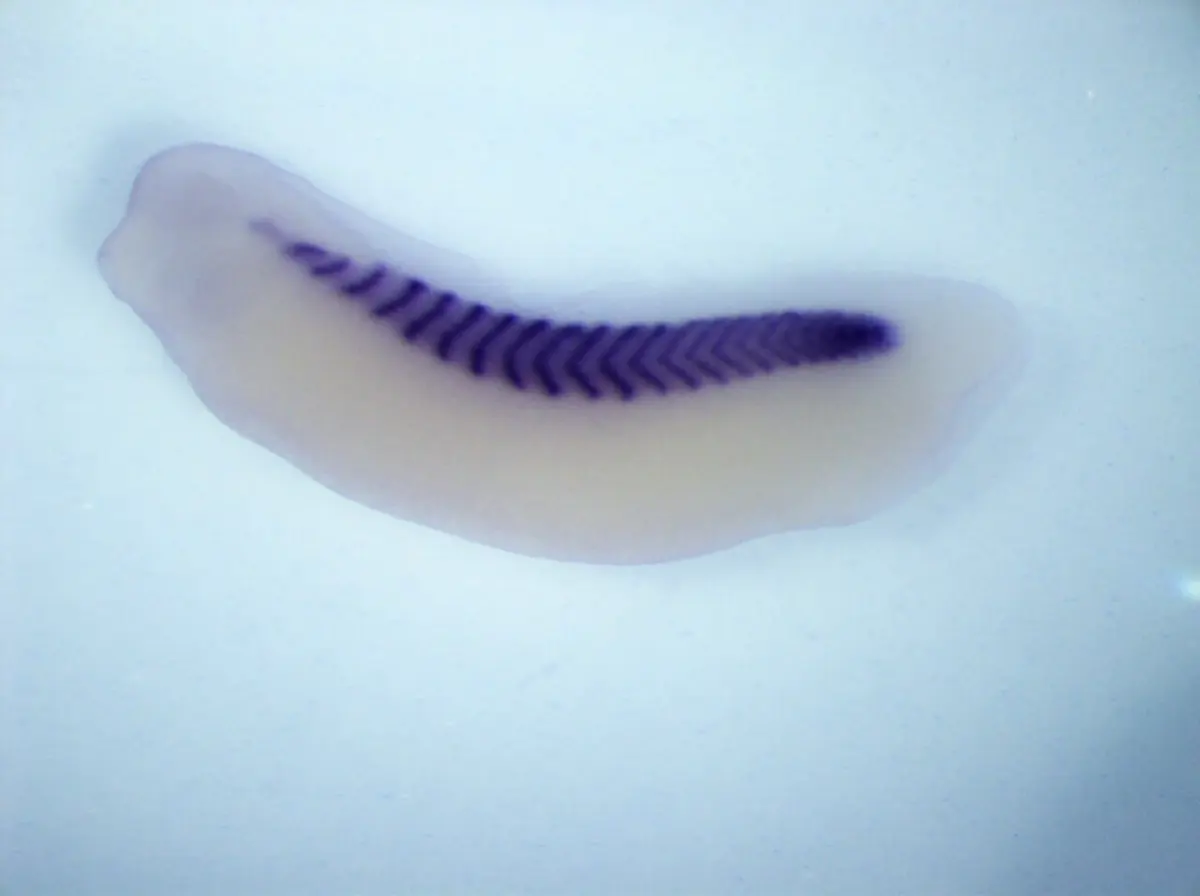
- vii.It divides along the body into a row of blocks, the somites (mesodermal segments).
- viii.A narrow cavity, the myocoel, appears in each somite.
- ix.Each somite then separates into three parts:
- an outer, thin dermatome, which becomes the dermis of the skin;
- a middle, thick myotome, which develops into the muscles of the body and limbs;
- an inner, thin sclerotome, whose cells move round the notochord and neural tube and form the vertebrae (the backbone).
Which part of a somite forms the vertebrae?
Pick an answer to see why.
From embryo to frog
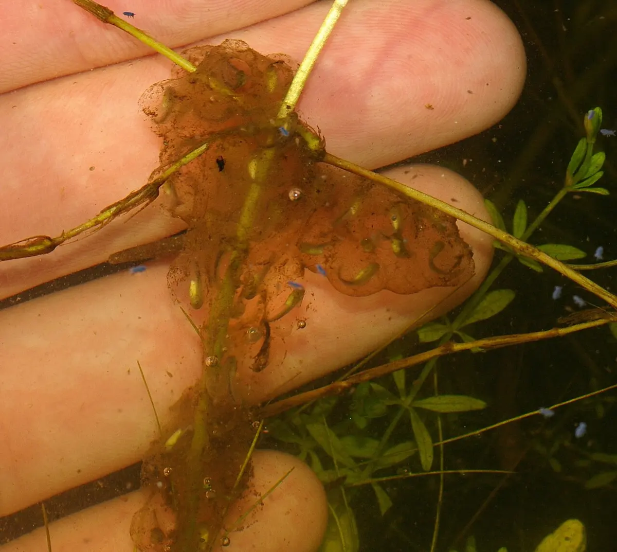
The neurula lengthens into a tailbud embryo, with a head, a tail bud, gill swellings and suckers under the head. In a few days to two weeks, sooner in warm water, it wriggles out of its jelly: the young tadpole hatches. A frog’s development is indirect: the tadpole has gills, a tail and a mouth for scraping plants, and lives only in water. It grows, then changes into a frog by metamorphosis: the hind legs grow, then the front legs; the gills give way to lungs; the gut shortens for a diet of insects; and the tail is absorbed. The young frog (froglet) climbs out onto land.
Exercise
This exercise covers this chapter and the last. The questions on gametogenesis (A1–A7, B1 and B2) are in Unit 15; the rest are here.
A. Choose the best alternative
- What does the formation of the gray crescent signify in the frog’s fertilization?
Pick an answer to see why.
- Which of the following describes the role of the blastocoel in the blastula stage of development?
Pick an answer to see why.
- What is the major event of epiboly in the frog’s gastrulation?
Pick an answer to see why.
- A frog embryo is exposed to a chemical that blocks the signaling molecules involved in forming mesoderm. What effect would this most likely have on coelom formation?
Pick an answer to see why.
B. Answer the following questions
Identify figures A and B above. Write any four differences between A and B.
Answer
Describe the entire process of cleavage in the frog with diagrams.
Answer
In the development of the frog, an embryonic stage appears with a fluid-filled cavity. What is this stage called? Describe briefly how it forms.
Answer
What is gastrulation? What are its major events? Describe them.
Answer
Which germ layer contributes to the formation of the nervous system in amphibians? Describe the process by which the neural tube forms.
Answer
What is a schizocoel? How is it formed? Describe the entire process.
Answer
Look at the diagram above and answer: i. Which embryonic stage is shown? ii. Label A, B and C. iii. Write any two differences between A and B.
Answer
Printable PDFs
- Study notes 5 pages
- Worksheet 4 pages Answer key
Quiz yourself: Life Processes and the Human Body, Grades 5–12 →
Practice every question in the book, and keep track of the ones you miss →
A companion to your textbook, not a replacement for it: study from the book your school uses, and where the two differ, follow your teacher. Found a mistake? Please tell us.