
Bite into a piece of chicken leg and you are eating four different materials at once. The skin is a sheet of cells packed edge to edge. The meat is long cells that can shorten. The white, chewy strip at the end that won’t come off the bone is a rope of protein fibers, and the bone itself is a mineral that living cells have built round themselves. Running through all of it, too fine to see, are nerve cells.
Every animal from a jellyfish to an elephant is built from just these four kinds of material, the four animal tissues: epithelial, connective, muscular and nervous tissue. Each kind has a shape that suits its job so closely that a biologist can look at a thin slice of any organ under a microscope and say what each part does. This unit is a tour of the four, the cells in each, and the places in your own body where you can find them.
Cells: Servier Medical Art (smart.servier.com), CC BY 4.0; stomach and body: BodyParts3D, © The Database Center for Life Science, CC BY 4.0
Animal tissue
The word tissue comes from the French tissu, a woven cloth. It was first used for the body’s materials by a young French doctor, Xavier Bichat, who noticed that organs are made of a few kinds of “fabric” used again and again.
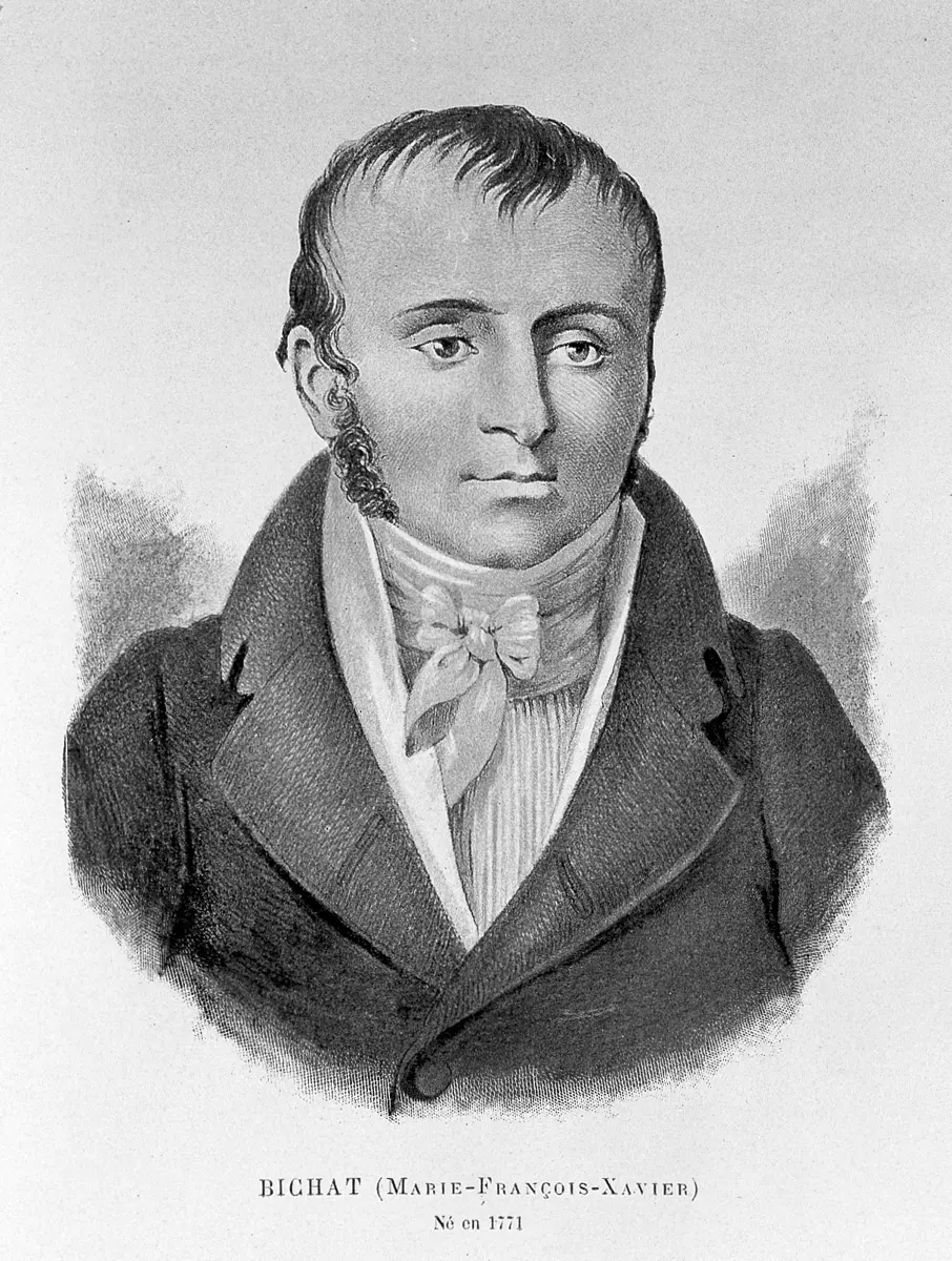
The study of tissues is histology (Greek histos, a web or tissue; logos, study). The name was coined in 1819 by the German anatomist August Mayer. The Italian Marcello Malpighi (1628–1694), the first to study animal structure with a microscope (he discovered the capillaries), is often called the founder of microscopic anatomy and of histology.
An animal tissue is a group of cells of common origin (from the same layer of the embryo), together with the material they put out round themselves, working together at a particular function. The cells of one tissue are usually similar in structure, but some tissues hold several kinds of cell: blood has red cells, white cells and platelets.
The material round the cells is the matrix (the extracellular matrix, ECM). It has two possible parts: a ground substance (a watery gel of proteins and sugars) and protein fibers set in it. How much matrix a tissue has is one of the clearest ways to tell the tissues apart:
- Connective tissue has all three components: cells, plenty of matrix, and fibers in the matrix.
- Epithelial, muscular and nervous tissue are mostly cells, with very little matrix between them, and no fibers of their own in it.
Types of animal tissue
By their origin, structure, place and function, animal tissues are of four types:
| Tissue | Main job | Comes from |
|---|---|---|
| Epithelial | covers, lines, protects, secretes | all three germ layers |
| Connective | binds, supports, transports | mesoderm |
| Muscular | contracts, moves | mesoderm |
| Nervous | carries messages, controls | ectoderm |
True tissues first appear in the Coelenterata (Hydra, jellyfish, sea anemones), which are said to have the tissue grade of organization. Their body has an outer and an inner layer of epithelium, cells that contract (muscle-like cells in the epithelium) and a nerve net. With the Platyhelminthes (flatworms) comes a third germ layer, the mesoderm, and with it true muscle and connective tissue and the first organs.
Which of these is true of connective tissue but not of the other three tissues?
Pick an answer to see why.
6.1 Epithelial tissue
Epithelial tissue, or epithelium (Greek epi, upon; thele, nipple: the word first named the thin skin of the lips), is the tissue of surfaces. It does four things:
- it covers: the skin over the body, the outer layer of organs;
- it lines: the inside of the gut, the airways, blood vessels and body cavities;
- it forms glands: every gland, from a sweat gland to the liver, grows from an epithelium;
- it secretes: mucus, sweat, enzymes and hormones.
Features of epithelium
- Cells packed close together, with almost no matrix between them. Epithelium has the most cells for its size of any tissue, and its little matrix has no fibers.
- A free surface and a basement membrane. One side of the sheet faces a space (the outside, or a cavity); the other side sits on a thin mat of proteins and sugars called the basement membrane (mostly a fine collagen and a sticky protein, laminin), which joins it to the connective tissue below. In a single-layered epithelium every cell sits on it; in a many-layered one, only the bottom layer does.
- No blood vessels (it is avascular). Food and oxygen reach the cells by diffusion from capillaries in the connective tissue under the basement membrane, and wastes leave the same way. This is one reason an epithelium is thin.
- Cells joined by cell junctions, so that the sheet holds together and controls what passes between its cells.
- Quick to renew. Cells lost from the surface are replaced by cell division in the deeper layers: the lining of your intestine is replaced every few days.
- It comes from all three germ layers: the skin’s epidermis from ectoderm, the lining of blood vessels and body cavities from mesoderm, the lining of the gut and airways from endoderm.
Cell junctions
The cells of an epithelium are joined to each other by cell junctions, small patches where proteins of the two cell membranes lock together. There are four kinds between cells:
- Tight junction (zonula occludens, a belt that closes). A belt round the top of each cell where the two membranes are stitched together so tightly that nothing leaks between the cells: whatever crosses the sheet must go through the cells, which choose what to let through. In the intestine and the urinary bladder they keep the gut’s contents and urine from seeping into the body.
- Adherens junction (zonula adherens, a belt that sticks). A belt just below the tight junction, joining the cells through proteins anchored to the actin threads inside each cell.
- Desmosome (macula adherens, a spot that sticks). Small round “spot welds” whose plates are tied to tough keratin threads inside the cells. They hold cells together against pulling and rubbing, so they are many in the skin and the heart muscle.
- Gap junction. Clusters of tiny protein channels that connect the cytoplasm of two cells, so ions and small molecules pass directly from cell to cell. They let the cells of the heart beat together and let cells pass signals.
The bottom of each cell is fixed to the basement membrane by hemidesmosomes (half-desmosomes) and other cell-to-matrix junctions.
Functions of epithelium
Besides covering, lining, forming glands and secreting, epithelium works in:
- protection of the tissues beneath from drying, rubbing, germs and chemicals (the skin);
- movement of substances over its surface, by cilia (mucus in the airways, the egg in the oviduct);
- absorption (the intestine) and excretion (the kidney tubules);
- exchange and diffusion of gases (the air sacs of the lungs) and filtration (the kidney);
- sensation (the smell cells in the nose, the taste buds).
Why must an epithelium be thin enough for food to diffuse through it?
Pick an answer to see why.
Which junction stops substances leaking between neighboring cells of the gut lining?
Pick an answer to see why.
Types of epithelial tissue
Epithelium is named by two things: how many layers of cells it has, and the shape of the cells at its free surface. By the number of layers there are two types: simple (one layer) and compound (many layers).
The shapes are:
- squamous (Latin squama, a scale): flat, thin cells, wider than tall, with a flat nucleus;
- cuboidal: cells about as tall as wide, with a round nucleus in the middle;
- columnar: cells taller than wide, with an oval nucleus near the base.
1. Simple epithelium
Simple epithelium has a single layer of cells, all resting on the basement membrane. Being thin, it is found where substances must pass through: where things are absorbed, secreted, filtered or exchanged. It does little to protect. By the shape of its cells, it is of four types: simple squamous, simple cuboidal, simple columnar and pseudostratified epithelium.
Servier Medical Art (smart.servier.com), CC BY 4.0
i. Simple squamous epithelium
Simple squamous epithelium is a single layer of thin, flat, many-sided (polygonal) cells, each with a flattened, oval nucleus that makes a small bump. Seen from above, the cells fit together edge to edge like the tiles of a floor or the stones of a courtyard, so it is also called pavement, tessellated (tiled) or mosaic epithelium. Being the thinnest of all tissues, it is found wherever things must pass through quickly:
- the air sacs (alveoli) of the lungs: exchange of gases;
- Bowman’s capsule of the kidney: filtering the blood (ultrafiltration);
- the thin descending limb of the loop of Henle: letting water out of the kidney tubule;
- the walls of capillaries: exchange between blood and tissues;
- the lining of body cavities and the covering of organs inside them.
Two linings of simple squamous epithelium have names of their own. Both come from the mesoderm; they differ in where they are:
- Endothelium lines the heart (the endocardium), all blood vessels (the tunica interna of arteries and veins, and the whole wall of capillaries) and the lymph vessels.
- Mesothelium lines the body cavities and covers the organs in them: the parietal epithelium on the body wall, the visceral (splanchnic) epithelium over the gut and other organs, and the mesenteries that hang the gut. In us these form the peritoneum, the pleura round the lungs and the pericardium round the heart.
ii. Simple cuboidal epithelium
Simple cuboidal epithelium is a single layer of cube-like cells, about as tall as they are wide (many-sided seen from above), each with a large, round, central nucleus. Its cells have more cytoplasm than squamous cells, room for the machinery to secrete and absorb. It may be:
- non-ciliated (no cilia): lining the kidney tubules (reabsorbing and secreting), the follicles of the thyroid gland (making its hormone), the small ducts of the salivary glands and pancreas (carrying their juices), the back of the iris, and the front of the lens of the eye; it also covers the surface of the ovary;
- ciliated (with cilia on the free surface): in the smallest bronchioles of the lungs, where its cilia keep moving mucus and trapped dust upward;
- brush-bordered (with a dense fringe of microvilli): in the proximal convoluted tubule (PCT) of the kidney’s nephrons, where the microvilli give a huge surface for reabsorbing water, glucose and salts from the filtrate.
iii. Simple columnar epithelium
Simple columnar epithelium is a single layer of tall, pillar-like cells, taller than wide, each with an elongated, oval nucleus near its base. Tall cells have room for much machinery, so this epithelium secretes and absorbs. It may be:
- non-ciliated: lining the stomach (secreting mucus and gastric juice), the intestines and the gall bladder (which stores bile and absorbs water from it). Among the columnar cells of the intestine are goblet cells, which secrete mucus;
- ciliated: lining the oviducts (fallopian tubes), where the beating cilia catch the egg released from the ovary and move it toward the uterus, and the small bronchi; and in the brain’s cavities (the ventricles) and the central canal of the spinal cord (the neurocoel), where cilia keep the cerebrospinal fluid (CSF) moving;
- brush-bordered: lining the small intestine, whose cells carry about 3,000 microvilli each, multiplying the surface for absorbing digested food.
A cell has a round, central nucleus and is as tall as it is wide. It lines a kidney tubule and has a dense fringe of microvilli. It is:
Pick an answer to see why.
iv. Pseudostratified epithelium
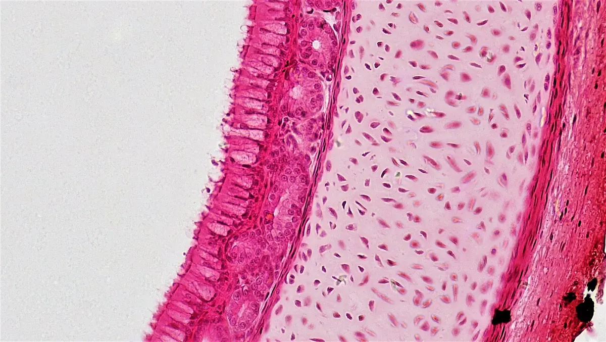
In pseudostratified epithelium (Greek pseudo, false; stratified, layered) the cells are of different heights. Every cell rests on the basement membrane, so it is really a single layer, but only the tall columnar cells reach the free surface. The short basal cells, low and rounded, stay at the bottom; they are stem cells that divide to replace the others. Because the nuclei sit at different levels, a section looks as if it had several layers: hence “false layered”.
Most of the tall cells carry cilia, and scattered among them are goblet cells, which secrete mucus. So this is a ciliated pseudostratified columnar epithelium with goblet cells, built for cleaning air: dust and germs breathed in stick to the mucus, and the cilia beat it upward like a moving carpet, toward the throat, where it is swallowed or coughed out.
It lines the nasal cavity, the trachea and the bronchi, down to the larger bronchioles. (A non-ciliated form lines parts of the male urethra and the large ducts of some glands.)
2. Compound epithelium
Compound epithelium has two or more layers of cells. Only the bottom layer rests on the basement membrane, and only the top layer has a free surface. The cells of the bottom layer are cuboidal or columnar and keep dividing; their daughter cells are pushed up, change shape, and replace the cells worn away from the top. The top cells may be squamous, cuboidal, columnar or rounded, and may be specialized for a job.
Many layers make it thick and slow for substances to cross, so compound epithelium does little secreting or absorbing. Its main job is protection where a surface is rubbed, scraped or stretched. It is of two types: stratified epithelium, which resists wear, and transitional epithelium, which stretches.
Servier Medical Art (smart.servier.com), CC BY 4.0
I. Stratified epithelium
Stratified epithelium has many layers and is named by the shape of the cells in its top layer: stratified squamous, stratified cuboidal or stratified columnar. Its cells are constantly worn off the surface and replaced from below, so it protects the tissues under it against rubbing, drying and germs.
i. Stratified squamous epithelium. Many layers of cells: the bottom ones cuboidal or columnar, the middle ones many-sided, and the top layers flat (squamous). The most common compound epithelium. It is of two kinds, depending on whether its top cells fill with keratin, a tough, waterproof protein:
A. Non-keratinized (stratified squamous) epithelium. The top cells stay alive and moist, with their nuclei, and the surface is smooth and wet. It lines places that are rubbed but kept moist: the mouth (buccal cavity), pharynx, esophagus (gullet), anal canal, vagina, and the front of the eye (the cornea).
B. Keratinized (stratified squamous) epithelium. As cells are pushed up, they fill with keratin, lose their nucleus and die, so the top layers are flat, dry, dead scales of keratin. This is the epidermis of the skin. The dead layer makes the surface waterproof, resists scratching and germs, protects the living layers and keeps the body from drying out.
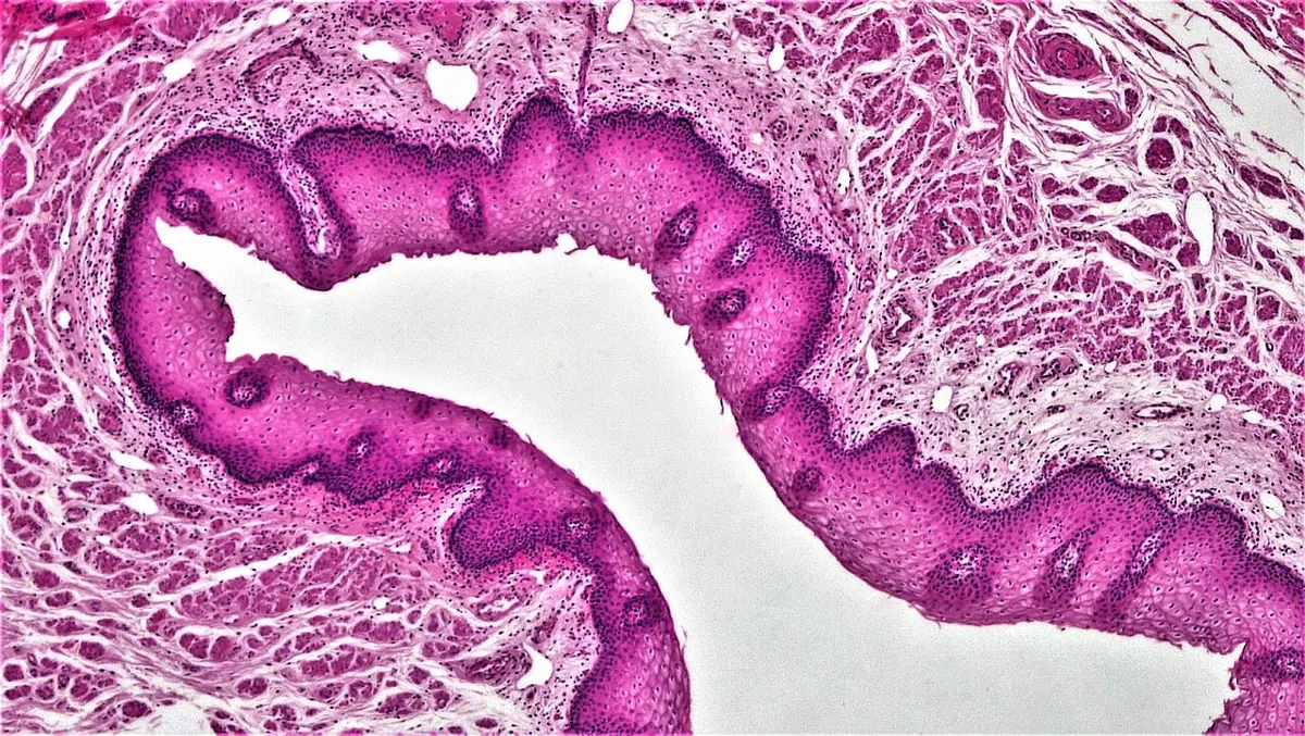
Keratin is a tough protein found only in vertebrates. Their skin cells make it and fill with it, and it builds many structures that grow from the skin: the dry scales and claws of reptiles and the shell plates (scutes) of tortoises; the feathers, beaks, claws and leg scales of birds; and the hair, horns, hoofs, nails and claws of mammals, as well as the spines of a porcupine and the scales of a pangolin.
ii. Stratified cuboidal epithelium. Usually just two or three layers, with cuboidal cells at the top. A rare type, found in the ducts of the larger glands: sweat glands, salivary glands and mammary glands, where it gives a tougher lining than a single layer.
iii. Stratified columnar epithelium. A few layers of small, many-sided or cuboidal cells under a top layer of columnar cells. Uncommon, found mainly where one kind of epithelium meets another: the conjunctiva of the eye, part of the male urethra, the large ducts of salivary and mammary glands, and small patches of the pharynx and anal canal.
II. Transitional epithelium
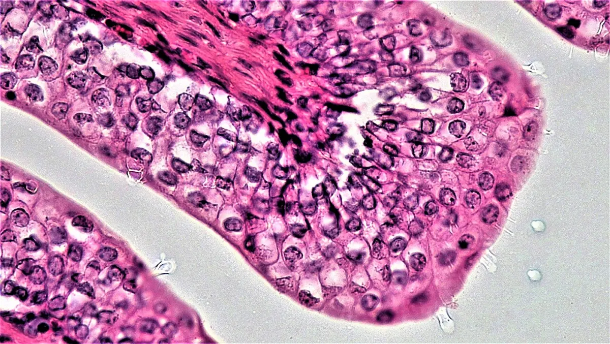
Transitional epithelium lines organs that fill up and empty, and have to stretch a great deal without tearing or leaking: the pelvis of the kidney, the ureters, the urinary bladder and the upper part of the urethra. Because it lines the urinary tract it is also called urothelium, and because it changes shape, plastic epithelium.
It has about five or six layers of cells. When the bladder is empty and relaxed, the bottom cells are cuboidal or columnar and the top cells are large, rounded and dome-shaped, bulging into the space. When the bladder fills and stretches, the cells slide over each other and flatten: the epithelium becomes thinner, with only two or three layers, and the top cells become squamous. Its top cells are joined by tight junctions and covered with a thick membrane that stops urine soaking back into the body. The bottom layer always rests on its basement membrane, stretched or not.
A surface is lined by many layers of cells whose top cells are flat, dead and full of keratin. Where is it?
Pick an answer to see why.
Why is the lining of the urinary bladder transitional and not stratified squamous?
Pick an answer to see why.
Modified (specialized) epithelium
Many epithelia have cells specialized for a particular job, usually by changes at their free surface. Six kinds are described: ciliated, brush-bordered, neurosensory, pigmented, germinal and glandular epithelium.
1. Ciliated epithelium
Ciliated epithelium carries cilia on the free surface of its cells. By the kind of cilia, it is of two kinds:
I. Kinociliated epithelium (Greek kinein, to move) has true, moving cilia, each with the 9 + 2 pattern of microtubules: nine doublets (pairs) of microtubules round the edge and two single microtubules in the center. Their beating moves things along: mucus in the airways, the egg in the oviduct, and CSF in the cavities of the brain and spinal cord.
II. Stereociliated epithelium carries stereocilia (Greek stereos, solid, stiff). In spite of the name, these are not true cilia: they are very long, branched microvilli with a core of actin threads and no microtubules (so some books call them “0 + 0”), and they cannot beat. They line the epididymis, where they absorb fluid while the sperms are stored and mature there, and in the inner ear they form the hair bundles of the sound-sensing hair cells.
2. Brush-bordered epithelium
Brush-bordered epithelium carries a dense fringe of microvilli on its free surface, fine finger-like folds of the cell membrane. Under the microscope they look like the bristles of a brush. They increase the surface for absorption many times over. Found in the proximal tubules of the kidney (cuboidal) and the small intestine (columnar).
3. Neurosensory epithelium
In neurosensory epithelium, some cells are sense cells (receptors) with fine sensory hairs on the free surface, connected to nerve fibers; they lie among supporting cells. Examples are the olfactory epithelium high in the nose, whose cells detect smells, the taste buds of the tongue, and the hair cells of the inner ear.
4. Pigmented epithelium
In pigmented epithelium, the cells contain grains of a dark pigment (melanin). The best example is the retinal pigment epithelium, the single layer of cells at the back of the retina: it absorbs the stray light that passes through the light-sensing cells, so it isn’t reflected back to blur the image, and it looks after those cells.
5. Germinal epithelium
Germinal epithelium lines the seminiferous tubules of the testes, where its cells divide to form the sperms. (The layer called germinal epithelium on the surface of the ovary keeps an old name but does not form eggs.)
6. Glandular epithelium
Glandular epithelium is made of cells specialized to secrete: to make substances and pour them out. A cell or a group of such cells is a gland. Every gland grows from an epithelium. Glands are classified in five ways.
I. By ducts:
- Exocrine glands pour their secretion through a duct onto a surface or into a cavity: sweat glands, salivary glands, the gastric glands of the stomach and the intestinal glands. They secrete enzymes, mucus, sweat, oil and milk. Also called ducted glands.
- Endocrine glands are ductless; they release hormones straight into the blood: the pituitary, pineal, thyroid and adrenal glands.
- Heterocrine (mixed) glands are partly exocrine and partly endocrine. The pancreas pours pancreatic juice (with enzymes) through its duct into the intestine, while its islets of Langerhans release the hormones insulin and glucagon into the blood. The testes and ovaries are heterocrine too: they release gametes and hormones.
II. By the kind of secretion:
- Serous glands secrete a thin, watery fluid, often with enzymes: the sweat glands, the tear (lacrimal) glands and the parotid salivary glands.
- Mucous glands secrete thick, slimy mucus: the goblet cells and the glands of the palate.
- Mixed glands secrete both: the submandibular and sublingual salivary glands.
III. By how the secretion leaves the cell:
- Merocrine glands release their secretion by exocytosis: small packets fuse with the cell membrane and empty out, and the cell loses nothing of itself. Most glands are merocrine: the salivary glands, the pancreas and the ordinary sweat glands.
- Apocrine glands release their secretion by pinching off the top of the cell, with a little cytoplasm; the cell then repairs itself. The fat droplets of milk leave the cells of the mammary glands this way; the scent glands of the armpit are called apocrine glands.
- Holocrine glands release the whole cell: the cell fills with its product, then breaks apart and dies, and is replaced by a new one from below. Example: the sebaceous (oil) glands of the skin.
IV. By the number of cells:
- Unicellular glands are single cells: the mucus-secreting goblet cells of the intestine and airways.
- Multicellular glands are made of many cells: the sweat (sudoriferous) glands, the liver and the thyroid.
V. By shape: a multicellular exocrine gland has a secretory part, the cells that make the secretion, and a duct that carries it away. If the duct is unbranched, the gland is simple; if it branches, the gland is compound (with many secretory units). The secretory part is either tubular (a tube) or alveolar (acinar) (a flask-shaped sac; acinus, a grape), or a mixture of the two.
i. Simple glands (an unbranched duct):
| Kind | Shape | Example |
|---|---|---|
| A. Simple tubular | a straight tube | intestinal glands (crypts of Lieberkühn) |
| B. Simple branched tubular | a tube branching at its end | gastric glands, uterine glands |
| C. Simple coiled tubular | a tube coiled at its end | sweat glands |
| D. Simple alveolar (acinar) | a single flask | rare in adults: the mucous glands of a frog’s skin (some books give the seminal vesicles) |
| E. Simple branched alveolar (acinar) | several flasks on one duct | sebaceous glands of the skin |
ii. Compound glands (a branched duct):
| Kind | Example |
|---|---|
| A. Compound tubular | the Brunner’s glands of the duodenum, the bulbourethral glands; many books also list the liver |
| B. Compound alveolar (acinar) | the salivary glands, the mammary glands |
| C. Compound tubulo-alveolar (tubulo-acinar) | the pancreas (exocrine part), the mammary glands |
Sebaceous glands release the whole cell, which breaks open and dies. They are:
Pick an answer to see why.
The pancreas is a heterocrine gland because:
Pick an answer to see why.
6.2 Connective tissue
Connective tissue connects, binds and supports the other tissues and organs, and carries materials round the body. It is the most abundant and most widely spread tissue in the body, and it comes from the mesoderm.
Unlike the other three tissues, connective tissue has fewer cells, spread apart in a large amount of matrix, with fibers in the matrix: the cells and fibers seem to float in it. The matrix and the fibers are both made and given out by the cells. Its job is to join tissue to tissue, building up organs, organ systems and the whole body, and (as blood and lymph) to carry materials from place to place.
By whether it binds, supports or circulates, connective tissue is of three types: proper, supportive (skeletal) and fluid connective tissue.
1. Proper connective tissue
Proper connective tissue binds tissues and organs together, fills the spaces between them and wraps them. It wraps muscle, binds bone to bone and muscle to bone, and wraps nerves too: every nerve has a sheath of connective tissue round it and round each bundle of fibers inside it. (Inside the brain and spinal cord, though, neurons are held together not by connective tissue but by their own supporting cells, the neuroglia.) Proper connective tissue has three components: cells, matrix and fibers.
I. Cells
Proper connective tissue has few cells, but of several kinds:
Fibroblasts are the main cells. They are flat, long and irregular, with branching processes, and they make the fibers and the matrix. (When they are resting, they are called fibrocytes.)
Mast cells are oval cells packed with granules. When the tissue is injured or meets something it is allergic to, they release their granules, which contain:
- heparin, an anticoagulant, which stops blood clotting inside the vessels;
- histamine, which widens (dilates) small blood vessels and makes them leaky, so that more blood and fluid reach the spot: the redness and swelling of inflammation, and many of the signs of allergy;
- serotonin, which acts on blood vessels, narrowing some of them (plentiful in the mast cells of rats and mice; ours hold little).
Mast cells look like the basophils of blood, and do much the same job, but they are a separate kind of cell, made in the bone marrow and settling in the tissues.
Macrophages (here called histiocytes) are large, amoeba-like cells that develop from monocytes of the blood. They are phagocytic: they move about, engulf and digest germs, dead cells and dust.
Lymphocytes, a kind of white blood cell, also leave the blood and wander in the tissue.
Plasma cells develop from B lymphocytes. They secrete antibodies (immunoglobulins), which stick to foreign substances (antigens) and to germs and toxins, marking them for destruction. Their nucleus has its dense material arranged like the spokes of a wheel, so they are also called cartwheel cells.
II. Matrix
The matrix (ground substance) is a watery mixture of proteins combined with sugars (glycoproteins and proteoglycans) and hyaluronic acid, a long sugar chain that holds water. With more water and less of these, the matrix is a thin, clear jelly; with less water, a thicker, cloudy gel.
III. Fibers
The fibers are made of insoluble proteins. There are three kinds:
| Fiber | Protein | Color (fresh) | What it is like | What it does |
|---|---|---|---|---|
| Collagen (white) fibers | collagen | white | thick, wavy, unbranched, in parallel bundles; very strong; do not stretch | resist pulling; let a part move only as far as it should |
| Elastic (yellow) fibers | elastin | yellow | thin, branched, single, forming a network; stretch to about 1.5 times their length | stretch and spring back (recoil) |
| Reticular fibers | reticulin (a fine form of collagen) | colorless | very thin, delicate, branched, forming a network | make the soft supporting framework (stroma) of the liver, spleen, lymph nodes and bone marrow |
Which cell of connective tissue makes its fibers and its matrix?
Pick an answer to see why.
Types of proper connective tissue
By its matrix, proper connective tissue is of two types: loose connective tissue, with a soft jelly-like matrix and fibers loosely woven, and dense connective tissue, packed with fibers.
I. Loose connective tissue
In loose connective tissue, cells and fibers lie scattered in a jelly-like matrix. It is of two types: areolar tissue and adipose tissue.
i. Areolar tissue
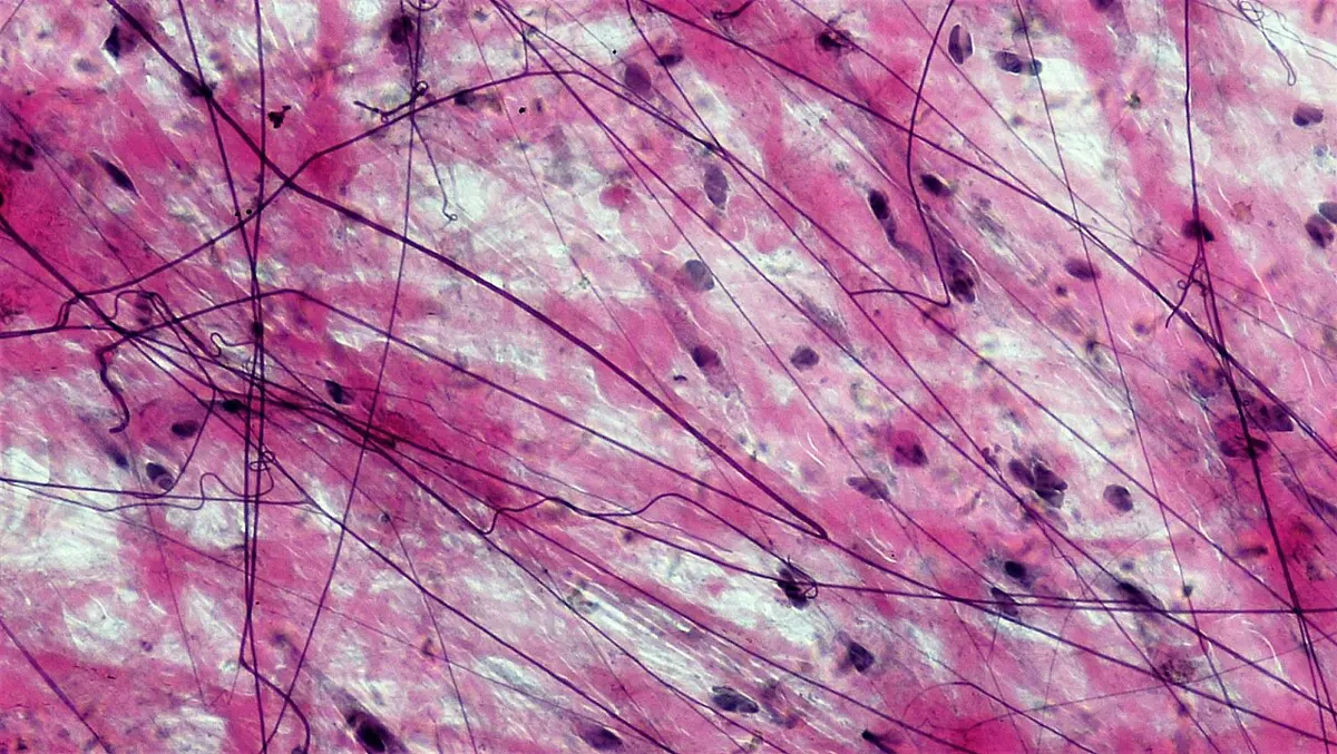
Areolar tissue (Latin areola, a little open space) is the commonest connective tissue. It is a loose, soft network of collagen and elastic fibers running in all directions, with plenty of open, matrix-filled spaces between them, and all five kinds of cell: fibroblasts, mast cells, macrophages, lymphocytes and plasma cells.
It lies under the skin, joining the skin to the muscles beneath (so your skin can be pinched up and slides over them), and between organs, round blood vessels and nerves, and under the epithelium that lines the gut (in the submucosa). Because it fills the spaces and packs organs in, it is called packing tissue. It also holds tissue fluid, and its wandering cells fight infection at the first place germs reach.
ii. Adipose tissue
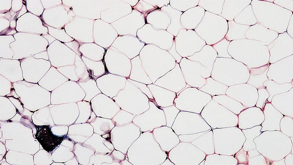
Adipose tissue is areolar tissue in which most of the cells have become fat cells (adipocytes): large round or oval cells, each storing fat. It is also called fatty tissue.
In a white fat cell, one big droplet of fat fills almost the whole cell and pushes the nucleus and a thin rim of cytoplasm to one side (a “signet ring”). In a brown fat cell, the fat is in many small droplets among many mitochondria, which give it its brown color and burn the fat to make heat.
- White adipose tissue (white or yellowish fat) is the body’s main store of fat: under the skin, round the kidneys, in the belly and buttocks of people, the hump of a camel, the blubber of whales and seals, and the fat bodies of a frog.
- Brown adipose tissue keeps warm those who most need it: newborn babies (between the shoulder blades and round the neck and kidneys) and hibernating mammals such as bats and marmots, which use it to warm themselves up as they wake. Adults keep a little of it.
Fat has several jobs, depending on where it is:
- a store of food for hard times: the camel’s hump, the frog’s fat bodies (which feed it through its winter sleep), the fat in the belly;
- an insulator that keeps heat in: the fat under the skin (panniculus adiposus) and the whale’s blubber, which also helps it float;
- a cushion that protects: the fat round the kidneys and behind the eyeballs, and in the soles and palms.
Bone marrow is part of this story. Red bone marrow, which makes blood cells, is gradually filled with fat cells as we grow up, and becomes yellow bone marrow. In an emergency, such as a heavy loss of blood, yellow marrow can turn back into red marrow and make blood cells again.
II. Dense connective tissue
In dense connective tissue, the fibers are packed closely together with few cells (mostly fibroblasts) and little matrix between them. It is strong and tough. By which fibers it has, it is of three types: dense fibrous (white fibrous), elastic (yellow fibrous) and reticular tissue.
i. Dense fibrous (white fibrous) connective tissue
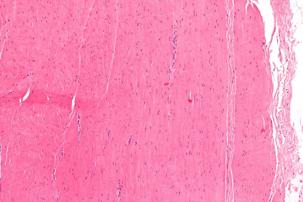
This is made mainly of collagen fibers in thick bundles, with rows of fibroblasts between them. It is white, very strong and does not stretch. It occurs in three forms:
A. White fibrous tissue (sheets and capsules). The collagen bundles run in different directions, so it resists pulling in any direction. It makes the strong sheath round bone (periosteum) and cartilage (perichondrium), the tough white of the eye (sclera), the capsules of organs such as the kidney, the deep layer of the skin (dermis), and the fibrous joints between the bones of the skull (sutures), where it lets no movement at all.
B. Tendon. The collagen bundles all lie parallel, in the direction of pull, with rows of fibroblasts squeezed between them. A tendon attaches muscle to bone. It is so strong that a tendon as thick as a finger could hold up a small car.
C. Ligament. Like tendon, parallel bundles of collagen, but with some elastic fibers among them, so it can give a little. A ligament attaches bone to bone at a joint, holding the joint together and letting it bend only as far as it should.
ii. Elastic (yellow fibrous) connective tissue
Elastic connective tissue is made mainly of elastic fibers, so it is yellow and can stretch and spring back. It is found where a part must stretch and recoil again and again: in the walls of large arteries (which swell at each heartbeat and recoil to keep the blood moving), in the lungs, between the cartilage rings of the trachea and bronchi, in the vocal cords of the larynx, and in a few ligaments.
iii. Reticular tissue
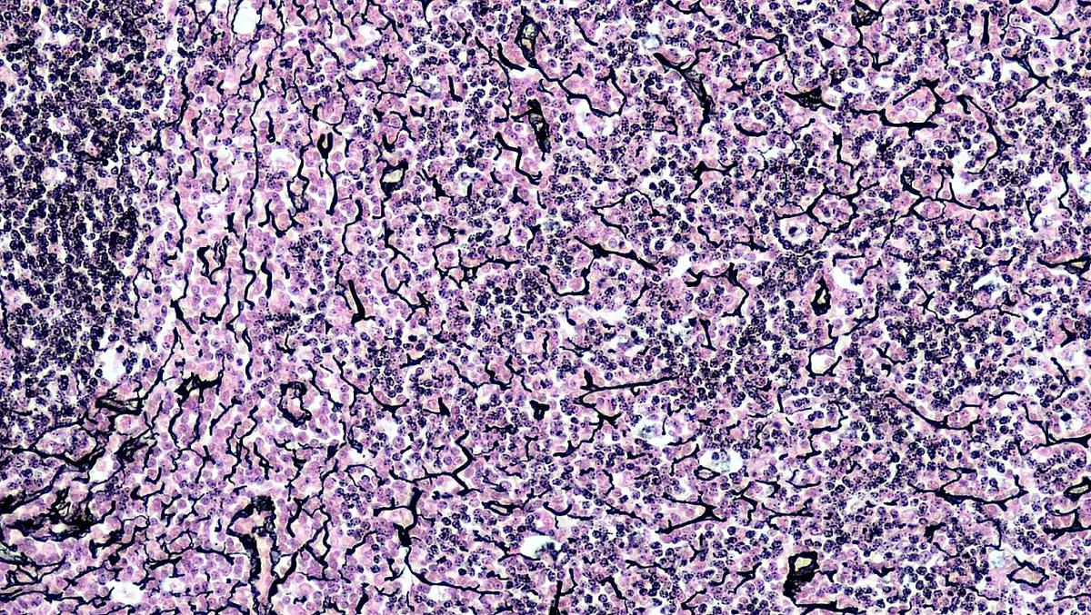
Reticular tissue is a delicate network of reticular fibers with reticular cells (a kind of fibroblast) lying on them. It forms the soft inner framework (stroma) that holds the cells of the liver, spleen, lymph nodes, bone marrow and other lymphoid organs, and a fine web round smooth muscle cells and fat cells.
A tendon is very strong but does not stretch. Its fibers are:
Pick an answer to see why.
Which tissue would you expect in the wall of the aorta, which swells at each heartbeat and springs back?
Pick an answer to see why.
2. Supportive connective tissue
Supportive (skeletal) connective tissue forms the framework of the body and of organs. Its matrix is firm or hard. There are two types: cartilage and bone.
I. Cartilage
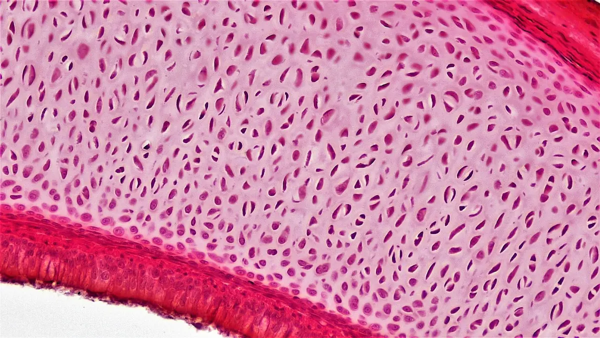
Cartilage is a firm, flexible connective tissue. Its matrix is a rubbery gel of proteins and sugars called chondrin, made by its cells. Its young, dividing cells are chondroblasts; they become mature cells, chondrocytes, which sit in small fluid-filled spaces (lacunae) in the matrix, singly or in groups of two, three or four (the daughters of one cell). Its matrix may also hold collagen or elastic fibers.
Cartilage is covered by a sheath of dense fibrous tissue, the perichondrium, except at the surfaces of joints. It has no blood vessels (it is avascular): food and oxygen diffuse to its cells through the matrix from vessels in the perichondrium. For this reason injured cartilage heals very slowly. It works as a shock absorber and makes smooth, slippery surfaces where bones meet.
By its fibers, cartilage is of four types: hyaline, calcified, fibrocartilage and elastic cartilage.
i. Hyaline cartilage
Hyaline cartilage (Greek hyalos, glass) is clear and bluish-white, like frosted glass, because its fibers are collagen fibers too fine to see under an ordinary microscope. It is the most common cartilage, also called gristle. It is found:
- on the ends of long bones, where they meet at joints (articular cartilage), as a smooth, slippery shock absorber;
- at the front ends of the ribs, joining them to the breastbone (costal cartilage), where it lets the chest move as we breathe;
- in the nose, making the nasal septum and the soft lower part of the nose, which holds its shape;
- in most of the larynx (voice box);
- as the C-shaped rings of the trachea and the plates in the bronchi, which keep the airways from collapsing. (The bronchioles have no cartilage.)
- as the whole skeleton of sharks, skates and rays (Chondrichthyes) and of lampreys and hagfishes (Cyclostomata), and as the first skeleton of every vertebrate embryo, most of which is later replaced by bone.
ii. Calcified cartilage
Calcified cartilage is hyaline cartilage whose matrix has been hardened with calcium salts. It is hard and rigid, but not bone. In a frog it forms the suprascapula of the shoulder girdle and the pubis of the hip girdle. In people, the deepest layer of cartilage at a joint and the cartilage at a growing bone’s ends calcify before they are replaced by bone.
iii. Fibrocartilage
Fibrocartilage has thick bundles of collagen fibers running through its matrix, with rows of cartilage cells between them, so it is also called white fibrous cartilage. It has no perichondrium. It is the toughest cartilage, able to stand both squeezing and pulling. It forms the intervertebral discs between the backbones, the pubic symphysis where the two hip bones meet in front, and the menisci of the knee.
iv. Elastic cartilage
Elastic cartilage has a network of elastic fibers in its matrix, so it is also called yellow fibrous cartilage. It bends easily and springs back. It forms the pinna (the outer ear) of mammals, the epiglottis (the flap that closes the larynx when we swallow), part of the larynx, and the Eustachian tube.
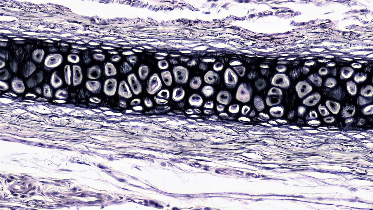
Which cartilage would you expect between two backbones, which must withstand the body’s weight pressing down and twisting?
Pick an answer to see why.
II. Bone
Bone is the hardest connective tissue and the strongest; the only thing in the body harder is the enamel of the teeth. It is rigid, and, unlike cartilage, it is full of blood vessels.
Its matrix is made of two things:
- organic (about 25 to 30% of its weight): mostly collagen fibers, together called ossein, which make bone tough and slightly springy, so that it doesn’t shatter;
- inorganic (about 60 to 70%): crystals of calcium phosphate (with some calcium carbonate and magnesium), which make it hard.
The rest, about 10%, is water.
Its cells:
- osteoblasts (bone-forming cells) lie on the surfaces of bone, under the periosteum and lining its cavities; they lay down new matrix;
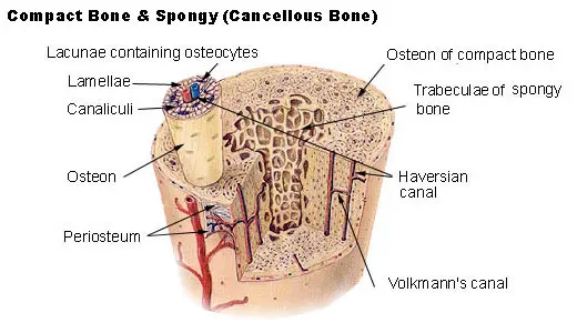
- osteocytes, the mature bone cells, are osteoblasts that have walled themselves in. Each sits alone in its lacuna, a small space in the hard matrix, and sends many fine branches through tiny channels, the canaliculi (“little canals”), to touch the branches of its neighbors; food, oxygen and messages pass along these;
- osteoclasts, large cells with many nuclei, dissolve bone. With the osteoblasts they keep rebuilding the skeleton all our lives: an adult’s skeleton is entirely replaced about every ten years.
Bone is covered by periosteum, a sheath of white fibrous tissue, except at the joints, where it is capped by cartilage. Its main job is to support the body as the endoskeleton of bony fishes, amphibians, reptiles, birds and mammals. It also protects the brain, heart and lungs, gives muscles something to pull on, stores calcium and phosphate, and holds the marrow that makes blood cells.
Types of bone by how they form
Bones form in two ways in the embryo:
- Cartilage bones (replacing bones, endochondral bones) are first made as small models of hyaline cartilage, which is then gradually replaced by bone (ossification). Most of the skeleton is formed this way: the limb bones, the vertebrae, the ribs, and the bones of the base of the skull.
- Membrane bones (investing or dermal bones) form directly in a sheet of fibrous connective tissue, with no cartilage first: the flat bones of the roof and sides of the skull, most of the face and the jaw, and most of the clavicle (collar bone).
- Sesamoid bones form inside a tendon, where it passes over a joint: the patella (kneecap) is the largest; the small fabella sits behind the knee of some people.
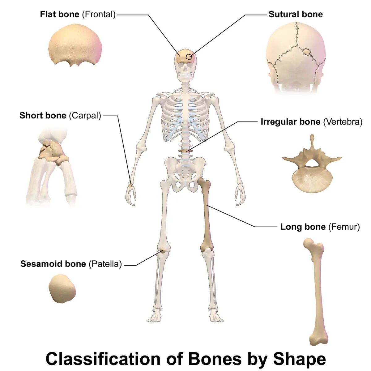
Types of bone by shape
- Flat bones are thin and flat, often curved: the bones of the skull’s roof (frontal, parietal, occipital), the sternum, the ribs, the scapula, and the ilium of the hip.
- Irregular bones have complex shapes: the vertebrae, many facial bones, and the ear ossicles.
- Short bones are about as wide as they are long: the carpals (wrist) and tarsals (ankle).
- Long bones are longer than wide, with a shaft and two ends: the bones of the limbs (other than the carpals and tarsals), such as the femur, humerus and the bones of the fingers.
A long bone: epiphysis and diaphysis
A long bone, such as the femur, has two parts:
A. Epiphysis (plural epiphyses). Each end of the bone is swollen, and is made inside of spongy bone (also called cancellous or trabecular bone): a honeycomb of thin bony plates and bars (trabeculae), with no osteons, covered by a thin shell of compact bone. The spaces between the trabeculae are filled with red bone marrow, the myeloid tissue where stem cells make blood cells (hematopoiesis). The trabeculae lie along the lines of stress, so the end of the bone is light but strong.
B. Diaphysis. The shaft in the middle is a thick tube of hard compact bone (also called cortical bone), round a central marrow cavity filled with yellow bone marrow, which is mostly fat. Its stem cells can make fat, cartilage and bone; in an emergency, part of it can turn back into red marrow and make blood cells. Compact bone is built of osteons (Haversian systems).
In children, a plate of hyaline cartilage, the growth plate, separates each epiphysis from the shaft. The bone grows in length there. In the late teens the plates turn to bone and the bones stop growing longer. Doctors read a child’s age from an X-ray of the hand by how far its growth plates have closed.
Compact bone in section
The structure of compact bone can be seen in a thin transverse section (T.S.) of the shaft.
In an amphibian (a frog), from outside in: the periosteum; the osteoblasts under it; lamellae (layers of matrix with osteocytes between them) running round the bone as simple rings, with no osteons; osteoblasts on their inside (endosteum); and the marrow cavity.
In a mammal, from outside in:
- Periosteum, with osteoblasts under it.
- Outer circumferential lamellae: a few rings running round the whole bone, with no osteons.
- Haversian systems (osteons), packed side by side, making most of the wall, with interstitial lamellae (pieces of old osteons) filling the spaces between them.
- Inner circumferential lamellae round the marrow cavity, with no osteons.
- Endosteum, with osteoblasts, lining the marrow cavity.
- Bone marrow.
The Haversian system (osteon) is a long, narrow cylinder of bone, running along the length of the bone, about 0.2 mm across. In its center is a Haversian canal, carrying a small artery, a vein, a lymph vessel and a nerve. Round the canal lie concentric lamellae, rings of matrix like the rings of a tree trunk (usually about 4 to 20 of them), whose collagen fibers spiral in a different direction in each ring, which makes the osteon very strong. Between the lamellae are the lacunae with their osteocytes, linked to each other and to the central canal by the canaliculi, so that every osteocyte is fed from the canal even through solid mineral. The Haversian canals are joined to each other, and to the blood vessels of the periosteum and the marrow, by canals that cross at right angles, the Volkmann’s canals.
Osteons were first described by the English doctor Clopton Havers in 1691. They are characteristic of the compact bone of mammals (and of large birds and reptiles), and are not found in amphibians.
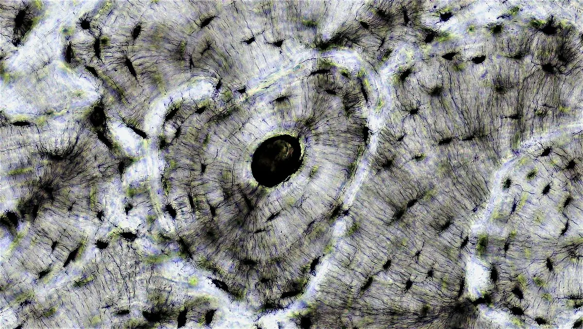
In a section of bone, the fine channels that link osteocytes to each other and to the central canal are:
Pick an answer to see why.
Spongy bone, with red marrow, is found mainly in:
Pick an answer to see why.
3. Fluid connective tissue
Fluid connective tissue has its cells in a liquid matrix (plasma), with no fibers (though one of the proteins dissolved in it, fibrinogen, turns into fibers when blood clots). It flows inside vessels and carries materials round the body, so it is also called vascular or mobile connective tissue or circulating fluid. It transports food, gases, wastes and hormones; it helps keep the body’s temperature, water and pH steady; and it defends the body against disease. Its cells are made by hematopoiesis. There are two kinds: blood and lymph.
I. Blood
Blood flows inside the heart and blood vessels: arteries, capillaries and veins. It makes up about 7 to 8% of the body’s weight: a man of 70 kg has about 5 to 5.5 liters of blood, and a woman of 60 kg about 4 to 4.5 liters.
It is heavier and thicker than water, tastes salty (about 0.9% salts), and is slightly alkaline, with a pH of about 7.4 (from 7.35 to 7.45). It resists changes in its pH: it is a buffer, mainly because of the bicarbonate (HCO₃⁻) and carbonic acid (H₂CO₃) in it, kept in a ratio of about 20 to 1. Blood is made of two parts, the liquid plasma and the blood cells.
Blood cells: S. J. Holmes, The Biology of the Frog (1906), public domain
i. Plasma
Plasma is the pale yellow liquid part of blood, about 55% of its volume. It is about 90 to 92% water, about 7% plasma proteins, and about 1 to 2% other dissolved substances.
The plasma proteins are:
- albumins, the most plentiful, made in the liver; they hold water in the blood by osmosis (keep up the blood’s osmotic pressure) and carry some substances;
- globulins: alpha and beta globulins carry substances such as iron, fats and hormones; gamma globulins are the antibodies (immunoglobulins), which protect against infection;
- fibrinogen, made in the liver, which turns into threads of fibrin when blood clots. (Plasma with its fibrinogen removed, the liquid left after blood clots, is called serum.)
Plasma carries digested food (glucose, amino acids, fats), gases, wastes such as urea and lactic acid, hormones, salts, vitamins, and drugs and toxins taken in.
ii. Blood cells (formed elements)
The blood cells, also called blood corpuscles (“little bodies”) or formed elements, make up about 45% of the volume of blood. In the embryo they are made first in the yolk sac, then in the liver and spleen; from before birth onward, in the red bone marrow (myeloid tissue). Red cells, granulocytes, monocytes and platelets are made in the marrow; lymphocytes are made there too, but multiply and mature in the lymphoid tissue: the lymph nodes, thymus, spleen and tonsils. Making blood cells is hematopoiesis.
There are three types: red blood cells, white blood cells and platelets.
Cells: Servier Medical Art (smart.servier.com), CC BY 4.0
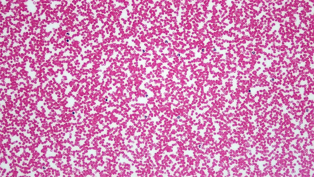
A. Red blood cells (RBCs), or erythrocytes (Greek erythros, red), are red because they are filled with hemoglobin, the iron-containing red protein that carries oxygen.
- Shape: in mammals, round (circular) biconcave discs, thinner in the middle than at the edge; in fishes, amphibians, reptiles and birds, oval and biconvex, bulging in the middle where the nucleus is.
- Size: a human red cell is about 7.5 µm across and 2 µm thick. Amphibians have the largest red cells of all vertebrates (in some salamanders over 70 µm long); mammals have the smallest.
- Contents: the red cells of fishes, amphibians, reptiles and birds have a nucleus, mitochondria and other organelles. A mammal’s red cell loses its nucleus and organelles as it matures. With no mitochondria, it gets its energy by anaerobic respiration (glycolysis), and so it uses none of the oxygen it carries.
- Job: to carry oxygen from the lungs to the tissues as oxyhemoglobin, and part of the carbon dioxide back as carbaminohemoglobin (most of the carbon dioxide travels in the plasma as bicarbonate).
- Number: in a healthy man about 5 million per mm³ (4.5 to 5.5 million), in a woman about 4.5 million per mm³. There are about 600 red cells for every white cell.
- Life: about 120 days in people. Old red cells are broken down in the spleen and liver.
- Made from erythroblasts by erythropoiesis: in a human fetus in the liver and spleen; after birth in the red bone marrow. In a tadpole they are made in its kidneys, liver and spleen; in an adult frog, in the spleen and bone marrow.
The biconcave shape gives a red cell a large surface for its volume, so oxygen diffuses in and out quickly, and makes it flexible, so it can fold to squeeze through capillaries narrower than itself. With no nucleus, it has more room for hemoglobin: each cell carries about 270 million molecules of it.
A healthy man has about 13 to 17 g of hemoglobin in every 100 mL of blood, a woman about 12 to 15 g. A person whose hemoglobin is below about 13 g (men) or 12 g (women) per 100 mL is said to have anemia, and tires easily. In Nepal anemia is common: the 2022 Demographic and Health Survey found 34% of women aged 15 to 49 anemic, and 43% of children aged 6 to 59 months. Lack of iron in the diet, hookworm, and blood lost in childbirth and menstruation are its commonest causes. (Nepal DHS 2022.)
B. White blood cells (WBCs), or leukocytes (Greek leukos, white), are colorless cells with a nucleus, and no hemoglobin. They defend the body: some engulf germs (phagocytes), some make antibodies, some cause allergic reactions. They are larger than red cells, and their shape changes as they move, like an amoeba. The largest are the monocytes; the smallest the small lymphocytes.
- Number: a total count (TC) of about 4,000 to 11,000 per mm³ in a healthy adult. The share of each kind, in percent, is the differential count (DC); doctors use both to spot infections.
- Life: from a few hours (neutrophils) to many years (some lymphocytes, which “remember” germs).
- Made from stem cells in the bone marrow (leukopoiesis).
They can squeeze out through the walls of capillaries, between the cells, to reach a place of infection: this is diapedesis. Too few white cells is leukopenia; too many, leukocytosis, as in an infection. Leukemia is a cancer of the blood-forming cells, in which the marrow makes huge numbers of abnormal white cells.
By whether their cytoplasm has granules that show up when stained, white cells are of two types: granulocytes and agranulocytes.
a. Granulocytes have many granules in their cytoplasm and a nucleus divided into lobes. They are named by the dyes their granules take up:
| Granulocyte | Granules stain with | Nucleus | Share of WBCs | Job |
|---|---|---|---|---|
| Neutrophils | both dyes: fine, pale lilac granules | 3 to 5 lobes | 60 to 70%, the most numerous | the first phagocytes to reach an infection; pus is mostly dead neutrophils |
| Eosinophils (acidophils) | the acid dye eosin: large, red-orange granules | usually 2 lobes | 2 to 4% | fight parasitic worms; increase in allergies and worm infections |
| Basophils | basic dyes: large, dark blue granules that hide the nucleus | 2 to 3 lobes, S-shaped | 0.5 to 1%, the fewest | release histamine and heparin: inflammation and allergy |
b. Agranulocytes have no visible granules, and an unlobed nucleus:
1. Lymphocytes have a large, round nucleus that fills most of the cell. They make up about 20 to 30% of the white cells. They make the body’s specific defense against germs (immunity). They are of two kinds:
- B lymphocytes (B cells) mature in the bone marrow. They turn into plasma cells, which make antibodies. (The letter first stood for the bursa of Fabricius, the organ in birds where these cells were discovered; in mammals, it is the bone marrow.)
- T lymphocytes (T cells) are made in the bone marrow and travel to the thymus to mature, before they go out into the blood. Some, the helper T cells, carry a marker called CD4 and direct the other defense cells: they are the cells HIV attacks; others (cytotoxic T cells, CD8) kill infected cells directly.
2. Monocytes have a large kidney-shaped or horseshoe-shaped nucleus. They make up about 2 to 8% of the white cells, and are the largest. They are phagocytes: they leave the blood and become the macrophages of the tissues.
C. Platelets (thrombocytes) are small, colorless, oval or irregular pieces of cytoplasm, without a nucleus, about 2 to 3 µm across: the smallest of the formed elements. In mammals they are not whole cells but fragments pinched off giant cells of the bone marrow (megakaryocytes). There are about 150,000 to 400,000 per mm³ of blood (0.15 to 0.4 million), and each lives about 7 to 10 days. Their job is to stop bleeding: they stick together to plug a small break in a vessel, and release substances that start blood clotting.
In other vertebrates (fishes, frogs, lizards and birds), the same job is done by small, oval, nucleated cells called thrombocytes; only in mammals are they fragments, the platelets.
Blood clotting
When a blood vessel is cut, blood clots within a few minutes and stops the bleeding. Clotting happens in three steps:
- Damaged tissue and platelets release thromboplastin (tissue factor), which, with the help of other clotting factors and calcium ions (Ca²⁺), forms prothrombinase (also called thrombokinase).
- Prothrombinase, with Ca²⁺, changes prothrombin, a plasma protein made in the liver with the help of vitamin K, into thrombin.
- Thrombin changes the soluble plasma protein fibrinogen into insoluble threads of fibrin.
The fibrin threads form a mesh across the wound, and red cells, white cells and platelets are caught in it: this mesh with its trapped cells is the clot. It then shrinks, squeezing out the clear yellow serum.
A blood smear shows a white cell with large, red-orange granules and a nucleus of two lobes. Which cell is it, and when might there be more of them than usual?
Pick an answer to see why.
Why does a mammal’s red blood cell use none of the oxygen it carries?
Pick an answer to see why.
II. Lymph
When blood flows through capillaries, some of its plasma seeps out between the cells, carrying food and oxygen to them: this is tissue fluid (interstitial fluid). Most of it goes back into the capillaries, but some is left in the tissues. This extra fluid drains into thin, blind-ended tubes, the lymph capillaries, and is then called lymph.
Lymph flows through lymph vessels that join into larger ones, passing through lymph nodes, which filter it and add lymphocytes to it. The vessels finally empty through the thoracic duct and the right lymphatic duct into the subclavian veins, in the neck, so the fluid returns to the blood. Lymph and its vessels and nodes make up the lymphatic system.
Lymph forms in the tissues all over the body; the liver and the intestines give a large share of it. In the small intestine, the lymph capillaries (lacteals) take up digested fats, which make the lymph there milky.
Lymph is like plasma but has less protein; its cells are mostly lymphocytes, and it has no red blood cells and no platelets. It is much the same as the tissue fluid (interstitial fluid, part of the extracellular fluid) from which it forms. Its jobs are to return to the blood the fluid and proteins that leak from the capillaries, to carry absorbed fats, and to fight infection: swollen lymph nodes in the neck during a sore throat are lymph nodes busy fighting germs.
6.3 Muscular tissue
Muscular tissue (muscle) is the tissue that contracts. Its cells, the muscle cells or myocytes, are long and thin, so they are called muscle fibers, with very little matrix between them. They are filled with two contractile proteins in fine threads (myofilaments): thin filaments of actin and thick filaments of myosin. When a nerve impulse or another signal excites a muscle fiber, its actin and myosin slide past each other and the fiber shortens: this is contraction. When the signal stops, the fiber goes back to its length and comes to rest: relaxation.
The cell membrane of a muscle fiber is the sarcolemma (Greek sarx, flesh), and its cytoplasm the sarcoplasm. Muscle tissue moves the body from place to place (locomotion), moves its parts, moves things inside it (food, blood, urine) and makes heat. It develops from the mesoderm.
There are three types: skeletal (striped), smooth (unstriped) and cardiac muscle.
Servier Medical Art (smart.servier.com), CC BY 4.0
1. Skeletal (striped) muscle
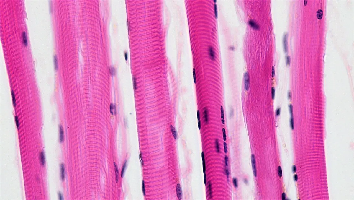
Skeletal muscle, also called striped, striated or voluntary muscle, is made of long, cylindrical, unbranched fibers with cross stripes (striations) of dark and light bands.
Each fiber is a single, very long cell with many nuclei lying just under the sarcolemma. It forms in the embryo when many small cells (myoblasts) fuse end to end, so it is a syncytium (a mass of cytoplasm with many nuclei). It may be several centimeters long: the longest fibers, in the thigh, run most of the muscle’s length.
Inside a fiber: the sarcomere
A fiber is packed with many myofibrils, thin threads that run its whole length, and each myofibril has alternate dark and light bands, lined up with those of its neighbors, which give the whole fiber its stripes.
- The dark band, or A band, is where the thick myosin filaments lie; its length is the length of the myosin filaments. (Anisotropic: it splits a beam of polarized light in two.)
- The light band, or I band (isotropic: it leaves polarized light as it is), has only thin actin filaments.
- Across the middle of each I band runs a thin dark line, the Z line (Z disc; in older books Krause’s membrane), to which the actin filaments are fixed.
- In the middle of the A band is a paler zone, the H zone, where there is only myosin, no actin. In the middle of the H zone is the M line, which holds the myosin filaments in place.
The stretch of myofibril from one Z line to the next is a sarcomere, the unit of contraction, about 2.5 µm long at rest. From each Z line the actin filaments reach toward the middle of the sarcomere; the myosin filaments lie between them in the middle, overlapping the ends of the actin filaments.
How a muscle contracts: the sliding filament theory
When a muscle contracts, its filaments do not get shorter. Instead, little heads on each myosin filament grip the actin filaments beside them, pull, let go and grip again, so that the actin filaments slide toward the middle of the sarcomere over the myosin. This is the sliding filament theory, put forward in 1954 by Hugh Huxley and Jean Hanson, and at the same time by Andrew Huxley and Rolf Niedergerke (the two Huxleys were not related). The energy comes from ATP, and the signal from calcium ions released inside the fiber.
As the actin slides in:
- the Z lines come closer together, so each sarcomere shortens;
- the I band (only actin) gets shorter;
- the H zone (only myosin) gets shorter, and may disappear when the actin filaments meet in the middle;
- the A band stays the same length, because the myosin filaments themselves do not change.
When the muscle relaxes, the filaments slide apart again and the bands return to their resting widths.
From fiber to muscle
Many myofibrils make one muscle fiber; many fibers are grouped into a bundle, a fascicle; and many fascicles make a whole muscle, such as the biceps or triceps. Each level is wrapped in connective tissue:
- the endomysium wraps each fiber;
- the perimysium wraps each fascicle;
- the epimysium wraps the whole muscle, and runs on into its tendon.
Skeletal muscle is about 75% water, 20% protein (mostly actin and myosin), and 5% other substances such as potassium, calcium, glycogen and fats.
Red and white muscle fibers
Skeletal muscle fibers are of two kinds:
- Red (slow) fibers are thin, with a rich blood supply, many mitochondria and much myoglobin (a red protein that stores oxygen), which gives them their color. They contract slowly and can keep working for a long time without tiring: endurance. They are many in the muscles that hold us upright, in the legs of long-distance runners, and in the flight muscles of birds that fly far.
- White (fast) fibers are thicker, with little myoglobin and few mitochondria; they get energy quickly from glycogen without oxygen. They contract fast and strongly, but tire quickly: sprints, jumps and lifts. A chicken, which flutters only in short bursts, has white breast meat, while a duck or pigeon that flies far has dark breast meat.
Every person’s muscles contain both kinds, mixed: sprinters tend to have more white fibers in their legs and marathon runners more red ones, partly from birth, and training changes them a little.
Skeletal muscle is attached to the bones, usually by tendons, and moves them; it is also found in the tongue, the upper part of the esophagus, the muscles that move the eyeball, and the diaphragm. It is under our control: voluntary. It contracts quickly and strongly but tires (fatigues) quickly.
2. Smooth (unstriped) muscle
Smooth muscle, also called unstriped, non-striated, visceral or involuntary muscle, is made of spindle-shaped cells, thick in the middle and tapering at both ends, each with a single, central nucleus and no cross stripes. Its actin and myosin filaments are not arranged in sarcomeres but criss-cross the cell, so no bands show. The cells lie side by side, the thick middle of one against the thin ends of its neighbors, forming sheets or layers.
It is of two types:
- Single-unit (visceral) smooth muscle: its cells are joined by gap junctions, so a signal passes from cell to cell and the whole sheet contracts together, as one unit, often in waves. It is found in the walls of the gut, the urinary bladder and the uterus.
- Multi-unit smooth muscle: each cell is supplied by a nerve and contracts on its own, independently of its neighbors, allowing fine control. It is found in the arrector pili muscles that raise the hairs (“goose bumps”), the iris of the eye, and the walls of large blood vessels and airways.
Smooth muscle lines the walls of hollow organs: the alimentary canal, where it pushes food along by peristalsis; blood vessels, which it narrows and widens; the airways, the bladder and the uterus. It is not under our control: involuntary. It contracts slowly and can stay contracted for a long time without tiring.
3. Cardiac muscle
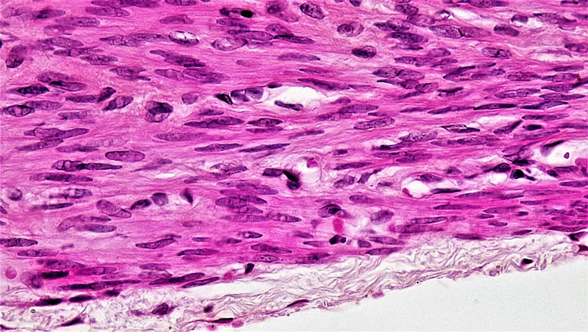
Cardiac muscle (heart muscle) is found only in the wall of the heart. Its fibers are cylindrical and striped, though the stripes are fainter than in skeletal muscle, and they branch and join each other, forming a network. Each fiber is made of separate cells (cardiac muscle cells, cardiomyocytes), joined end to end. Each cell has one, sometimes two, nuclei in its center. Where two cells meet, there is a dark cross line, the intercalated disc: a strong joint of desmosomes that keeps the cells from pulling apart at each beat, with gap junctions through which the signal to contract passes from cell to cell almost instantly, so that the whole heart beats together.
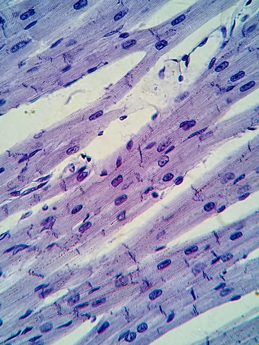
Cardiac muscle is like skeletal muscle in structure, as both are striped. It is like smooth muscle in function, as both are involuntary: cardiac and unstriped muscles both work without our control. But smooth muscle can tire after long work, and cardiac muscle never tires. It starts its own beat: special cardiac muscle cells in the heart’s pacemaker (the SA node) set off each beat by themselves, without a nerve, and nerves only speed the beat up or slow it down. It beats about 70 times a minute, about 100,000 times a day, and resting for a moment after each beat keeps it from tiring.
There is less cardiac muscle in the body than either of the other two kinds.
| Feature | Skeletal muscle | Smooth muscle | Cardiac muscle |
|---|---|---|---|
| Shape of fiber | long, cylindrical, unbranched | spindle-shaped, tapering | cylindrical, branched, network |
| Nuclei | many, at the edge (syncytium) | one, central | one (or two), central |
| Stripes | clear, dark and light bands | none | present, fainter |
| Intercalated discs | absent | absent | present |
| Control | voluntary | involuntary | involuntary, beats by itself |
| Speed and fatigue | fast; tires quickly | slow; can work for long periods | rhythmic; never tires |
| Where | attached to bones; tongue | walls of gut, vessels, bladder, uterus; iris; arrector pili | wall of the heart only |
Which feature is found only in cardiac muscle?
Pick an answer to see why.
During contraction of a skeletal muscle, which part of the sarcomere keeps the same length?
Pick an answer to see why.
6.4 Nervous tissue
Nervous tissue controls and coordinates the activities of the body. Its cells, the neurons, are specialized to receive stimuli and conduct nerve impulses: they are excitable (they respond to a stimulus) and conductive (they carry the signal on). They are long cells, with a long process, the axon or nerve fiber; there is very little matrix between them. Nervous tissue comes from the ectoderm. It makes up the brain, the spinal cord and the nerves.
Nervous tissue has two kinds of cells: neurons and neuroglia.
1. Neuroglia
Neuroglia (Greek glia, glue), or glial cells, are the cells of nervous tissue that don’t carry nerve impulses. They hold neurons in place, protect and feed them, form their myelin, clean up and keep their surroundings right. Unlike neurons, they can divide all through life. In the brain there are about as many glial cells as neurons.
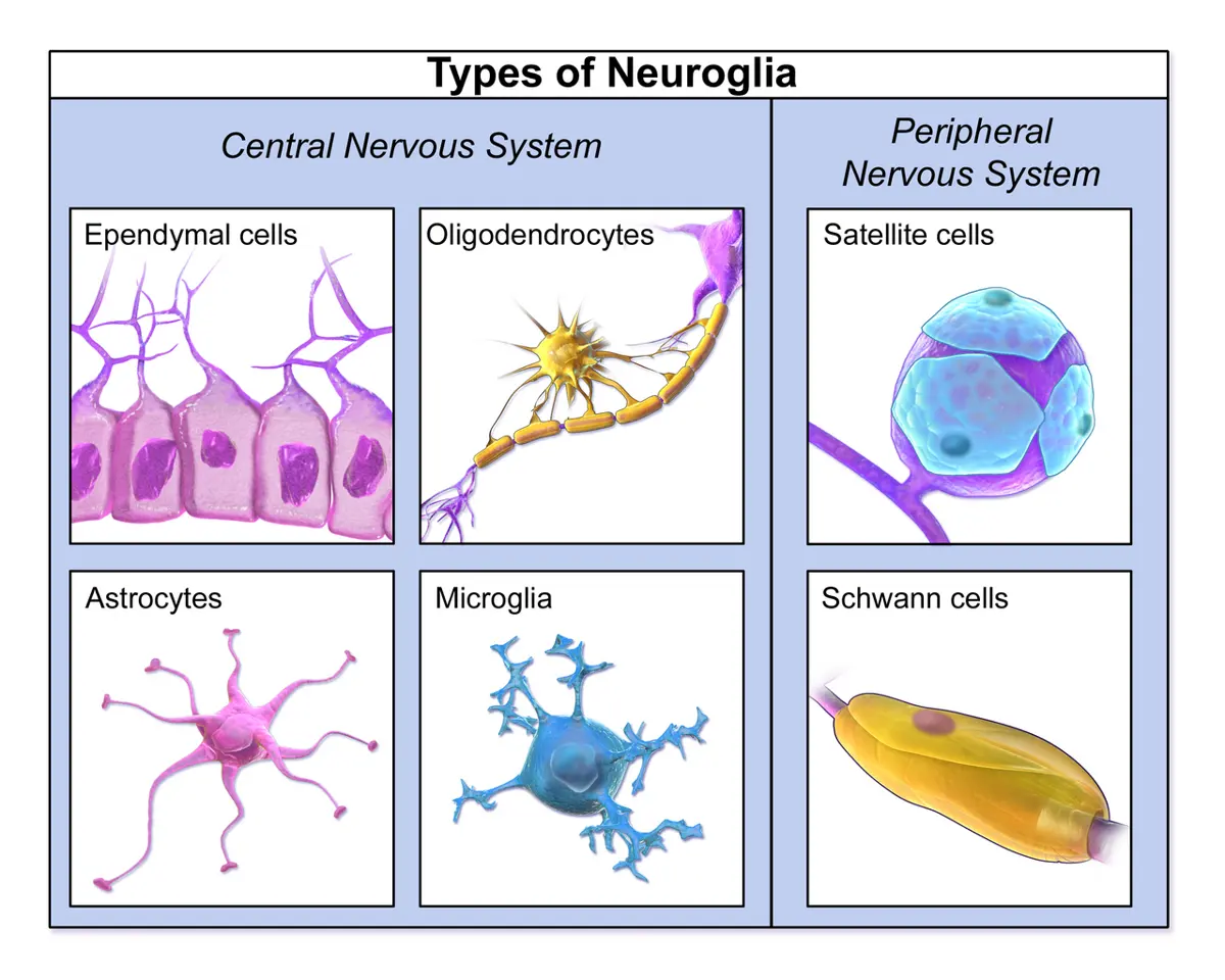
In the central nervous system (CNS), the brain and spinal cord, there are four kinds:
- Astrocytes are star-shaped, with many processes. Their processes wrap the blood capillaries of the brain and hold neurons to them; they control what passes from the blood to the neurons (the blood–brain barrier), keep the fluid round the neurons right, and fill in damaged areas with a scar after an injury.
- Oligodendrocytes have a few processes, each of which wraps round a stretch of an axon to form its myelin sheath in the CNS. One oligodendrocyte myelinates parts of many axons.
- Microglia are small cells that are the macrophages of the CNS: they move about and engulf germs and the remains of dead neurons.
- Ependymal cells line the ventricles (cavities) of the brain and the central canal of the spinal cord. Some have cilia that keep the cerebrospinal fluid (CSF) moving, and together with blood capillaries they form the choroid plexuses, which make the CSF.
In the peripheral nervous system (PNS), the cranial and spinal nerves and their ganglia, there are two kinds:
- Satellite cells surround the cell bodies of neurons in the ganglia, supporting them and controlling their surroundings, like the astrocytes of the brain.
- Schwann cells wrap round the axons to form their myelin sheath in the PNS, each Schwann cell covering one stretch of one axon. They also help a cut nerve fiber to regrow.
Neuroglia, like neurons, develop from the ectoderm, except the microglia, which come from the mesoderm: they are descended from the same blood-making cells as the macrophages.
2. Neurons
Neurons (nerve cells) are the structural and functional units of nervous tissue and of the whole nervous system. They are the body’s longest cells: the axon of a neuron in the spinal cord can reach to the foot, about a meter long.
A neuron has two parts: the cell body and its processes (neurites).
I. Cell body
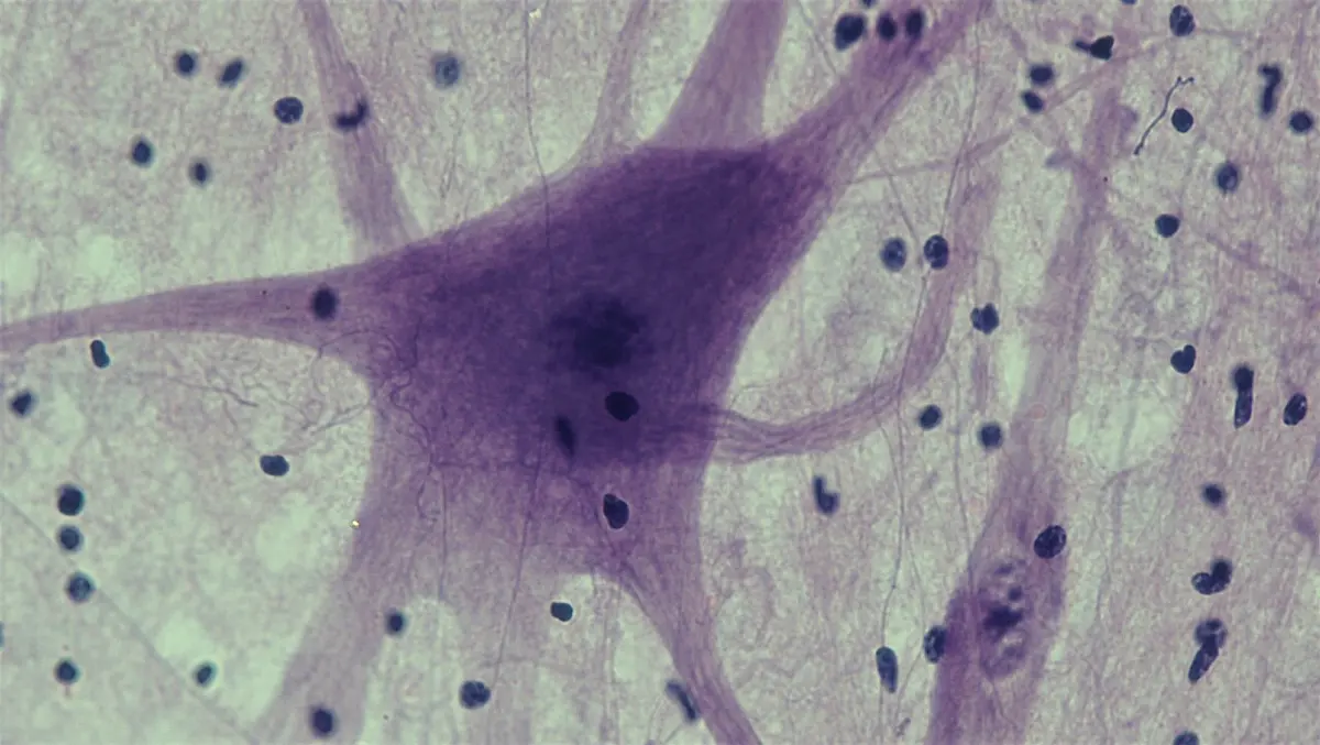
The cell body is also called the cyton, soma or perikaryon. It is often star-shaped, with a large round nucleus and a clear nucleolus, and its cytoplasm has the usual organelles. Scattered in it are Nissl bodies (Nissl granules), groups of rough endoplasmic reticulum and ribosomes, which make proteins for the long cell. Stained, they look like the spots of a tiger’s coat, so they are also called tigroid bodies. Cell bodies are gray, and are found in the brain, the spinal cord and the ganglia (clusters of cell bodies outside them).
II. Processes (neurites)
The processes that grow out of the cell body are of two kinds, dendrites and the axon.
i. Dendrites (Greek dendron, tree; also called dendrons) are usually many, short and much branched. They are extensions of the cell body and contain the same cytoplasm, with Nissl bodies. They receive messages from other neurons or from sense cells, and carry impulses toward the cell body: so they are the neuron’s afferent (incoming) processes.
ii. The axon is a single, usually long process, which grows out from a cone-shaped part of the cell body, the axon hillock. The axon hillock and the axon have no Nissl bodies. Its membrane is the axolemma, and its cytoplasm the axoplasm. It may give off a few side branches (collaterals), and at its end it divides into many fine branches, the telodendria, each ending in a small swelling, the axon terminal (synaptic knob, end bulb). The axon carries impulses away from the cell body to another neuron, a muscle or a gland: it is the neuron’s efferent (outgoing) process.
In the PNS, many axons are wrapped along their length by a row of Schwann cells; in the CNS, by the processes of oligodendrocytes. In a myelinated fiber, each Schwann cell wraps itself round the axon many times, like a bandage, so that the axon is covered by many layers of the Schwann cell’s own membrane: this layered, fatty covering is the myelin sheath. Myelin is mostly lipid (fats and phospholipids), with some protein, and is white. Outside the myelin, the outermost layer of the Schwann cell, with its nucleus and the rest of its cytoplasm, forms a thin sheath round the whole fiber, the neurilemma (sheath of Schwann). The gaps between neighboring Schwann cells, where the axon is bare, are the nodes of Ranvier (neurofibril nodes). (Fibers in the CNS are myelinated by oligodendrocytes, and have no neurilemma.)
Neuron: Pearson Scott Foresman, Public domain, via Wikimedia Commons
The myelin sheath insulates the axon. An impulse can’t cross the myelin, so it jumps from node to node: this is saltatory conduction (Latin saltare, to jump), and it is much faster. A thick myelinated fiber can carry an impulse at up to about 120 meters per second, while a thin non-myelinated one carries it at about 1 meter per second (0.5 to 2), slower than a cyclist. These are typical speeds, and the race below uses them; its nodes are drawn 5 cm apart so they can be seen, where real ones are about a millimeter apart, so a real impulse makes about a thousand jumps on its way to the foot. The nodes of Ranvier are found only on myelinated fibers.
At the end of the axon, each axon terminal meets the next cell at a synapse: a tiny gap across which chemical messengers (neurotransmitters) carry the signal on to a dendrite or the cell body of another neuron. Where an axon meets a muscle fiber, the junction is the neuromuscular junction, and the specialized part of the muscle fiber is the motor end plate.
Types of neurons
By their job (the direction they carry impulses):
- Sensory (afferent) neurons carry impulses from the sense organs (receptors) to the CNS.
- Motor (efferent) neurons carry impulses from the CNS to the effectors: muscles and glands.
- Interneurons (relay, association or adjustor neurons) lie inside the CNS, between sensory and motor neurons, and connect them. Most of the neurons of the brain are interneurons.
By myelin:
- Myelinated (medullated) fibers have a myelin sheath with nodes of Ranvier. They are white and conduct fast. Most of the fibers of the nerves to the muscles, and of the brain’s white matter, are myelinated.
- Non-myelinated (non-medullated) fibers have no myelin sheath and no nodes, though in the PNS they still lie in grooves of Schwann cells. They are gray and conduct slowly: for example, many fibers that carry dull, aching pain, and the fibers that control the gut and the blood vessels. Some are long: the length of a fiber does not depend on whether it has myelin.
By the number of processes from the cell body:
- Apolar neurons have no distinct axon or dendrites, only short, similar processes. They are found in the nerve net of coelenterates such as Hydra.
- Unipolar neurons have a single process from the cell body, which works as both dendrite and axon. They are found in invertebrates, such as insects, and in the early embryo of vertebrates.
- Pseudounipolar neurons (“false unipolar”) start as bipolar neurons whose two processes later join near the cell body, so a single process leaves the cell body and then divides like a T into two branches: one runs out to the sense organ (the peripheral branch) and the other into the spinal cord (the central branch). These are the sensory neurons of the dorsal root ganglia of the spinal nerves.
- Bipolar neurons have two processes, from opposite ends of the cell body: one dendrite and one axon. They are found in the retina of the eye, the olfactory epithelium of the nose and the inner ear.
- Multipolar neurons have many dendrites and one axon. They are the commonest neurons: the motor neurons and most interneurons of the brain and spinal cord.
Gray and white matter
Nervous tissue looks gray or white to the naked eye, depending on what it contains:
- Gray matter is made mostly of the cell bodies of neurons, their dendrites and non-myelinated fibers, with neuroglia. It forms the outer layer (cortex) of the brain and the inner, H-shaped core of the spinal cord.
- White matter is made mostly of myelinated axons, which the fatty myelin makes white. It forms the inner part of the brain and the outer part of the spinal cord.
A nerve is a bundle of axons outside the CNS, wrapped in connective tissue: the endoneurium round each fiber, the perineurium round each small bundle (fascicle), and the epineurium round the whole nerve. Many fascicles together make a nerve.
Which glial cell forms the myelin sheath of the axons in the spinal nerves?
Pick an answer to see why.
A neuron in the dorsal root ganglion has one process leaving its cell body, which then divides into a branch to the skin and a branch to the spinal cord. It is:
Pick an answer to see why.
Why does an impulse travel faster along a myelinated fiber than along a non-myelinated one of the same thickness?
Pick an answer to see why.
Comparing the four tissues
Exercise
A. Tick the correct alternatives
Who coined the term tissue?
Pick an answer to see why.
What is meant by a tissue?
Pick an answer to see why.
Which of the following is not an animal tissue?
Pick an answer to see why.
Which animal tissue is formed from all three germ layers?
Pick an answer to see why.
Which statement about epithelial tissue is incorrect?
Pick an answer to see why.
Which one of the following is a mesothelium?
Pick an answer to see why.
A basement membrane is a characteristic feature of:
Pick an answer to see why.
The lining (mucosal epithelium) of the intestine is made of:
Pick an answer to see why.
Which tissue is found in the skin?
Pick an answer to see why.
Which epithelium forms the inner lining of the vagina?
Pick an answer to see why.
Plastic epithelium is:
Pick an answer to see why.
Which animal tissue is the most abundant?
Pick an answer to see why.
All three components, cells, matrix and fibers, are present in:
Pick an answer to see why.
Which cells of areolar tissue can move about and engulf foreign particles?
Pick an answer to see why.
Which tissue has cells filled with fat globules?
Pick an answer to see why.
The suture joints between skull bones are made of:
Pick an answer to see why.
Which fibers are most abundant in a tendon?
Pick an answer to see why.
A tendon connects muscle to bone. What does a ligament connect?
Pick an answer to see why.
What are ligaments made of?
Pick an answer to see why.
Which one of these is avascular (has no blood vessels)?
Pick an answer to see why.
The Haversian system is a characteristic feature of the bone of:
Pick an answer to see why.
Human red blood cells are:
Pick an answer to see why.
The function of erythrocytes is:
Pick an answer to see why.
Which pair of white blood cells are the main phagocytes of the blood?
Pick an answer to see why.
A cancer of the blood-forming cells in which far too many white blood cells are made is:
Pick an answer to see why.
Which muscle cells are syncytial (many nuclei in one cell)?
Pick an answer to see why.
The epimysium is the covering of a skeletal muscle’s:
Pick an answer to see why.
The muscle responsible for moving the tongue in speech is:
Pick an answer to see why.
Which muscle has no stripes?
Pick an answer to see why.
The arrector pili are made of:
Pick an answer to see why.
Which muscle has intercalated discs?
Pick an answer to see why.
The wall of the heart is cardiac muscle. Cardiac muscle is:
Pick an answer to see why.
Which tissue holds the neurons together in the brain and spinal cord?
Pick an answer to see why.
Which tissue’s main cells don’t divide by mitosis once mature?
Pick an answer to see why.
Which cells are the macrophages of the CNS?
Pick an answer to see why.
In which part of a neuron are Nissl bodies found?
Pick an answer to see why.
Bipolar neurons are found in the:
Pick an answer to see why.
Gray matter is found in the:
Pick an answer to see why.
Which cells produce cerebrospinal fluid?
Pick an answer to see why.
Which animal tissue has the longest cells?
Pick an answer to see why.
B. Answer the following questions
Explain in short the levels of organization shown in the figure of the cell, smooth muscle tissue, the stomach, the digestive system and the human body.
Answer
Describe the different types of simple epithelium in animals, with diagrams.
Answer
Write the similarities and differences between simple and compound epithelium.
Answer
What is tessellated epithelium? How is it different from simple cuboidal and simple columnar epithelium?
Answer
Discuss the rare and uncommon types of epithelium, with their structure, function and location.
Answer
Is there an epithelium known as plastic epithelium? If so, explain it.
Answer
Summarize the modified (specialized) epithelia.
Answer
What is glandular epithelium? Describe the different types of glands in animals.
Answer
Mention the types and functions of connective tissue.
Answer
Give an account of areolar tissue.
Answer
Compare and contrast cartilage and bone.
Answer
Describe the Haversian system.
Answer
Write a short note on formed elements.
Answer
Describe the structure of voluntary muscle.
Answer
Draw a well-labeled diagram of a multipolar neuron. (No description is needed.)
Answer
Printable PDFs
- Study notes 8 pages
- Worksheet 4 pages Answer key
Quiz yourself: Living Things and Their Structure, Grades 5–12 →
Practice every question in the book, and keep track of the ones you miss →
A companion to your textbook, not a replacement for it: study from the book your school uses, and where the two differ, follow your teacher. Found a mistake? Please tell us.