
Every day your two kidneys, each about the size of a bar of soap, filter about 180 liters of fluid out of your blood: enough to fill a large water drum. Yet you pass only about one and a half liters of urine. The other 178 liters or so are quietly taken back, along with almost every grain of sugar, nearly all the salt and most of the useful chemicals, while the poisons are left behind. All of it is done by about a million microscopic tubes in each kidney, each one a filter and a sorting machine.
This unit is about how animals get rid of the nitrogen from the proteins they eat, how the human kidney is built, how a single tube in it makes urine, and the other organs that help.
The excretory system
Excretion is the removal from the body of the metabolic wastes made in its own cells. The excretory system is the group of organs that does this. The wastes include nitrogenous compounds (ammonia, urea, uric acid, creatinine), excess water, inorganic salts, carbon dioxide, and used-up hormones and drugs.
Excretion is essential for homeostasis: keeping the composition of the body fluids and tissues steady (their water, salts and pH), whatever the animal eats or drinks.
Types of nitrogenous excretory products
When animals break down proteins (and nucleic acids), the amino groups (–NH₂) are removed from the amino acids (deamination) and become ammonia. Ammonia is very toxic, so it must be removed quickly or turned into something safer. Which form an animal gets rid of depends mainly on how much water it can spare.
1. Ammonia (ammonotelism)
Ammonia is highly toxic and highly soluble in water, so it must be washed out at once in a large volume of water. It suits animals that live in water and lose it straight from their body surface or gills. Getting rid of nitrogen mainly as ammonia is ammonotelism.
Ammonotelic animals: protozoans (Amoeba), sponges, cnidarians (Hydra), tapeworms, leeches, most aquatic arthropods (crabs, prawns), most aquatic mollusks, bony fishes (freshwater ones, and most marine ones too), tadpoles of frogs and toads, and tailed amphibians that live in water (newts).
2. Urea (ureotelism)
Urea is much less toxic than ammonia and less soluble, so it can stay in the body for some time and be passed out in a smaller volume of water. It is made from ammonia and carbon dioxide in the liver. Getting rid of nitrogen mainly as urea is ureotelism.
Ureotelic animals: mammals including humans, adult frogs and toads, cartilaginous fishes (sharks and rays, the elasmobranchs, which keep a lot of urea in their blood to match the saltiness of the sea), and turtles.
3. Uric acid (uricotelism)
Uric acid is the least toxic, and it is almost insoluble in water, so it can be stored and passed out as a paste or solid with very little water. It suits animals that live in dry places and must save water, and animals whose young grow inside a hard-shelled egg, where any toxic ammonia would build up. Getting rid of nitrogen mainly as uric acid is uricotelism.
Uricotelic animals: insects, land snails, lizards and snakes, and birds. (The white part of a pigeon’s dropping is uric acid.)
4. Amino acids (aminotelism)
A few aquatic animals pass some of their nitrogen out as amino acids themselves, without breaking them down: some mollusks such as the freshwater mussel Unio and the pond snail Limnaea, and some echinoderms such as the starfish (Asterias). This is aminotelism. Animals can’t store spare amino acids, and these ones lose them directly into the water around them.
Dual excretion
Some animals switch between two waste products as their conditions change:
- The earthworm passes out ammonia when it is well fed and in wet soil, and more urea when it is short of water or food.
- Lungfishes pass out ammonia in water, but make urea while they lie dried up in mud during a drought.
- The African clawed toad (Xenopus) is ammonotelic in water and turns ureotelic if kept out of it.
- Crocodiles pass out both ammonia and uric acid.
| Ammonia | Urea | Uric acid | |
|---|---|---|---|
| Toxicity | highest | less | least |
| Solubility in water | very high | high | almost insoluble |
| Water needed to pass it out | a great deal | moderate | very little |
| Typical animals | aquatic invertebrates, bony fishes, tadpoles | mammals, adult amphibians, sharks | insects, reptiles, birds |
Why do birds pass out uric acid instead of urea?
Pick an answer to see why.
A tadpole is ammonotelic, but the adult frog is ureotelic. What best explains the change?
Pick an answer to see why.
Excretory organs in the human body
The human excretory (urinary) system has:
- a pair of kidneys, which make urine;
- a pair of ureters, which carry it to
- the urinary bladder, which stores it;
- the urethra, which carries it out of the body.
Servier Medical Art (smart.servier.com), CC BY 4.0
Turn the body round and see where the kidneys really are: against the back wall of the abdomen, either side of the backbone, partly behind the lowest ribs.
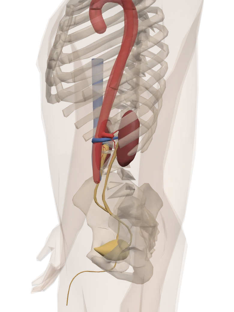
A model of a real body. Tap Show in 3D to turn it and tap its parts.
KidneysTwo bean-shaped organs, each about the size of your fist, at the back of the belly on either side of the backbone. They filter the blood and take out urea, uric acid, extra salt and water, and harmful chemicals as urine.
UretersTwo thin tubes that carry the urine from the kidneys down to the urinary bladder.
Urinary bladderA stretchy bag that stores the urine until it is passed out.
UrethraThe tube through which the urine stored in the bladder is passed out of the body.
Renal arteriesBring blood, with its wastes, from the aorta to each kidney.
Renal veinsTake the cleaned blood from each kidney to the inferior vena cava, and on to the heart.
AortaThe largest artery. It carries oxygenated blood from the heart’s left ventricle to the whole body, the kidneys too.
Inferior vena cavaCarries deoxygenated blood from the trunk and legs, the kidneys’ blood too, back to the heart’s right auricle.
Body and organs: BodyParts3D, The Database Center for Life Science (CC BY 4.0)
1. Kidneys
External features
The kidneys are a pair of dark red, bean-shaped organs lying against the back wall of the abdomen, one on each side of the backbone, at about the level of the last thoracic to the third lumbar vertebrae (T12 to L3). They are covered on the front by the peritoneum and lie behind the peritoneal cavity: they are retroperitoneal. The right kidney lies a little lower than the left, pushed down by the large right lobe of the liver.
Each kidney is about 11 cm long, 5.5 cm wide and 3 cm thick, and weighs about 150 g in a man and 135 g in a woman (sizes vary with age and body size). The human kidney is a metanephros, the third and most advanced kind of kidney to form in the embryo of reptiles, birds and mammals.
Each kidney is wrapped in three protective layers:
- the renal capsule, innermost: a thin, tough, white, fibrous skin stuck to the kidney;
- the adipose capsule, the middle layer of fat, which cushions it;
- the renal fascia, outermost: a sheet of fibrous tissue that anchors the kidney to the back wall of the abdomen.
Each kidney has an outer convex border and an inner concave border with a notch, the hilum (hilus). Through the hilum the ureter leaves the kidney, and the renal artery, renal vein, lymph vessels and nerves enter or leave it.
Internal structure
A longitudinal section (L.S.) of the kidney shows two distinct regions:
- the outer cortex, darker, making up about one-third of the kidney’s tissue;
- the inner medulla, lighter and striped, making up about two-thirds. It is divided into 8–18 cone-shaped renal pyramids, whose stripes are the straight tubules and vessels inside them. The tip of each pyramid, the renal papilla, points inwards.
The cortex dips down between the pyramids as the renal columns (columns of Bertini). Each papilla opens into a cup, a minor calyx; minor calyces join into two or three major calyces, which join into the funnel-shaped renal pelvis. The pelvis narrows into the ureter at the hilum.
Inside, each kidney is made of about one million nephrons (uriniferous tubules), the kidney’s functional units, packed in connective tissue with blood vessels. The cortex holds the Malpighian bodies (Bowman’s capsules and glomeruli) and the convoluted tubules (PCT and DCT); the medulla holds the loops of Henle, the collecting ducts and the ducts of Bellini. The nephrons make the urine.
Servier Medical Art (smart.servier.com), CC BY 4.0
The human kidney is said to be retroperitoneal. What does this mean?
Pick an answer to see why.
Which is the right order for urine leaving a nephron?
Pick an answer to see why.
2. Ureters
From the hilum of each kidney the pelvis narrows into a whitish, narrow, muscular tube, the ureter, about 25–30 cm long. The ureters run down on either side of the backbone and open separately, close to each other, into the back of the urinary bladder.
The ureter’s wall has three layers: an outer fibrous layer, a middle smooth muscle layer, and an inner mucosa lined with transitional epithelium. Waves of contraction of the muscle (peristalsis) push urine down to the bladder in spurts, even when you lie down or stand on your head. Each ureter passes slantwise through the bladder wall, and a fold of mucous membrane at its opening acts like a valve: as the bladder fills and its pressure rises, the opening is pressed shut, so urine cannot flow back from the bladder up into the ureter.
3. Urinary bladder
The urinary bladder is a hollow, pear-shaped muscular bag on the floor of the pelvic cavity. It stores urine for a while.
- Its wall has an outer fibrous layer, a thick middle layer of smooth muscle with loose connective tissue, and an inner lining of transitional epithelium. The muscle layer is called the detrusor muscle (“the muscle that pushes out”).
- When the bladder is empty its lining lies in folds, the rugae, which flatten out as it fills.
- It is supplied by sympathetic and parasympathetic nerves.
- On the inside of its base is a smooth triangle, the trigone (trigonum vesicae), with three openings at its corners: the two ureters’ openings and the opening of the urethra.
A normal adult bladder comfortably holds about 500 ml, and can stretch to about 1000 ml, which is uncomfortable and can be harmful. The urge to pass urine (micturition) begins when it holds about 300 ml.
4. Urethra
The urethra is a thick-walled tube that carries urine out from the bladder.
- In a woman it is short, about 4 cm, and opens between the clitoris and the vaginal opening. It carries only urine. (Being short, it lets bacteria reach the bladder more easily, which is why urinary infections are commoner in women.)
- In a man it is about 18–20 cm long and has three parts: the prostatic urethra (about 3 cm, through the prostate gland), the membranous urethra (about 1–2 cm, the shortest and narrowest part, through the floor of the pelvis) and the spongy (penile) urethra (about 15 cm, through the penis). It carries both urine and semen, so it is a urinogenital duct.
The urethra has two rings of muscle (sphincters) that keep it closed:
- the internal sphincter, of smooth muscle, at the neck of the bladder, under involuntary control;
- the external sphincter, of striated muscle, a little further down, under voluntary control.
Both relax to let urine out.
Structure of nephron (uriniferous tubule)
A kidney contains about one million thin, long, much-coiled tubes called nephrons (uriniferous tubules), packed in connective tissue with blood vessels, lymph vessels and nerves. Each makes only a tiny amount of urine, but together they make it all: the nephron is the structural and functional unit of the kidney. Each nephron is about 3–5.5 cm long and has two main parts, the Malpighian body and the renal tubule.
1. Malpighian body (renal corpuscle)
The Malpighian body, about 0.2 mm across, is the filter. It has two parts.
a. Bowman’s capsule, named after Sir William Bowman, is the swollen, double-walled cup at the start of the nephron.
- Its outer (parietal) layer is a sheet of flat, squamous epithelium.
- Its inner (visceral) layer lies against the capillaries of the glomerulus. Its cells, the podocytes (“foot cells”), wrap the capillaries with many foot-like branches (pedicels) that interlock like fingers. The narrow gaps between the feet, the filtration slits (slit pores), let fluid through.
- Between the two layers is the capsular (urinary) space, which catches the filtrate and leads into the tubule.
b. Glomerulus. The Bowman’s capsule is pushed in by a tuft of capillaries, the glomerulus.
- Blood enters it through a wide afferent arteriole, which breaks up into a knot of capillaries; these join again into a narrower efferent arteriole, which leaves the capsule. (The glomerulus is a capillary bed between two arterioles, not between an arteriole and a venule.)
- The capillary wall (endothelium) has many pores (fenestrae) about 0.07–0.1 µm across.
- Filtration of the blood, under pressure, happens here.
Servier Medical Art (smart.servier.com), CC BY 4.0, arterioles redrawn
2. Renal tubule
The renal tubule is the rest of the nephron after the Malpighian body.
a. Proximal convoluted tubule (PCT). Bowman’s capsule leads into a coiled tube, the PCT, in the cortex. It is lined by cuboidal epithelium whose cells have a brush border of microvilli on their free surface, which gives a huge area for taking substances back, and many large mitochondria, which supply ATP for active transport. The PCT is the main site of selective reabsorption.
b. Loop of Henle (LOH). The PCT leads into a U-shaped loop, most of which lies in the medulla.
- The descending limb has a short thick part and a long thin part. The thin part is lined by squamous epithelium, the thick part by cuboidal epithelium. The descending limb is permeable to water.
- The ascending limb has a thin part (squamous epithelium) and a thick part (cuboidal epithelium). It is impermeable to water. Salt (NaCl) leaves the thin part passively, and the thick part pumps NaCl out actively.
c. Distal convoluted tubule (DCT). A second coiled part after the loop, back in the cortex, lined by cuboidal epithelium (without a brush border). Here sodium ions and water are taken back and potassium ions are secreted, under the control of hormones. The last, short and straight part of the DCT is the connecting (collecting) tubule, which joins a collecting duct.
d. Collecting duct (CD). The DCTs of many nephrons drain into a bigger tube, the collecting duct, lined by cuboidal and columnar epithelium, which runs down through the medulla. Collecting ducts join into larger ducts of Bellini, which open at the renal papillae into the calyces and so into the renal pelvis.
Servier Medical Art (smart.servier.com), CC BY 4.0
Types of nephron
| Cortical nephron | Juxtamedullary nephron | |
|---|---|---|
| Position | almost wholly in the cortex; Malpighian body in the outer cortex | Malpighian body in the cortex next to the medulla (juxta, beside) |
| Share of all nephrons | about 80–85% | about 15–20% |
| Size | smaller | larger |
| Loop of Henle | short, dipping only a little way into the medulla | long, reaching deep into the medulla |
| Vasa recta | absent (only a network of peritubular capillaries round the tubule) | present: long, straight capillary loops beside the loop of Henle |
| Main role | adjusting the plasma volume when water intake is normal | making concentrated urine and saving water when water intake is low |
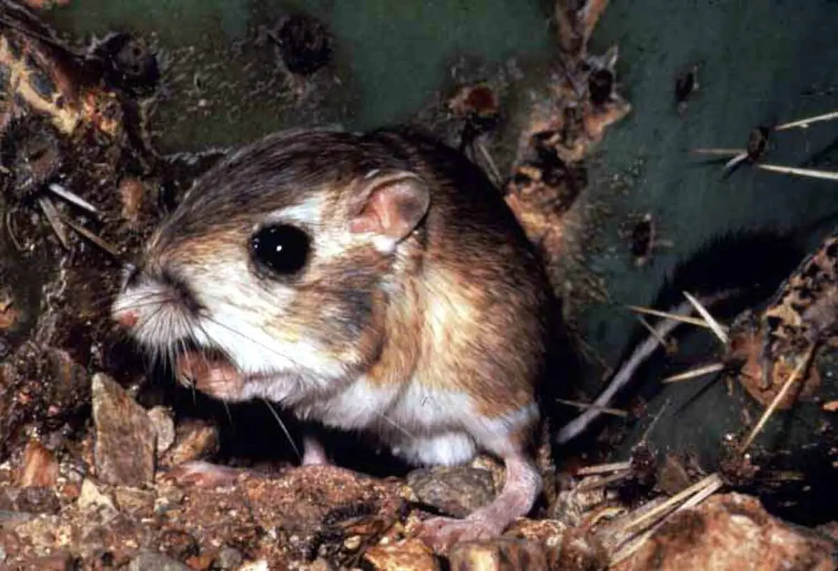
The kangaroo rat of North America’s deserts makes urine far more concentrated than ours. What would you expect to find in its kidneys?
Pick an answer to see why.
Mechanism (physiology) of urine formation
Urine is made in three steps: ultrafiltration in the Malpighian body, selective reabsorption in the tubules, and tubular secretion.
1. Ultrafiltration (glomerular filtration)
Ultrafiltration is a purely physical process: blood is filtered under high pressure in the glomerulus, and water and small dissolved substances of the plasma are pushed out into Bowman’s capsule.
The filtration membrane the fluid must cross has three layers:
- the endothelium of the glomerular capillaries, with its pores;
- the basement membrane under it;
- the visceral layer of Bowman’s capsule, the podocytes, with their filtration slits.
Everything in the plasma passes except blood cells and most proteins, which are too big.
The pressure for filtration comes from the heart, and the kidney keeps it high in the glomerulus because the afferent arteriole is wider than the efferent arteriole: more blood enters the glomerulus than can easily leave it, so blood is squeezed in its capillaries, like water in a hose whose outlet is pinched.
Three pressures act on the fluid:
- Glomerular hydrostatic pressure (the blood pressure in the glomerular capillaries), about 55 mmHg: pushes fluid out into the capsule.
- Colloid osmotic pressure of the blood, about 30 mmHg: the plasma proteins (mainly albumin), which cannot leave, draw water back into the capillaries.
- Capsular hydrostatic pressure, about 15 mmHg: the fluid already in the capsule pushes back against filtration.
In kilopascals: 7.3 − (4.0 + 2.0) = 1.3 kPa. (It can range from about 8 to 15 mmHg.)
This net filtration pressure drives large amounts of water and small dissolved substances out of the blood. The fluid formed, the glomerular (nephric) filtrate, crosses the capillary endothelium, the basement membrane and the slits between the podocytes’ feet into the capsular space. It contains useful substances (glucose, amino acids, vitamins, salts) and wastes (urea, uric acid, creatinine, a little ammonia): it is plasma without its proteins.
The volume of filtrate formed by both kidneys in one minute is the glomerular filtration rate (GFR): about 125 ml/min in a healthy adult, or about 180 liters a day. Since all the blood plasma is only about 3 liters, the kidneys filter it about 60 times a day.
2. Selective reabsorption
As the filtrate flows along the tubule, selected useful substances (and most of the water) are taken back into the blood of the capillaries round the tubule, by active transport (using ATP) or passive transport (diffusion and osmosis).
In the PCT, the main site of reabsorption, about 65–70% of the filtrate is taken back:
- The filtrate entering the PCT is isotonic to the blood plasma.
- About 65% of the Na⁺ is reabsorbed by active transport.
- Glucose, amino acids, water-soluble vitamins, lactic acid and HCO₃⁻ (bicarbonate) are reabsorbed actively; normally all the glucose and amino acids are taken back here.
- About 65–70% of the water follows by osmosis, however much or little the person drinks. This is obligatory reabsorption of water.
- Ions such as K⁺, Cl⁻, Ca²⁺ and Mg²⁺ are reabsorbed, mostly by diffusion.
- About 50% of the urea is reabsorbed by diffusion.
Because water and solutes leave together, the fluid leaving the PCT is still isotonic.
In the loop of Henle:
- The descending limb is very permeable to water but hardly at all to salts. Water moves out into the salty fluid of the medulla (and from there into the vasa recta), so the filtrate becomes more and more hypertonic as it goes down.
- The ascending limb is impermeable to water but lets salts out: Na⁺, K⁺ and Cl⁻ leave, passively in the thin part and by active transport in the thick part. So the filtrate becomes more and more hypotonic (dilute) as it goes up, while the salt pumped out makes the medulla’s tissue fluid very concentrated.
- This salty medulla in turn draws water out of the descending limb and, later, out of the collecting duct. The loop works as a counter-current multiplier, building the saltiness of the medulla step by step towards the papillae.
In the DCT:
- Na⁺ is reabsorbed under the control of the hormone aldosterone (from the adrenal cortex).
- Ca²⁺ is reabsorbed under the control of parathormone (from the parathyroid glands).
- Water is reabsorbed only when antidiuretic hormone (ADH) is present. This reabsorption, which depends on the body’s needs, is facultative reabsorption of water.
- The filtrate entering the DCT is hypotonic; with ADH present, water leaves it and it becomes isotonic by the end.
In the collecting duct:
- More water is reabsorbed, under the control of ADH: when the body is short of water, ADH makes the duct walls permeable, and water is drawn out into the salty medulla, so a small volume of concentrated urine is made. Without ADH (after drinking a lot) the walls stay waterproof and a large volume of dilute urine is passed.
- Na⁺ is reabsorbed under the control of aldosterone.
- Some urea, Cl⁻ and HCO₃⁻ are also reabsorbed. (The urea leaking out of the lower collecting duct adds to the saltiness of the medulla.)
Which of these is not true of the water taken back in the PCT?
Pick an answer to see why.
After a hot day’s work with little to drink, Bishal’s urine is small in volume and dark. Which hormone has been at work?
Pick an answer to see why.
3. Tubular secretion
Tubular secretion is the movement of unwanted substances from the blood (in the capillaries round the tubule, through the tissue fluid) into the filtrate, mostly by active transport. It removes substances that were not filtered enough, and helps control the blood’s pH and potassium level. It happens in the PCT, DCT and collecting duct:
- In the PCT: metabolic wastes such as creatinine and hippuric acid, H⁺ and NH₃ (ammonia), and foreign substances such as drugs (penicillin), dyes and pigments are secreted.
- In the DCT and collecting duct: K⁺, H⁺ and NH₄⁺ are secreted. Secreting H⁺ (and reabsorbing HCO₃⁻) keeps the blood from becoming too acidic.
- Urea diffuses into the thin segment of the loop of Henle from the medulla’s tissue fluid.
Tubular secretion is the main way of making urine in animals whose kidneys have small or no glomeruli. Several marine bony fishes, such as the toadfish, the goosefish and the pipefishes, have no glomeruli at all (they are aglomerular): living in salt water, they must save water, and they make their urine entirely by secretion.
Finally, the urine, made concentrated in the collecting ducts, flows through the ducts of Bellini into the calyces and the renal pelvis, and down the ureter to the bladder.
Urine
Urine is the liquid waste made by the kidneys.
- Composition: about 95% water and 5% dissolved substances: mostly urea, with creatinine, uric acid, salts (electrolytes such as Na⁺, K⁺, Cl⁻), a little ammonia, and broken-down hormones and pigments. The nitrogenous wastes are the largest part of the solutes. Normal urine has no glucose and no protein.
- Volume: about 1.5–2 liters a day for a healthy adult drinking normally.
- Color: clear, pale to deep yellow, from the pigment urochrome (urobilin), made from the breakdown of hemoglobin.
- Smell: fresh urine has a faint, aromatic smell (from small volatile substances). If it stands, bacteria break its urea down into ammonia, which gives the smell of stale urine.
- pH: slightly acidic, about 6, but it can vary from about 4.5 to 8 with the diet (more acidic on a meat-rich diet, more alkaline on a vegetable one).
- Specific gravity: about 1.005–1.030, higher than that of water (1.000) because of its dissolved substances: dilute urine after drinking a lot, concentrated after sweating.
Micturition
Micturition (urination) is the passing of urine out of the body by the contraction of the bladder, through the urethra. It is controlled by a reflex, the micturition reflex, which the brain can allow or hold back.
- While the bladder fills, sympathetic nerves keep the detrusor muscle relaxed and the internal sphincter closed, so urine collects.
- When the bladder holds about 300 ml, stretch receptors in its wall send impulses through sensory nerves to the spinal cord and the brain. You feel the urge to pass urine.
- When the time is right, the micturition center in the pons lets the reflex go ahead. Parasympathetic motor nerves make the detrusor muscle contract and the internal sphincter relax.
- The external sphincter, under voluntary control, is relaxed, and urine is passed out.
A baby passes urine as soon as the bladder fills, by the reflex alone; learning to hold it (“toilet training”) is learning voluntary control of the external sphincter.
Which nerves make the detrusor muscle contract during micturition?
Pick an answer to see why.
Role of other organs in excretion
Besides the kidneys, other organs help remove wastes.
- Skin. It has two kinds of glands.
- Sweat glands make sweat, which removes water, NaCl, a little urea, lactic acid and some amino acids. Sweating is mainly for cooling, so its excretory role is small.
- Sebaceous glands make an oily, wax-like sebum, which keeps the skin and hair soft and carries out some sterols, waxes and other fatty substances.
- Lungs. They remove carbon dioxide and some water vapor, the wastes made when food is oxidized in the tissues (cellular respiration), in every breath out.
- Liver.
- It makes urea from ammonia, which the kidneys then remove.
- It breaks down old red blood cells, and their hemoglobin’s pigments, bilirubin and biliverdin, are sent out in the bile to leave the body with the feces. It also sends out cholesterol, used steroid hormones, some vitamins and many drugs in the bile.
- Intestine. The wall of the large intestine passes excess calcium, iron and some other salts into the gut, to leave with the feces. (The undigested food in the feces, and the bad-smelling indole and skatole made from it by gut bacteria, leave by egestion.)
Exercise
A. Tick the correct alternatives
- Which of the following nitrogenous compounds is the most toxic?
Pick an answer to see why.
- Which of the following animals excretes amino acids without deamination?
Pick an answer to see why.
- A frog’s tadpole is:
Pick an answer to see why.
- Terrestrial insects are:
Pick an answer to see why.
- Which of the following is the position of the human kidney?
Pick an answer to see why.
- Which of the following is not correct about the human kidney?
Pick an answer to see why.
- Into which part do the ducts of Bellini open?
Pick an answer to see why.
- Which part of the nephron is responsible for the obligatory reabsorption of water?
Pick an answer to see why.
- What causes the yellowish color of urine?
Pick an answer to see why.
- Which of the following substances is most completely reabsorbed from the glomerular filtrate during urine formation?
Pick an answer to see why.
- Which of the following is a characteristic of juxtamedullary nephrons compared with cortical nephrons?
Pick an answer to see why.
- The muscular layer of the urinary bladder that contracts during micturition is the:
Pick an answer to see why.
B. Answer the following questions in brief
What is excretion? What are the types of animals on the basis of the nitrogenous wastes they expel?
Answer
Classify the following animals into groups according to their mode of excretion: pigeon, lizard, rohu, shark, earthworm, Limnaea, crocodile, crustacean, woodlouse, bat, cow, human, snake, dragonfly, common carp, tadpole, frog, sparrow, horse, crab, starfish, Unio.
Answer
Draw a neat, labeled diagram of the kidney.
Answer
Describe the structure of the human kidney in brief.
Answer
What is ultrafiltration? In which part of the kidney does it occur? Describe the process in detail.
Answer
What is tubular secretion? Which type of animals benefit most from this process?
Answer
Write a short note on urine. By which process is urine passed out? Describe the process in brief.
Answer
Observe the diagram below and answer the following questions.
Servier Medical Art (smart.servier.com), CC BY 4.0
- i.Label the parts A, B, C and D.
- ii.What are three differences between A and B?
- iii.What are the functions of C and D?
Answer
Observe the diagram below and answer the following questions.
Servier Medical Art (smart.servier.com), CC BY 4.0, arterioles redrawn
- a.Label the parts A, B, C, D, E, F and G.
- b.Write two differences between A and B.
- c.What is the function of C?
- d.What is the significance of G?
- e.Why is the blood pressure in C higher than in ordinary capillaries?
Answer
C. Answer the following questions in detail
Describe the human excretory organs, with a neat, labeled diagram.
Answer
What is the functional unit of the kidney called? How does it form urine? Describe the entire process.
Answer
Describe the structure of a nephron. What are the differences between cortical and juxtamedullary nephrons?
Answer
Printable PDFs
- Study notes 4 pages
- Worksheet 4 pages Answer key
Quiz yourself: Life Processes and the Human Body, Grades 5–12 →
Practice every question in the book, and keep track of the ones you miss →
A companion to your textbook, not a replacement for it: study from the book your school uses, and where the two differ, follow your teacher. Found a mistake? Please tell us.